Ch11 Notes
 Call Kai
 Learn
Learn Practice Test
 Spaced Repetition
 Match
Match Flashcards
 Knowt Play
1/95

There's no tags or description
Looks like no tags are added yet.
Name | Mastery | Learn | Test | Matching | Spaced | Call with Kai | Chat |
|---|
No analytics yet
Send a link to your students to track their progress
96 Terms

Pectoralis minor
Origin = anterior surfaces of ribs 3–5.
Insertion = coracoid process of scapula.
Action = draws scapula forward and downward

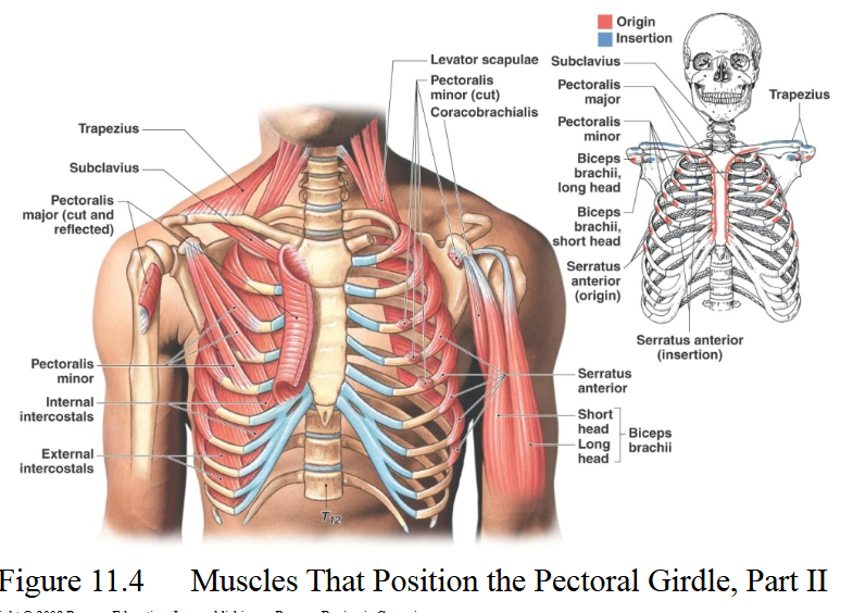
Serratus anterior
Fan-shaped muscle with serrated or sawtooth origins on the lateral rib cage
Origin = superior anterior ribs 1–8 or 9.
Insertion = anterior surface of vertebral border of scapula.
Action = upwardly rotates and protracts scapula and holds it against chest wall


Subclavius
Origin = costal cartilage of rib 1.
Insertion = inferior surface of clavicle.
Action = helps stabilize and depress pectoral girdle


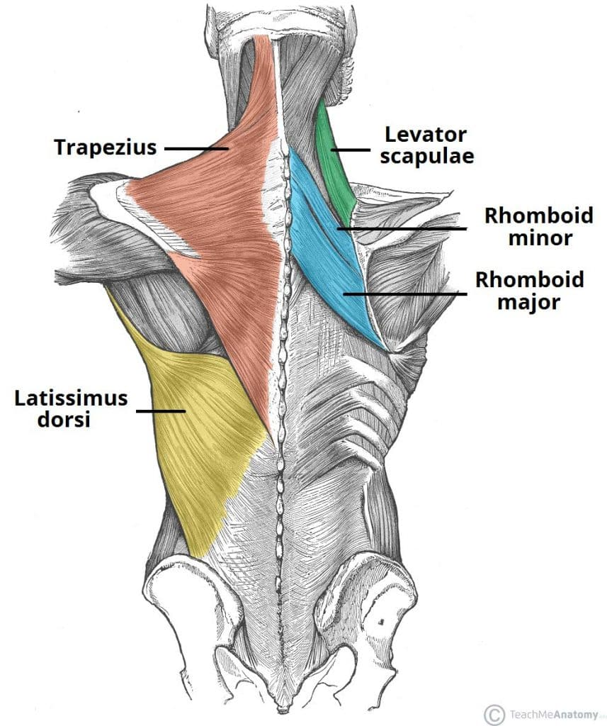
Trapezius
Upper fibers run inferiorly, middle fibers horizontally, and lower fibers superiorly to the scapula
Origin = occipital bone, ligamentum nuchae, and spinous processes of thoracic.
Insertion = clavicle, acromion, and scapular spine.
Action = stabilizes, raises, retracts, depresses, and rotates scapula


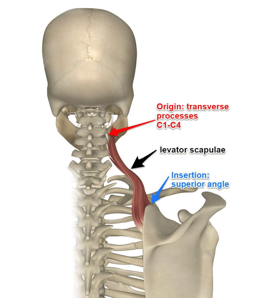
Levator scapulae
Located at back and side of neck deep to trapezius
Origin = transverse processes C1–C4.
Insertion = medial border of scapula near superior boarder.
Action = elevates scapula

Rhomboid minor
Origin = spinous processes C7–T1.
Insertion = medial border of scapula.
Action = stabilizes, adducts, and downwardly rotates scapula
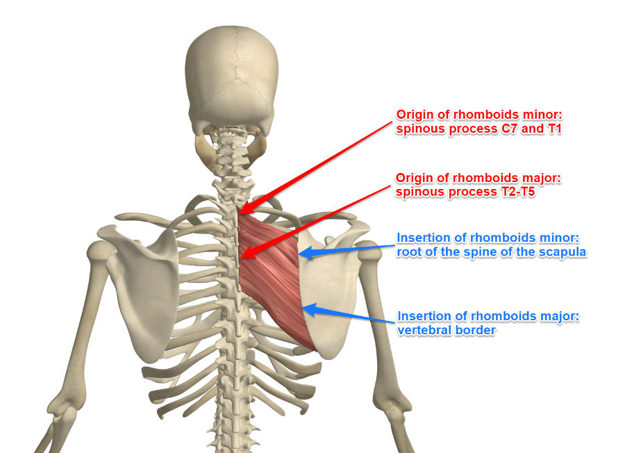

Rhomboid major
Origin = spinous processes of superior thoracic vertebrae.
Insertion = medial border of scapula.
Action = stabilizes, adducts, and downwardly rotates scapula

Rhomboid major and minor location
Both lie deep to trapezius
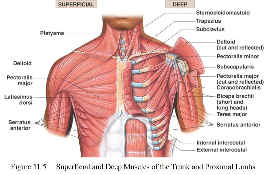
Pectoralis major
Origin = medial clavicle, body of sternum, ribs 2-6.
Insertion = crest of greater tubercle and lateral hip of intertubercular sulcus of humerus.
Action = primary mover of arm flexion. flexion, adduction, and medial rotation at shoulder

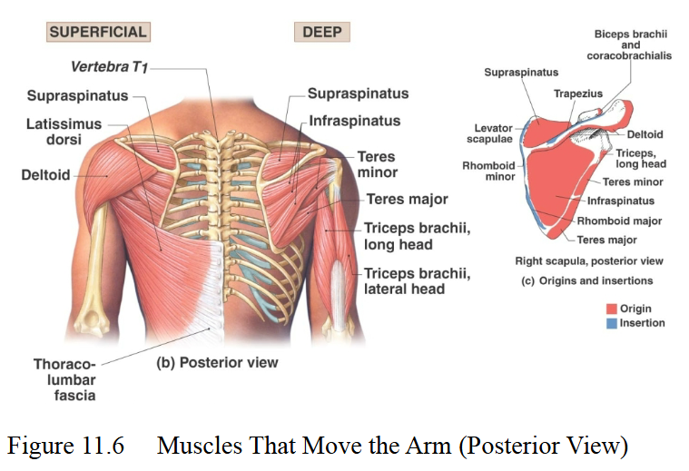
Latissimus dorsi
Origin = inferior thoracic and lumbar vertebrae, ribs 8–12, and thoracolumbar fascia.
Insertion = intertubercular sulcus of humerus.
Action = Prime mover of arm extension and adduction


Deltoid
Origin = clavicle, acromion, and scapular spine.
Insertion = deltoid tuberosity of humerus.
Action = primarily abducts arm
Prime mover of arm abduction when all fibers contract
Note: Deltoid is NOT a rotator cuff muscle
Deltoid fibers are opposites:
Anterior - Flex and medially rotate shoulder
posterior - Extend and laterally rotate sjoulder

Teres major
Origin = inferior angle of scapula.
Insertion = medial lip of intertubercular sulcus of humerus.
Action = extension and medially rotates shoulder


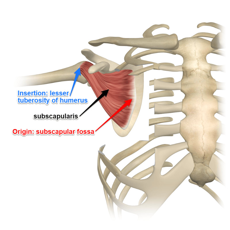
Subscapularis
Origin = subscapular fossa of scapula
Insertion = lesser tubercle of humerus
Action = medially rotates shoulder
Chief medial rotator of the humerus

Supraspinatus
Origin = supraspinous fossa of scapula.
Insertion = superior greater tubercle of humerus.
Action = shoulder abduction
Note: Deltoid is the prime mover while supraspinatus initiates abduction


Infraspinatus
Origin = infraspinous fossa of scapula.
Insertion = greater tubercle of humerus.
Action = laterally rotates shoulder


Teres minor
Origin = lateral border of scapula.
Insertion = greater tubercle of humerus.
Action = laterally rotates and adducts shoulder


Rotator cuff muscles
SITS muscles
Supraspinatus
infraspinatus
teres minor
subscapularis
Stabilizes the head of the humerus in the glenoid cavity during shoulder movement


Coracobrachialis
Origin = coracoid process of scapula.
Insertion = medial margin of shaft.
Action = flexes and adducts humerus
Note: Synergist of pectoralis major
A synergist muscle is a muscle that assists the primary moving muscle (the agonist) during a joint action by adding extra force or stabilizing the surrounding area

Elbow flexors location
Primarily found on the anterior aspect of the arm

Elbow extensors location
Primarily found on the posterior aspect of the arm


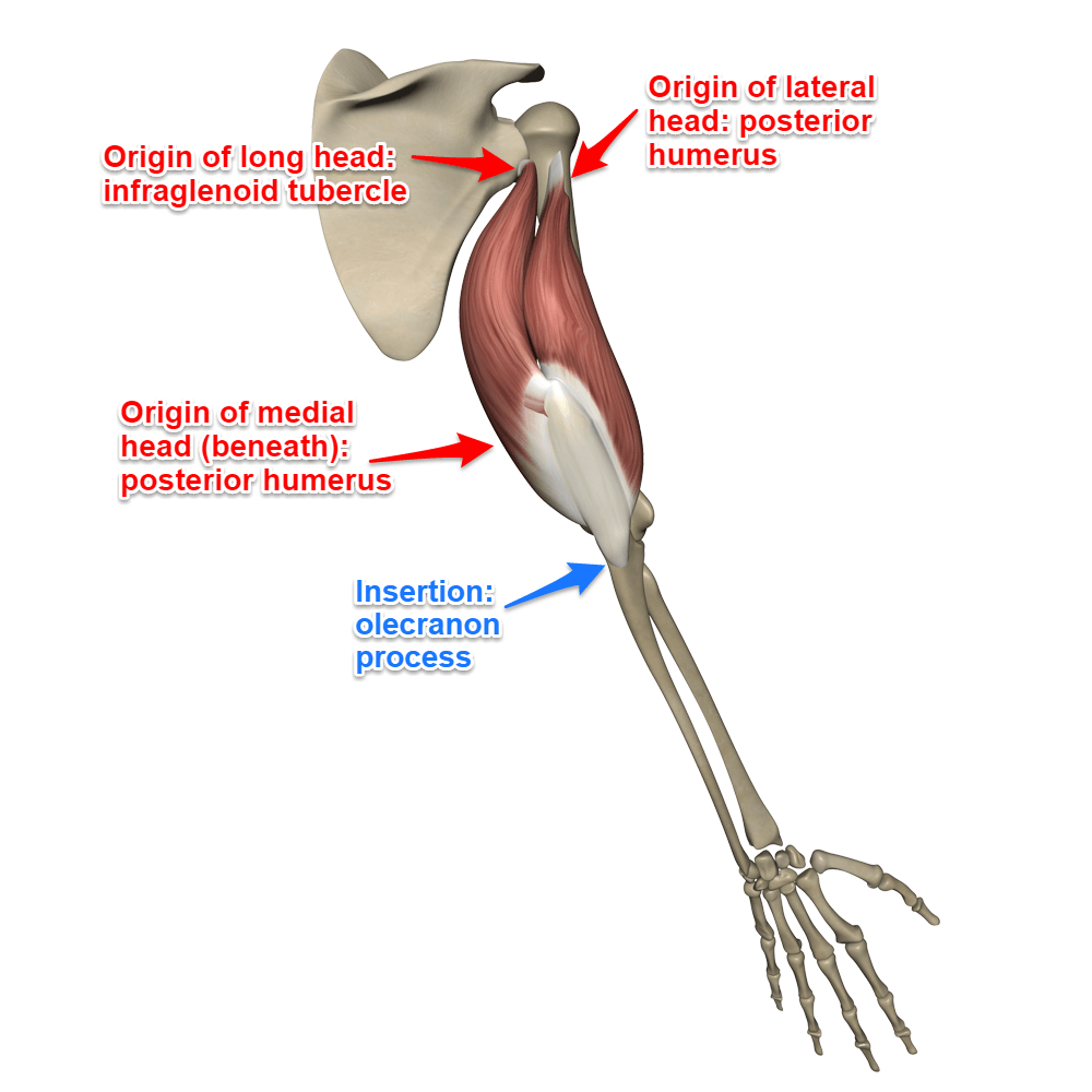
Triceps brachii
Origins:
Long head: infraglenoid tubercle of scapula
Lateral head: superior, lateral margin of humerus
Medial head: posterior surface of humerus, inferior to radial groove
Insertion: Olecranon process of ulna.
Action: Extends forearm at elbow; long head also extends and adducts arm at shoulder.
Note: Prime mover of forearm extension, especially the medial head

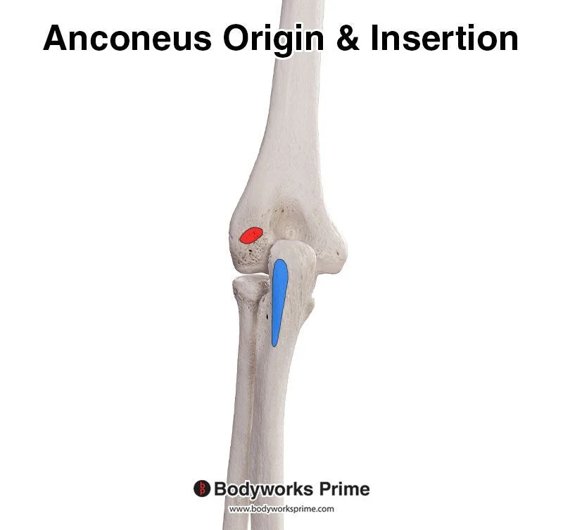
Anconeus
Origin = lateral epicondyle of humerus.
Insertion = lateral olecranon of ulna.
Action = extension at elbow

Biceps brachii
Origin =
short head: coracoid process
Long head: supraglenoid tubercle of scapula
Insertion = radial tuberosity.
Action = flexion at elbow and and supinates forearm
Note: There’s a short and long head of biceps brachii, both have different origins


Brachialis
Origin = anterior distal humerus.
Insertion = ulnar tuberosity.
Action = major forearm flexor


Brachialis vs biceps action
Brachialis lifts the ulna while biceps brachii lifts the radius

Brachioradialis
Superficial muscle on lateral forearm
Origin = lateral supracondylar ridge of humerus.
Insertion = base of styloid process of radius.
Action = flexion at elbow


Pronator teres
Origin = medial epicondyle of humerus and coronoid process of ulna.
Insertion = lateral radius.
Action = pronates forearm and weak elbow flexion


Flexor carpi radialis
Origin = medial epicondyle of humerus.
Insertion = bases of 2nd and 3rd metacarpals.
Action = flexes and abducts wrist


Palmaris longus
Origin = medial epicondyle of humerus.
Insertion = palmar aponeurosis and flexor retinaculum.
Action = flexion at wrist


Flexor carpi ulnaris
Origin = medial epicondyle, olecranon, and posterior ulna.
Insertion = pisiform, hamate, and base of 5th metacarpal.
Action = flexes and adducts wrist

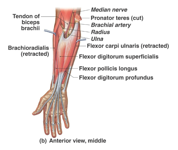
Flexor digitorum superficialis
Origin = medial epicondyle of humerus, coronoid process, and radius.
Insertion = middle phalanges of fingers 2–5.
Action = flexes fingers 2–5 and wrist

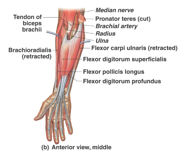
Flexor digitorum profundus
Origin = medial and posterior surfaces of ulna, coronoid process, and interosseous membrane.
Insertion = distal phalanges of fingers 2–5.
Action = flexes distal interphalangeal joints and assists wrist flexion


Extensor digitorum
Origin = lateral epicondyle of humerus.
Insertion = posterior surfaces of phalanges digits 2–5.
Action = extends fingers and wrist


Extensor carpi ulnaris
Origin = lateral epicondyle of humerus and posterior ulna.
Insertion = base of 5th metacarpal.
Action = extends and adducts wrist


Supinator
Origin = lateral epicondyle of humerus and ridge near radial notch
Insertion = proximal radius.
Action = supinates forearm

Shoulder medial rotators
Subscapularis
pectoralis major
latissimus dorsi
teres major
anterior deltoid

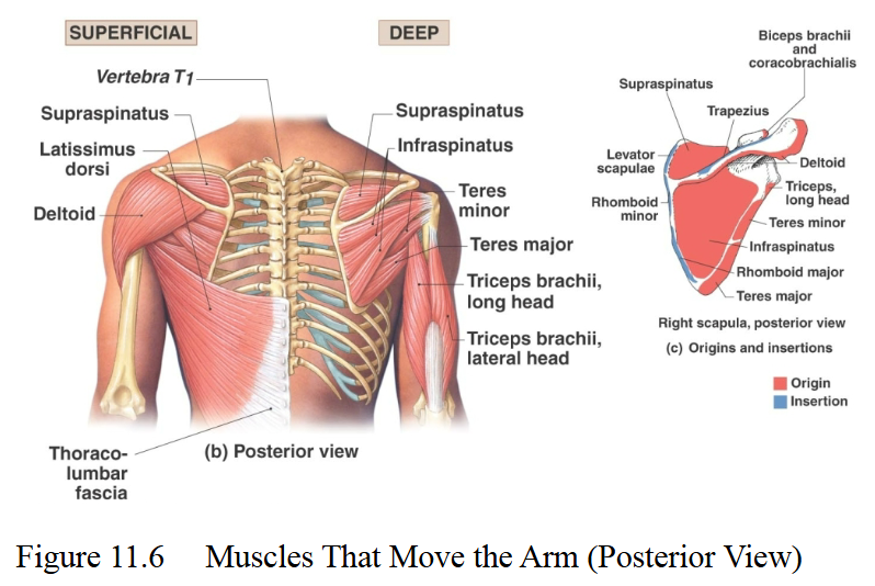
Shoulder lateral rotators
Infraspinatus
teres minor
posterior deltoid

Arm adductors
Pectoralis major
latissimus dorsi
teres major
coracobrachialis
teres minor

Arm flexors
Pectoralis major
anterior deltoid
coracobrachialis
biceps brachii

Arm extensors
Latissimus dorsi
posterior deltoid
teres major
long head of triceps brachii


Forearm pronators
Pronator teres and pronator quadratus

Forearm supinators
Biceps brachii and supinator
Note: Supinator assists biceps brachii during forceful supination and works alone during slow supination


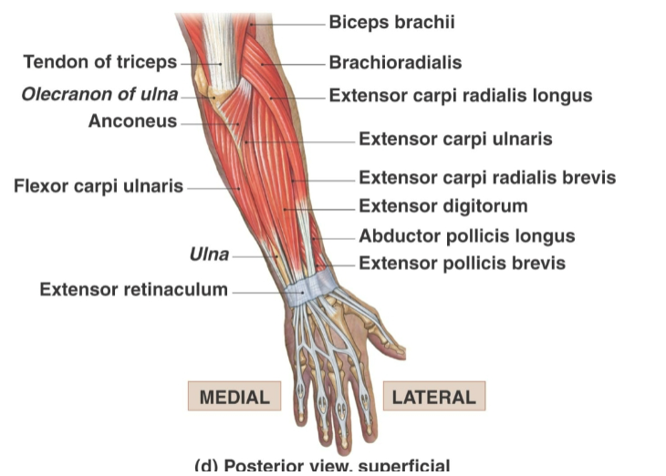
Wrist flexors
Flexor carpi radialis
flexor carpi ulnaris
and palmaris longus

Wrist extensors
Extensor carpi radialis longus
extensor carpi radialis brevis
extensor carpi ulnaris


Wrist abductors
Flexor carpi radialis
extensor carpi radialis: longus and brevis


Wrist adductors
Flexor carpi ulnaris
extensor carpi ulnaris

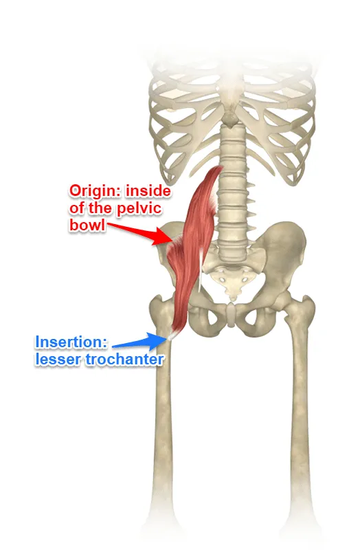
Iliacus
Iliopsoas muscles:
iliacus
Psoas major/minor
both insert through a common iliopsoas tendon on the femur
Origin = iliac fossa.
Insertion = lesser trochanter via iliopsoas tendon.
Action = prime mover of thigh flexion and flexes trunk

Psoas major
Iliopsoas muscles:
iliacus
Psoas major/minor
both insert through a common iliopsoas tendon on the femur
Origin = transverse processes, bodies, and discs of T12–L5.
Insertion = lesser trochanter via iliopsoas tendon.
Action = thigh flexion and flexes trunk


Sartorius
Longest muscle in the body
Sartorius joints cross both the hip and knee joints
helps you sit cross legged!
Origin = anterior superior iliac spine.
Insertion = medial surface of proximal tibia.
Action = flexes, abducts, and laterally rotates thigh and flexes knee.


Medial thigh muscles
Primarily adduct the thigh
Include:
Adductor longus, adductor brevis, and adductor magnus


Adductor longus
Origin = inferior ramus of pubis, near pubic symphysis.
Insertion = linea aspera of femur.
Action = adduction and flexion of hip


Pectineus
shortest of adductors
Origin = pectineal line and superior ramus of pubis. Insertion = line from lesser trochanter to linea aspera. Action = adducts, flexes, and medially rotates thigh
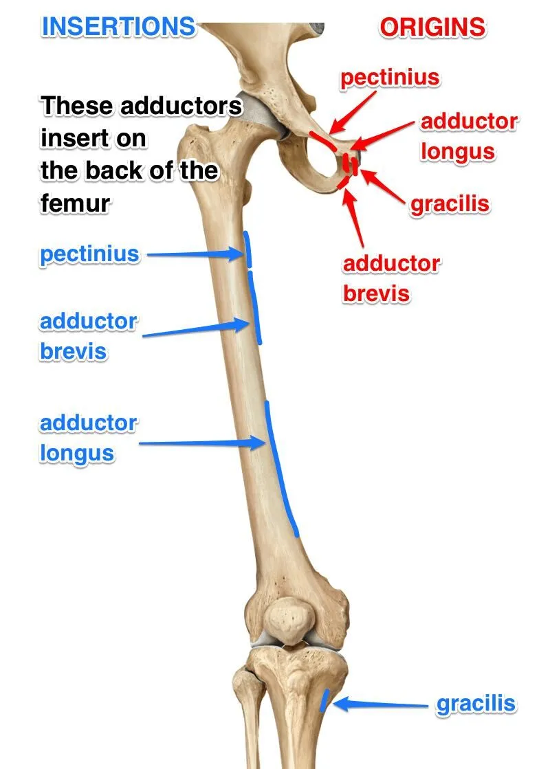

Gracilis
Origin = inferior ramus and body of pubis and adjacent ischial ramus. Insertion = medial tibia just inferior to medial condyle. Action = adducts and medially rotates thigh and flexes and medially rotates leg


Anterior thigh muscles
Flex the femur at the hip and extend the leg at the knee

Quadriceps femoris
Four-headed muscle group on front and sides of thigh
powerful knee extensors and prime movers in knee extension
includes: Rectus femoris, vastus lateralis, vastus medialis, and vastus intermedius

Rectus femoris
Origin = anterior inferior iliac spine and superior acetabular rim.
Insertion = tibial tuberosity via quadriceps tendon, patella, and patellar ligament.
Action = extends knee and flexes hip


Vastus lateralis
Largest head of the quadriceps femoris
Origin = greater trochanter, intertrochanteric line, and linea aspera.
Insertion = same as rectus femoris, tibial tuberosity via quadriceps tendon, patella, and patellar ligament.
Action = extends and stabilizes knee


Vastus medialis
Origin = entire length of linea aspera.
Insertion = same as rectus femoris. tibial tuberosity via quadriceps tendon, patella, and patellar ligament.
Action = extends knee and stabilizes patella


Vastus intermedius
Deep to and obscured by rectus femoris
Origin = anterior and lateral surfaces of proximal femoral shaft.
Insertion = same as rectus femoris. tibial tuberosity via quadriceps tendon, patella, and patellar ligament.
Action = extends knee


Tensor fasciae latae
Origin = anterior iliac crest and anterior superior iliac spine.
Insertion = iliotibial tract.
Action = flexes and abducts thigh, and medially rotates hip.
Steadies trunk on thigh by making iliotibial tract taut
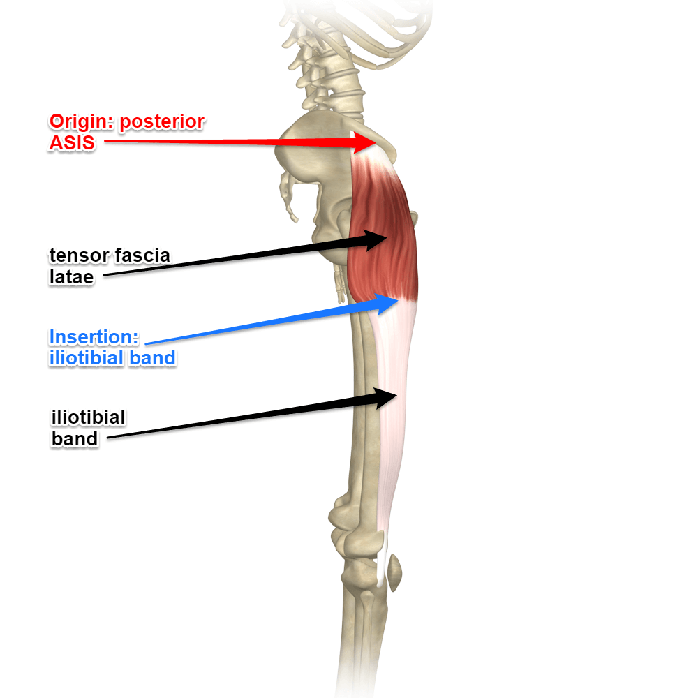

Iliotibial tract (IT band)
Thickened lateral portion of the Tensor fasciae latae

Posterior thigh muscles
Mostly extend the thigh and flex the leg during the backswing phase of walking
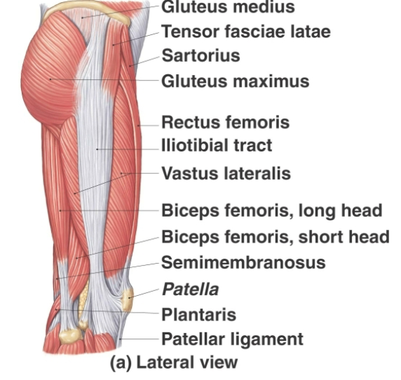
Gluteus medius
Important site for intramuscular injections at the ventral gluteal site
Origin = lateral ilium between anterior and posterior gluteal lines.
Insertion = lateral greater trochanter of femur.
Action = abducts and medially rotates hip

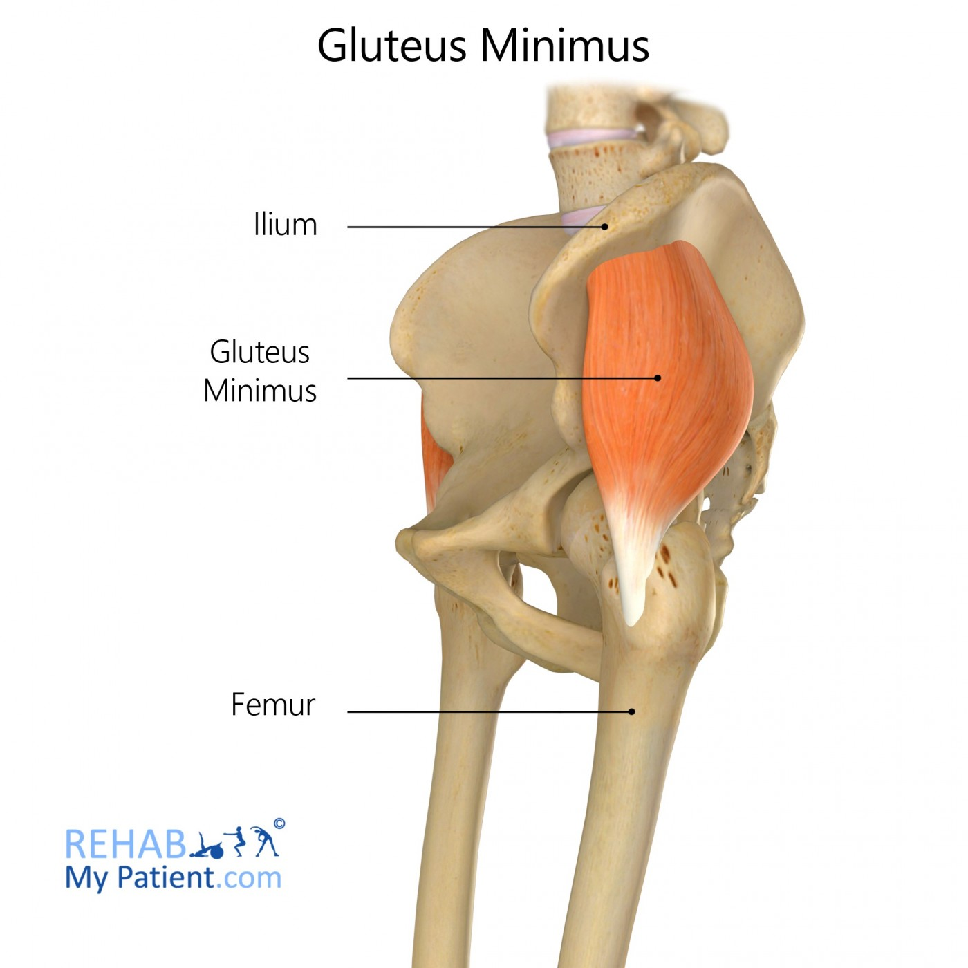
Gluteus minimus
Smallest and deepest of the gluteal muscles
Origin = lateral ilium between inferior and anterior gluteal lines.
Insertion = anterior border of greater trochanter.
Action = abducts and medially rotates thigh


Piriformis
Origin = anterolateral surface of sacrum.
Insertion = superior border of greater trochanter.
Action = laterally rotates extended thigh, abducts hip, and stabilizes hip


Obturator internus
Origin = margins of obturator foramen.
Insertion = medial surface of greater trochanter.
Action = laterally rotates extended thigh, abducts hip, and stabilizes hip


Superior gemellus
Gemelli:
Superior gemellus
Inferior gemellus
small lateral rotators associated with obturator internus
Origin = ischial spine.
Insertion = greater trochanter.
Action = laterally rotates extended thigh, abducts hip, and stabilizes hip


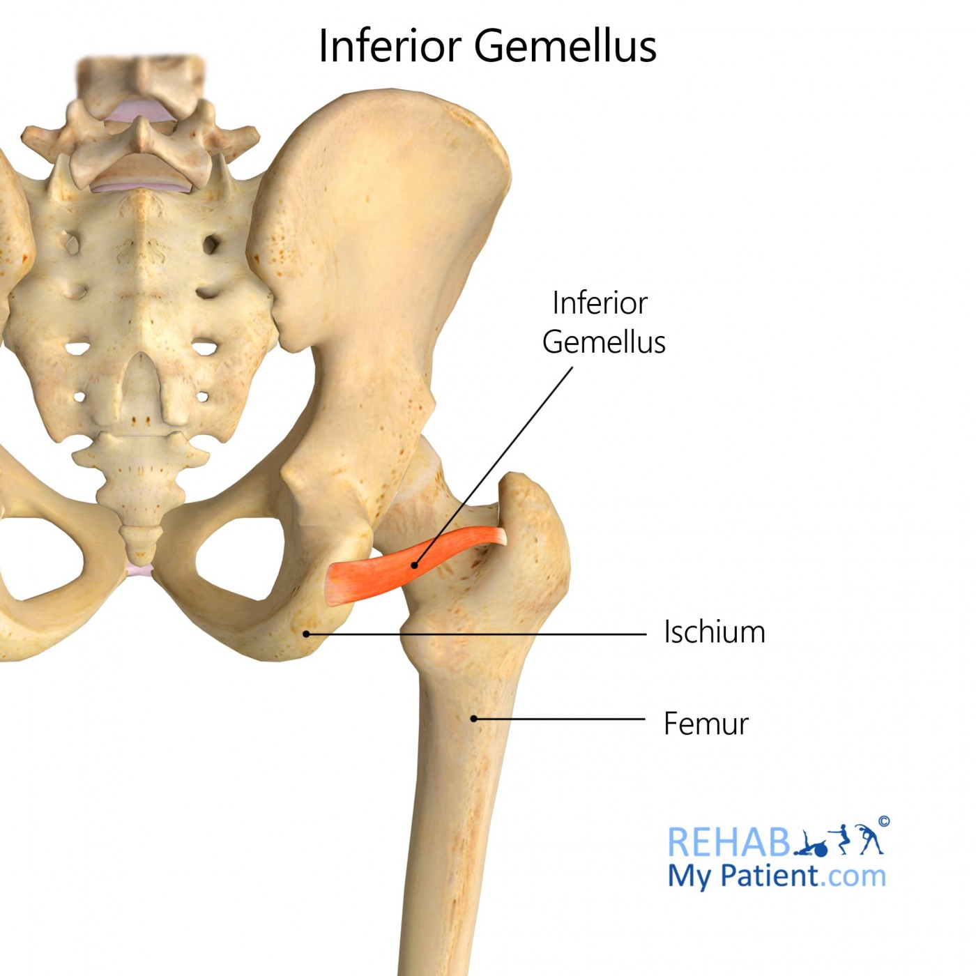
Inferior gemellus
Gemelli:
Superior gemellus
Inferior gemellus
small lateral rotators associated with obturator internus
Origin = ischial tuberosity.
Insertion = greater trochanter.
Action = laterally rotates extended thigh, abducts hip, and stabilizes hip
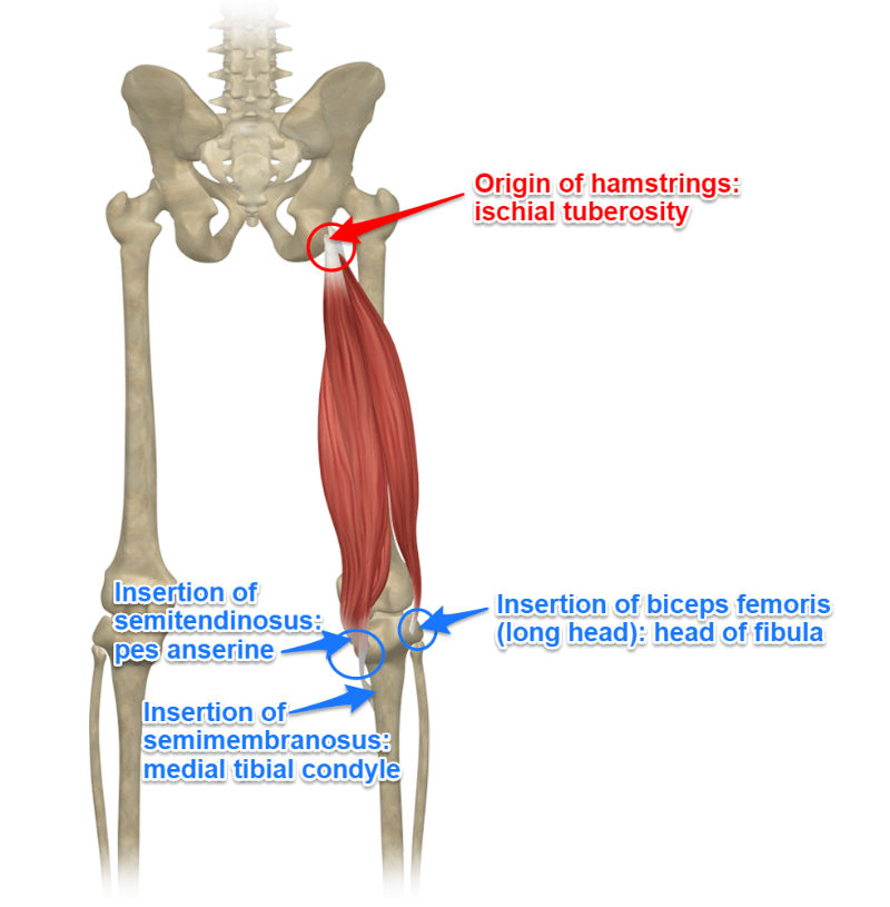
Hamstrings
Posterior compartment of the thigh
Prime movers of knee flexion and thigh extension
Includes:
Biceps femoris, semitendinosus, and semimembranosus

Biceps femoris
Most lateral muscle of the hamstring group
Includes Long head and short head
Origin =
long head: ischial tuberosity
short head: linea aspera/lateral supracondylar line
Insertion = head of fibula and lateral condyle of tibia.
Action = extends thigh, flexes knee, and laterally rotates leg


Semitendinosus
Origin = ischial tuberosity.
Insertion = medial upper tibial shaft.
Action = extends thigh, flexes knee, and medially rotates leg


Semimembranosus
Deep to semitendinosus
Origin = ischial tuberosity.
Insertion = medial condyle of tibia.
Action = extends thigh, flexes knee, and medially rotates leg

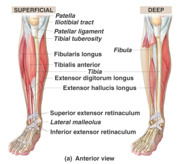
Anterior compartment of leg
Contains extensor muscles that extend toes and dorsiflex the foot
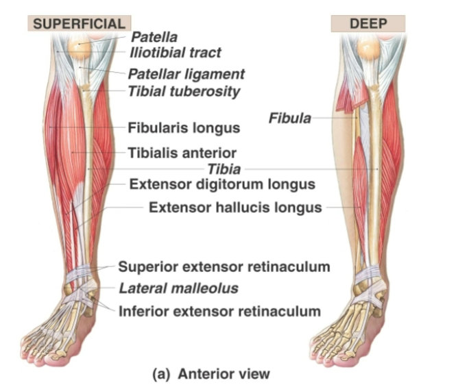
Lower leg compartment arrangement
Anterior lower-leg muscles are extensors
posterior lower-leg muscles are flexors

Tibialis anterior
prime mover in dorsiflexion
Origin = lateral condyle and upper two-thirds of tibial shaft.
Insertion = inferior medial cuneiform and first metatarsal.
Action = dorsiflexes and inverts foot


Extensor digitorum longus
Origin = lateral condyle of tibia and anterior surface of fibula.
Insertion = middle and distal phalanges of toes 2–5.
Action = extends toes 2–5 and dorsiflexes foot


Extensor hallucis longus
Origin = anteromedial fibular shaft.
Insertion = distal phalanx of great toe.
Action = extends great toe and dorsiflexes foot


Lateral compartment of leg function
Plantar flexes and everts foot and helps stabilize lateral ankle and foot arches

Fibularis longus
Origin = head and upper lateral fibula.
Insertion = first metatarsal and medial cuneiform.
Action = plantar flexes and everts foot and supports foot arches


Posterior compartment of leg function
Primarily produces plantar flexion of the ankle

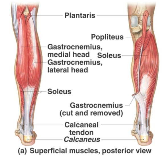
Triceps surae
Triceps surae muscles:
Gastrocnemius
soleus
form the posterior calf and insert through the calcaneal tendon
Prime movers of plantar flexion


Gastrocnemius
Superficial muscle of triceps surae with two prominent bellies forming proximal calf
Origin = medial and lateral condyles of femur.
Insertion = posterior calcaneus via calcaneal tendon.
Action = plantar flexes foot when knee is extended and flexes knee


Soleus
Broad, flat muscle deep to gastrocnemius
Acts as a postural muscle when standing
Origin = superior tibia, fibula, and interosseous membrane.
Insertion = posterior calcaneus via calcaneal tendon.
Action = plantar flexes foot


Plantaris
Origin = posterior femur above lateral condyle.
Insertion = via calcaneal tendon.
Action = assists knee flexion and plantar flexion


Tibialis posterior
Prime mover of foot inversion
Origin = interosseous membrane and adjacent tibia and fibula.
Insertion = navicular, cuneiforms, cuboid, and metatarsals 2–4.
Action = inverts and plantar flexes foot

Reference axis for toe abduction and adduction
Second toe is the reference axis for the foot


Hip flexors
Iliacus
psoas major
sartorius
rectus femoris
pectineus,
tensor fasciae latae
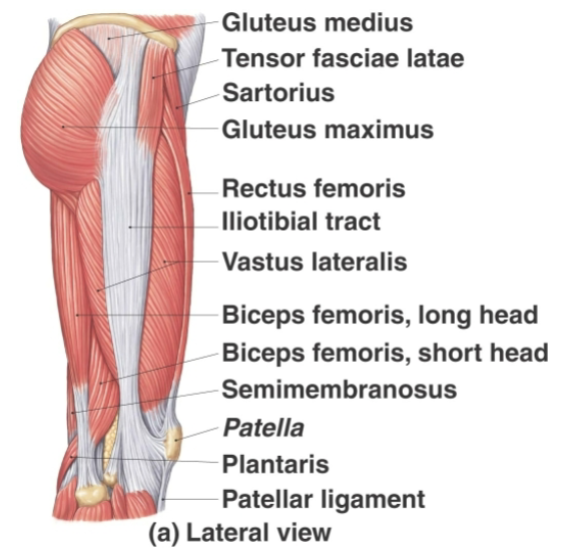
Hip abductors
Gluteus medius
gluteus minimus
tensor fasciae latae
superior fibers of gluteus maximus

Hip adductors
Adductor longus
adductor brevis
adductor magnus
pectineus
gracilis


Medial rotators of thigh
Gluteus medius
gluteus minimus
tensor fasciae latae
adductor longus
adductor brevis
pectineus
gracilis

Lateral rotators of thigh
Gluteus maximus
piriformis
obturator internus
obturator externus
gemelli
quadratus femoris


Knee flexors
Hamstrings
sartorius
gracilis
gastrocnemius
plantaris
popliteus

Foot dorsiflexors
Tibialis anterior
extensor digitorum longus
extensor hallucis longus
fibularis tertius
Foot plantar flexors
Gastrocnemius
soleus
fibularis longus
fibularis brevis
flexor digitorum longus
flexor hallucis longus
tibialis posterior


Foot invertors
Tibialis anterior
tibialis posterior
flexor digitorum longus
flexor hallucis longus

Foot evertors
Fibularis longus
fibularis brevis
fibularis tertius