pathways to defeat infection - week 6

0.0(0)
Studied by 0 people
call kaiCall Kai
learnLearn
examPractice Test
spaced repetitionSpaced Repetition
heart puzzleMatch
flashcardsFlashcards
GameKnowt Play
Card Sorting

1/33

encourage image

There's no tags or description

Looks like no tags are added yet.

34 Terms

1
New cards

where is the pathogen

  • outside cells

    • extracellular pathogens

  • inside cells

    • intracellular pathogens

the location of the pathogen decides which parts of the immune system can reach it


2
New cards

the team

innate immunity

  • Phagocytes: macrophages and neutrophils

  • Antimicrobial defensins

  • Eosinophils

  • Dendritic cells

  • Complement proteins

  • Mast cells

  • Interferons and NK cells


Adaptive immunity

  • B cells and antibodies

  • CD8 cytotoxic T cells

  • CD4 helper T cells: Th1 and Th2


innate acts first, adaptive is slower but specific - and they help each other


key terms:

  • Phagocytes: Cells that engulf and destroy microbes.

  • Defensins: Small antimicrobial peptides that damage microbial membranes.

  • Eosinophils: White blood cells that help defend against parasites.

  • Dendritic cells: Cells that capture antigens and help activate T cells.

  • Complement proteins: Blood proteins that help destroy or mark microbes for removal.

  • Interferons: Proteins that help cells defend against viral infections.

  • NK cells (natural killer cells): Cells that destroy infected or abnormal cells.

  • Cytotoxic: Able to kill cells.

  • Th1 and Th2: Types of CD4 helper T cells that coordinate different immune responses.


3
New cards

two ways to defeat infection

  • for extracellular pathogens

  • for intracellular pathogens


4
New cards

extracellular pathogens

Extracellular pathogens (most bacteria, fungi, parasites, toxins, and viruses before they enter cells) live in blood, tissue fluid and on mucosal surfaces – so soluble molecules and phagocytes can reach them

5
New cards

extracellular pathogens process

  1. recognition

  2. response


6
New cards

phagocytosis

RECOGNITION

Phagocytes engulf and digest bacteria and small particles.


Two main types:

  • Macrophages

    • Live in tissues; engulf and raise the alarm

  • Neutrophils

    • Arrive fast, in large numbers


phagocytes are the clean-up crew


<p>RECOGNITION</p><p>Phagocytes engulf and digest bacteria and small particles.</p><p><br>Two main types:</p><ul><li><p>Macrophages</p><ul><li><p>Live in tissues; engulf and raise the alarm</p></li></ul></li><li><p>Neutrophils</p><ul><li><p>Arrive fast, in large numbers</p></li></ul></li></ul><p></p><p>phagocytes are the clean-up crew</p><p></p>
7
New cards

phagocytosis in action

  1. Phagocytes express a range of pattern recognition
    receptors (PRRs)

  2. A microbe can bind directly to a macrophage via
    these pattern recognition receptors

  3. This activates the macrophage: it releases
    inflammatory mediators (e.g. cytokines) and starts
    phagocytosis


recognizing the microbe switches the phagocyte on - it engulfs and raises the alarm


8
New cards

what is complement?

RECOGNITION

  • ~30 proteins in blood and tissue fluid

  • named for ‘complimenting’ antibodies


Complement proteins acts as:

  • Activation enzymes – act on other complement proteins to activate them

  • Immune defence molecules – make holes in the surface of microbes

  • Control proteins – switch the system on when needed – and off when it is no longer needed


compliment punches holes, tags microbes and calls for help


<p>RECOGNITION</p><ul><li><p>~30 proteins in blood and tissue fluid</p></li><li><p>named for ‘complimenting’ antibodies</p></li></ul><p></p><p>Complement proteins acts as:</p><ul><li><p><span style="font-size: calc(var(--scale-factor)*13.73px);">Activation enzymes – act on other complement proteins to activate them</span></p></li><li><p><span style="font-size: calc(var(--scale-factor)*13.73px);">Immune defence molecules – make holes in the surface </span><span style="font-size: calc(var(--scale-factor)*13.71px);">of microbes</span></p></li><li><p><span style="font-size: calc(var(--scale-factor)*13.73px);">Control proteins – switch the system on when needed – </span><span style="font-size: calc(var(--scale-factor)*13.71px);">and off when it is no longer needed</span></p></li></ul><p></p><p>compliment punches holes, tags microbes and calls for help<br></p><p></p>
9
New cards

B cells vs T cells

RECOGNTION

  • recognition of antigen


B cells

  • ‘direct recognition’

  • Recognise pathogens in their native form

  • Have B cell receptors on the cell surface, allowing them to bind antigens

T cells

  • ‘indirect recognition

  • don’t recognize pathogens in native form

  • pathogen must be processed and presented on MHC class l or class ll


B cells see the whole pathogen; T cells see pieces on MHC


<p>RECOGNTION</p><ul><li><p>recognition of antigen</p></li></ul><p></p><p>B cells</p><ul><li><p>‘direct recognition’</p></li><li><p><span>Recognise pathogens in their native form</span></p></li><li><p><span>Have B cell receptors on the cell surface, allowing them to bind antigens</span><br></p></li></ul><p>T cells</p><ul><li><p>‘indirect recognition</p></li><li><p>don’t recognize pathogens in native form</p></li><li><p>pathogen must be processed and presented on MHC class l or class ll</p></li></ul><p></p><p>B cells see the whole pathogen; T cells see pieces on MHC</p><p></p>
10
New cards

Immunoglobulins: two forms

Membrane-bound antibodies

  • receptors on the surface of the B cell

  • bind foreign antigens encountered by the host


Soluble (secreted) antibodies

  • recreated by plasma cells

  • presented in serum and tissue fluid

  • mediate effector function to neutralise or eliminate antigen


same molecule, two jobs: a receptor on the B cell, or a free antibody in body fluids


<p>Membrane-bound antibodies</p><ul><li><p>receptors on the surface of the B cell</p></li><li><p>bind foreign antigens encountered by the host</p></li></ul><p></p><p>Soluble (secreted) antibodies</p><ul><li><p>recreated by plasma cells</p></li><li><p>presented in serum and tissue fluid</p></li><li><p>mediate effector function to neutralise or eliminate antigen</p></li></ul><p></p><p>same molecule, two jobs: a receptor on the B cell, or a free antibody in body fluids</p><p></p>
11
New cards

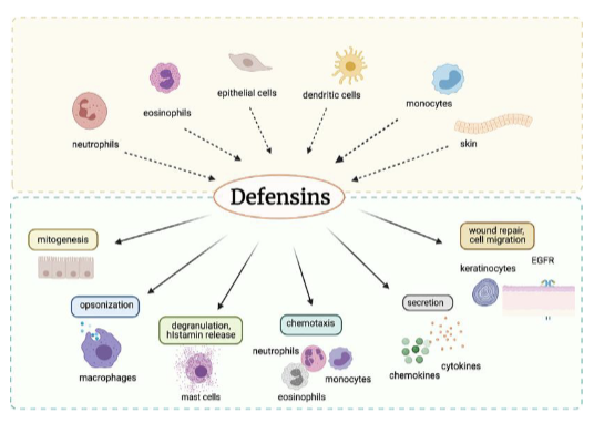
antimicrobial defensins

  • Small cationic peptides that act as host defence peptides

  • Act mainly by disrupting the membrane structure of bacterial cells

  • Active against bacteria, fungi and many enveloped and non- enveloped viruses

  • Immune cells contain these peptides to help kill phagocytosed bacteria

  • Form pore-like defects that let essential ions and nutrients leakout


Defensins are the body’s own antibiotics

<ul><li><p><span style="font-size: calc(var(--scale-factor)*13.73px);">Small cationic peptides that act as host defence peptides</span></p></li><li><p><span style="font-size: calc(var(--scale-factor)*13.73px);">Act mainly by disrupting the membrane structure of bacterial </span><span style="font-size: calc(var(--scale-factor)*13.71px);">cells</span></p></li><li><p><span style="font-size: calc(var(--scale-factor)*13.71px);">Active against bacteria, fungi and many enveloped and non- </span><span style="font-size: calc(var(--scale-factor)*13.73px);">enveloped viruses</span></p></li><li><p><span style="font-size: calc(var(--scale-factor)*13.73px);">Immune cells contain these peptides to help kill phagocytosed </span><span style="font-size: calc(var(--scale-factor)*13.71px);">bacteria</span></p></li><li><p><span style="font-size: calc(var(--scale-factor)*13.71px);">Form pore-like defects that let essential ions and nutrients leak</span><span style="font-size: calc(var(--scale-factor)*13.73px);">out</span></p></li></ul><p></p><p>Defensins are the body’s own antibiotics</p><p></p>
12
New cards

Antibody isotypes: recruiting other defences

Neutralisation (all isotypes, mainly IgG and IgA): antibodies block microbes and toxins from binding host cells

isotype-specific functions:

  • IgM IgG

    • compliment activation (via C1q)

  • IgG IgA

    • phagocyte binding (via FcγR or FcαR)

    • helps macrophages and neutrophils engulf and digest microbes

  • IgE

    • mast cell binding (via FcεR)

    • IgE on mast cells trigger release of inflammatory mediators)


different antibody classes recruit different helpers to remove the pathogen


<p><span style="font-size: calc(var(--scale-factor)*13.73px);">Neutralisation (all isotypes, mainly IgG and IgA): antibodies block microbes and toxins from binding host cells</span></p><p><span style="font-size: calc(var(--scale-factor)*13.73px);">isotype-specific functions:</span></p><ul><li><p>IgM IgG </p><ul><li><p>compliment activation (via C1q)</p></li></ul></li><li><p>IgG IgA</p><ul><li><p>phagocyte binding (via <span>FcγR</span> or <span>FcαR)</span></p></li><li><p><span>helps macrophages and neutrophils engulf and digest microbes</span></p></li></ul></li><li><p>IgE</p><ul><li><p>mast cell binding (via F<span>cεR)</span></p></li><li><p><span>IgE on mast cells trigger release of inflammatory mediators)</span></p></li></ul></li></ul><p></p><p>different antibody classes recruit different helpers to remove the pathogen</p><p></p>
13
New cards

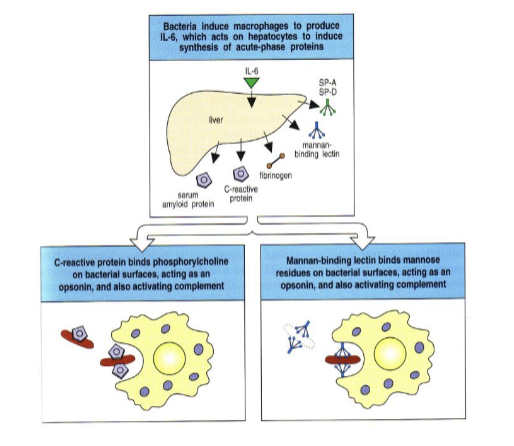
Opsonisation

  • Opsonisation is a way of marking the invading particle for the phagocyte

  • Main opsonins: complement C3b and IgG antibody – phagocytes have receptors for both

  • Acute-phase proteins (e.g. CRP) are another class: during infection, IL-6 triggers their release from the liver

  • Acute-phase proteins then coat the surface of the microbe,
    promoting phagocytosis


opsonization coats a microbe so phagocytes can grab it easily
<ul><li><p><span style="font-size: calc(var(--scale-factor)*13.73px);">Opsonisation is a way of marking the invading particle for the phagocyte</span></p></li><li><p><span style="font-size: calc(var(--scale-factor)*13.73px);">Main opsonins: complement C3b and IgG antibody – phagocytes have receptors for both</span></p></li><li><p><span style="font-size: calc(var(--scale-factor)*13.73px);">Acute-phase proteins (e.g. CRP) are another class: during </span><span style="font-size: calc(var(--scale-factor)*13.71px);">infection, IL-6 triggers their release from the liver</span></p></li><li><p><span style="font-size: calc(var(--scale-factor)*13.73px);">Acute-phase proteins then coat the surface of the microbe,</span><span><br></span><span style="font-size: calc(var(--scale-factor)*13.71px);">promoting phagocytosis</span></p></li></ul><p></p><p>opsonization coats a microbe so phagocytes can grab it easily<br></p>
14
New cards

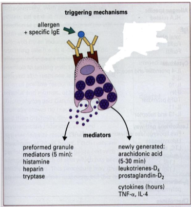
mast cell mediator release

  • Tissue-resident cell (connective tissue, skin, mucosa) with many granules rich in histamine and heparin.

  • High-affinity Fcε receptors (FcεRI) bind IgE

  • Cross-linking: if an antigen binds at least 2 IgE molecules on the mast cell surface, the mast cell is activated

  • C3a and C5a (complement) can also trigger mast cell activation


mast cells are alarm bells packed with histamine
<ul><li><p><span style="font-size: calc(var(--scale-factor)*13.73px);">Tissue-resident cell (connective tissue, skin, mucosa) with many granules rich in histamine and heparin.</span></p></li><li><p><span style="font-size: calc(var(--scale-factor)*13.73px);">High-affinity Fcε receptors (FcεRI) bind IgE</span></p></li><li><p><span style="font-size: calc(var(--scale-factor)*13.73px);">Cross-linking: if an antigen binds at least 2 IgE molecules on the mast cell surface, the mast cell is activated</span></p></li><li><p><span style="font-size: calc(var(--scale-factor)*13.73px);">C3a and C5a (complement) can also trigger mast cell activation</span></p></li></ul><p><br>mast cells are alarm bells packed with histamine</p>
15
New cards

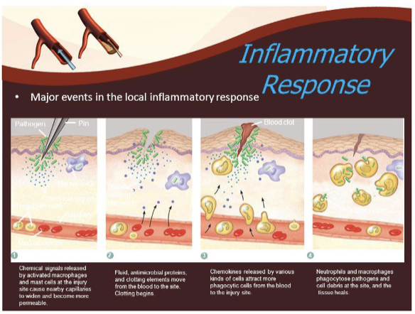
local inflammatory response

Inducers of inflammation (amplifying events):

  • Mast cell products – histamine (immediate release), leukotrienes (delayed release)

  • Prostaglandins – e.g. from macrophages and mast cells

  • Cytokines – e.g. from macrophages and T cells

  • Chemokines – attract leukocytes to the site 1

inflammation brings more blood, fluid and immune cells to the site of infection
<p><span style="font-size: calc(var(--scale-factor)*13.73px);">Inducers of inflammation (amplifying events):</span></p><ul><li><p><span style="font-size: calc(var(--scale-factor)*13.71px);">Mast cell products – histamine (immediate release), leukotrienes (delayed release)</span></p></li><li><p><span style="font-size: calc(var(--scale-factor)*13.73px);">Prostaglandins – e.g. from macrophages and mast cells</span></p></li><li><p><span style="font-size: calc(var(--scale-factor)*13.71px);">Cytokines – e.g. from macrophages and T cells</span></p></li><li><p><span style="font-size: calc(var(--scale-factor)*13.73px);">Chemokines – attract leukocytes to the site </span><span style="font-size: calc(var(--scale-factor)*10.11px);">1</span><br></p></li></ul><p>inflammation brings more blood, fluid and immune cells to the site of infection</p>
16
New cards

eosinophils

  • defence against parasites

  • Parasites such as worms are too large to be phagocytosed.

  • Eosinophils bind antibody-coated parasites via Fc receptors.

  • They release toxic granule proteins onto the parasite surface – extracellular digestion


too big to swallow? attack from outside


<ul><li><p>defence against parasites</p></li><li><p><span style="font-size: calc(var(--scale-factor)*13.71px);">Parasites such as worms are too large to be phagocytosed.</span></p></li><li><p><span style="font-size: calc(var(--scale-factor)*13.71px);">Eosinophils bind antibody-coated parasites via Fc receptors.</span></p></li><li><p><span style="font-size: calc(var(--scale-factor)*13.71px);">They release toxic granule </span><span style="font-size: calc(var(--scale-factor)*13.73px);">proteins onto the parasite surface – extracellular digestion</span></p></li></ul><p></p><p>too big to swallow? attack from outside</p><p></p>
17
New cards

where to T and B cells meet antigen

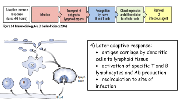
  1. Naive T cells are activated (primed) in secondary lymphoid organs – lymph nodes, spleen, mucosal lymphoid tissue

  2. Dendritic cells carry antigen from the infected tissue to the lymph node

  3. Effector T cells then leave and act in the infected tissue


adaptive responses start in lymph nodes
<ol><li><p><span style="font-size: calc(var(--scale-factor)*13.73px);">Naive T cells are activated (primed) in secondary lymphoid organs – lymph </span><span style="font-size: calc(var(--scale-factor)*13.71px);">nodes, spleen, mucosal lymphoid tissue</span></p></li><li><p><span style="font-size: calc(var(--scale-factor)*13.71px);">Dendritic cells carry antigen from the infected tissue to the lymph node</span></p></li><li><p><span style="font-size: calc(var(--scale-factor)*13.73px);">Effector T cells then leave and act in the infected tissue</span></p></li></ol><p><br>adaptive responses start in lymph nodes</p>
18
New cards

Th2 and help and B cell activation

  1. Pathogen enters the body

  2. Dendritic cell takes it up

  3. Shows it on MHC class II

  4. CD4 T cell becomes Th2 (makes IL-4)

  5. Meanwhile, a B cell binds the same pathogen

  6. Th2 helps the B cell make antibodies


Th2 cells help B cells make antibodies

<ol><li><p><span style="font-size: calc(var(--scale-factor)*15.89px);">Pathogen enters the body</span></p></li><li><p><span style="font-size: calc(var(--scale-factor)*15.89px);">Dendritic cell takes it up</span></p></li><li><p><span style="font-size: calc(var(--scale-factor)*15.89px);">Shows it on MHC class II</span></p></li><li><p><span style="font-size: calc(var(--scale-factor)*15.87px);">CD4 T cell becomes Th2 (makes IL-4)</span></p></li><li><p><span style="font-size: calc(var(--scale-factor)*15.89px);">Meanwhile, a B cell binds the same pathogen</span></p></li><li><p><span style="font-size: calc(var(--scale-factor)*15.89px);">Th2 helps the B cell make antibodies</span></p></li></ol><p></p><p>Th2 cells help B cells make antibodies<br></p>
19
New cards

Intracellular pathogens

Intracellular pathogens (viruses, M. tuberculosis, Listeria) hide inside host cells, out of reach of antibodies and complement – so the immune system must recognise and deal with the infected cell

20
New cards

intracellular pathogens

  • recognition

  • response


21
New cards

two key populations of T cells

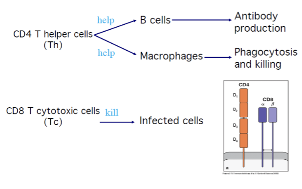
  • The CD4/CD8 co-receptors are very important – they ensure the correct type of T cell is activated.

  • CD4 helper T cells (Th)

    • Help B cells (antibody production) and macrophages (phagocytosis and killing)

  • CD8 cytotoxic T cells (Tc)

    • Kill infected cells

CD4 T cells are the helpers; CD8 T cells are the killers
<ul><li><p><span style="font-size: calc(var(--scale-factor)*13.73px);">The CD4/CD8 co-receptors are very important – they ensure the correct type of T cell is activated.</span></p></li><li><p><span style="font-size: calc(var(--scale-factor)*13.71px);">CD4 helper T cells (Th) </span></p><ul><li><p><span style="font-size: calc(var(--scale-factor)*13.71px);">Help B cells (antibody production) and macrophages (phagocytosis and killing)</span></p></li></ul></li><li><p><span style="font-size: calc(var(--scale-factor)*13.71px);">CD8 cytotoxic T cells (Tc)</span></p><ul><li><p><span style="font-size: calc(var(--scale-factor)*13.73px);">Kill infected cells</span><span><br></span></p></li></ul></li></ul><p>CD4 T cells are the helpers; CD8 T cells are the killers<br></p>
22
New cards

why do T cells need MHC presentation?

  • T cells don’t recognize pathogens in their native form

  • The pathogen must be processed and presented on MHC class I or class II for T cell activation


Why is this important?

  • We don’t want to kill cells that have deliberately taken up a pathogen – they need help

  • We only want to kill a cell if it has an infection in its cytosol

MHC is the cell’s displayed window _ T cells inspect what is shown there

<ul><li><p><span style="font-size: calc(var(--scale-factor)*13.73px);">T cells don’t recognize pathogens in their native form</span></p></li><li><p><span style="font-size: calc(var(--scale-factor)*13.71px);">The pathogen must be processed and presented on MHC class I or class II for T cell activation</span></p></li></ul><p><span><br></span><span style="font-size: calc(var(--scale-factor)*13.73px);">Why is this important?</span></p><ul><li><p><span style="font-size: calc(var(--scale-factor)*13.73px);">We don’t want to kill cells that have deliberately taken up a pathogen – they need help</span></p></li><li><p><span style="font-size: calc(var(--scale-factor)*13.71px);">We only want to kill a cell if it has an infection in its cytosol</span><span><br></span></p></li></ul><p>MHC is the cell’s displayed window _ T cells inspect what is shown there<br></p>
23
New cards

where is the pathogen?

  • Whether the pathogen is in the cytosol or in a vesicle determines whether it is presented on MHC class I or class II

  • This ensures that the right type of T cell is activated

where the pathogen lives decides which MHC - and which cell - responds

<ul><li><p><span style="font-size: calc(var(--scale-factor)*13.71px);">Whether the pathogen is in the cytosol or in a vesicle determines </span><span style="font-size: calc(var(--scale-factor)*13.73px);">whether it is presented on MHC class I or class II</span></p></li><li><p><span style="font-size: calc(var(--scale-factor)*13.73px);">This ensures that the right type of T cell is activated</span><span><br></span></p></li></ul><p>where the pathogen lives decides which MHC - and which  cell - responds<br></p>
24
New cards

MHC class l presents to CD8 T cells

  1. Virus has infected the cytosol of the cell

  2. Virus is degraded and presented on MHC class I

  3. Peptide recognised by the TCR, with CD8 as the co-receptor

  4. CD8 binds directly to the α3 domain of MHC class I

  5. CD8 cytotoxic T cell is activated and can kill the virally infected cell


MHC l + CD8 → kill the infected cell

<ol><li><p><span>Virus has infected the cytosol of the cell</span></p></li><li><p><span>Virus is degraded and presented on MHC class I</span></p></li><li><p><span>Peptide recognised by the TCR, with CD8 as the co-receptor</span></p></li><li><p><span>CD8 binds directly to the α3 domain of MHC class I</span></p></li><li><p><span>CD8 cytotoxic T cell is activated and can kill the virally infected cell</span></p></li></ol><p></p><p>MHC l + CD8 → kill the infected cell</p>
25
New cards

MHC class ll presents to CD4 T cells

  1. Vesicular pathogen – taken into the cell in a vesicle (e.g. M. tuberculosis in a macrophage phagosome)

  2. Pathogen is degraded and presented on MHC class II

  3. Peptide recognised by the TCR, with CD4 as the co-receptor

  4. CD4 binds directly to the β2 domain of MHC class II

  5. CD4 helper T cell is activated and makes cytokines that help the APC kill the pathogen it has ingested


MHC ll + CD4 → help the cell that swallowed the pathogen

<ol><li><p><span>Vesicular pathogen – taken into the cell in a vesicle (e.g. M. tuberculosis in a macrophage phagosome)</span></p></li><li><p><span>Pathogen is degraded and presented on MHC class II</span></p></li><li><p><span>Peptide recognised by the TCR, with CD4 as the co-receptor</span></p></li><li><p><span>CD4 binds directly to the β2 domain of MHC class II</span></p></li><li><p><span>CD4 helper T cell is activated and makes cytokines that help the APC kill the pathogen it has ingested</span></p></li></ol><p></p><p>MHC ll + CD4 → help the cell that swallowed the pathogen</p>
26
New cards

innate defence against intracellular pathogens

T cell responses take days to develop. Meanwhile, the innate system already limits infection inside cells:

Type I interferons (IFN-α, IFN-β)

  • Released by virus-infected cells

  • Make neighbouring cells resist viral replication

  • Increase MHC class I expression

  • Activate NK cells


Natural killer (NK) cells

  • Innate lymphocytes that kill infected cells

  • Detect cells that have lost MHC class I (‘missing self’) or show stress signals

  • Kill using perforin and granzymes – the same mechanism CD8 T cells use

  • Release IFN-γ, which activates macrophages


the innate system holds the line during the days before T cells arrive

27
New cards

T cell activation - putting it together

For a T cell to be activated, the peptide must be processed and presented on MHC class I or II


  • MHC class l + CD8 : ensures CD8 T cells respond to virally infected cells

  • MHC class ll + CD4 : ensures CD4 T cells respond when an APC needs help


Right MHC + right co-receptor + the right T cell responds


<p><span style="font-size: calc(var(--scale-factor)*15.17px);">For a T cell to be activated, the peptide must be processed and presented on MHC class I or II</span></p><p></p><ul><li><p>MHC class l + CD8 : ensures CD8 T cells respond to virally infected cells</p></li><li><p>MHC class ll + CD4 : ensures CD4 T cells respond when an APC needs help</p></li></ul><p></p><p>Right MHC + right co-receptor + the right T cell responds</p><p></p>
28
New cards

T cell activation - the three signals

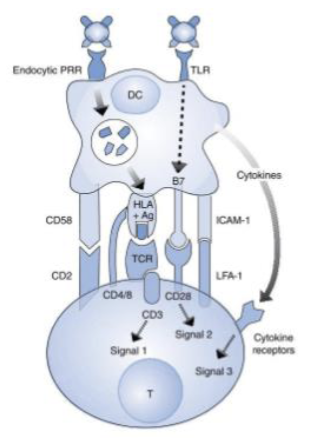
Signal 1

  • Recognition

    • The TCR recognises its specific peptide presented on MHC

Signal 2

  • Co-stimulation

    • CD28 on the T cell binds B7 (CD80/CD86) on the antigen- presenting cell

Signal 3

  • Polarising cytokines

    • Decide what type of T cell develops


No co-stimulation, no activation - a built-in safety check
<p><span>Signal 1 </span></p><ul><li><p><span>Recognition</span></p><ul><li><p><span>The TCR recognises its specific peptide presented on MHC</span></p></li></ul></li></ul><p>Signal 2</p><ul><li><p>Co-stimulation</p><ul><li><p>CD28 on the T cell binds B7 (CD80/CD86) on the antigen- presenting cell</p></li></ul></li></ul><p>Signal 3 </p><ul><li><p>Polarising cytokines</p><ul><li><p>Decide what type of T cell develops</p></li></ul></li></ul><p></p><p>No co-stimulation, no activation - a built-in safety check</p>
29
New cards

helper T cell subsets and cytokines

  • Th0 = naive CD$ T cell

  • signal 3 cytokines decide what it becomes


Th1

  • activates macrophages


Th2

  • helps B cells


cytokines decide what a helper T cell becomes


<ul><li><p>Th0 = naive CD$ T cell</p></li><li><p>signal 3 cytokines decide what it becomes</p></li></ul><p></p><p>Th1</p><ul><li><p>activates macrophages</p></li></ul><p></p><p>Th2</p><ul><li><p>helps B cells</p></li></ul><p></p><p>cytokines decide what a helper T cell becomes</p><p></p>
30
New cards

helper T cell subsets - Th1, Th2, Th17, and Treg

Th1 - intracellular

Th2 - parasites

Th17 - bacteria & fungi

treg - calms

<p>Th1 - intracellular</p><p>Th2 - parasites</p><p>Th17 - bacteria &amp; fungi</p><p>treg - calms</p>
31
New cards

CD8 cytoxic T cells

  • killing infected cells


  1. Naive CD8 T cells are activated by dendritic cells in the lymph node
    and proliferate

  2. Effector CD8 T cells travel to the infected tissue

  3. They recognise viral peptide on MHC class I and kill the infected cell by apoptosis (perforin and granzymes)


CD8 T cells make infected cells self-destruct - taking the virus down with them

<ul><li><p>killing infected cells</p></li></ul><p></p><ol><li><p><span>Naive CD8 T cells are activated by dendritic cells in the lymph node<br>and proliferate</span></p></li><li><p><span>Effector CD8 T cells travel to the infected tissue</span></p></li><li><p><span>They recognise viral peptide on MHC class I and kill the infected cell by apoptosis (perforin and granzymes)</span></p></li></ol><p></p><p>CD8 T cells make infected cells self-destruct - taking the virus down with them</p>
32
New cards

Th1 cells

Th1 cells activate macrophages so they can kill the microbes they
have ingested


two activating signals:

  • IFN-γ (cytokine)

  • CD40L (cell contact)

Result: activated macrophages fuse phagosomes with lysosomes and make more reactive oxygen and nitrogen species, killing intracellular bacteria

Th1 cells switch macrophages into killing mode

<p><span style="font-size: calc(var(--scale-factor)*15.17px);">Th1 cells activate macrophages so they can kill the microbes they</span><br><span style="font-size: calc(var(--scale-factor)*15.17px);">have ingested</span></p><p></p><p><span style="font-size: calc(var(--scale-factor)*15.17px);">two activating signals:</span></p><ul><li><p><span style="font-size: calc(var(--scale-factor)*18.05px);">IFN-γ (cytokine)</span></p></li><li><p><span style="font-size: calc(var(--scale-factor)*18.05px);">CD40L (cell contact)</span><br></p></li></ul><p><span style="font-size: calc(var(--scale-factor)*13.71px);">Result: activated macrophages fuse phagosomes with lysosomes and make more reactive oxygen and nitrogen species, killing </span><span style="font-size: calc(var(--scale-factor)*13.73px);">intracellular bacteria</span><span><br></span></p><p>Th1 cells switch macrophages into killing mode<br></p>
33
New cards

two pathways one team

Extracellular

where it lives: blood, tissue fluid, mucosa

Recognized by: PRRs, compliment, B cells

Main weapons: Antibodies, complete, phagocytes

Helper T cell: Th2 → B cells

Example: Streptococcus pneumoniae

Intracellular

where it lives: cytosol or vesicles

Recognized by: T cells via MHC l or ll

Main weapons: Interferins, NK cells, CD8 T cells

Helper T cell: th1 → macrophages

Example: Mycobacterium tuberculosis

34
New cards

overview

Location decides the strategy
Outside: antibodies & phagocytes.

Inside: target the infected cell


Innate first, adaptive next
Innate acts in hours; B and T cells take days but are specific.


The two arms work together
Antibodies help phagocytes; T cells help B cells and macrophages.

It matters clinically
Vaccines build antibodies; immune gaps open the door to infection.



