Unit 7 Cardiovascular/Hematology
 Call Kai
 Learn
 Practice Test
 Spaced Repetition
 Match
 Flashcards
 Knowt Play
1/128

There's no tags or description
Looks like no tags are added yet.
Name | Mastery | Learn | Test | Matching | Spaced | Call with Kai | Chat |
|---|
No analytics yet
Send a link to your students to track their progress
129 Terms
Cardiac Structures and Vessels
4 chambers
-right/ left atrium, right/left ventricle
Cardiac layers
-epicardium,myocardium, endocardium
Coronary artieries
Valves
-tricuspid,pulmonic, mitral, aortic
Flow of blood through heart
1- Superior and inferior vena cava
2- right atrium
3- tricuspid valve
4- right ventricle
5- pulmonic valve
6- pulmonary artery
7- lungs
8- pulmonary artery
9- left atrium
10- mitral valve
11- left ventricle
12- aortic valve
13- aorta
14- body

Cardiac conduction
Sinoatrial node
Atrioventricular node
Bundle of His
Right and left bundle branches
Purkinje Fibers
What is cardiac output
-amount of blood ejected from the left ventricle in 1 min
- stroke volume x heart rate= cardiac output
Hormones and the Heart
Epinephrine increases
-HR
-force of contraction
-cardiac output
-systolic blood pressure
Aldosterone
-regulates sodium and potassium
Atrial natriuretic peptide
-excretes sodium
Components of blood vessels
Arteries
Veins
Capillaries
Pathways of circulation
Pulmonary
-Right ventricle, lungs, left atrium
Systemic
-L ventricle, aorta, blood goes to body
Aging and the Cardiovascular system
-conduction less effective
-atherosclerosis
-resting blood pressure increases
-decreased heart rate
-vein valves incompetent
what to inspect during cardiovascular exam
-oxygenation, skin color
-extemities: hair, skin, nails, edema, color
-jugular vein distention
-capillary refill
-clubbing (diamond shape is normal)
what to palpate during cardiovascular exam
-point of maximum impulse
-extremity temperature
-edema
what to auscultate during cardiovascular exam
-heart sounds (S1 and S2)
-murmurs
-pericardial friction rub
lab tests for cardiovascular system
-Cardiac biom markers
-Lipids
-Electrolytes
-BNP
-PT/INR/aPTT
-D dimer
Diagnostic testing
-chest radiograph
-CT scan
-MRI and angiography
-stress test
Information for Electrocardiogram (ECG)
-records cardiac electrical activity
-12,15,18 leads
Signal averaged ECG
-risk for ventricular arrhythmias
Ambulatory ECG monitoring
-holter monitoring
Information about Echocardiogram
-ultrasound
-records motion: heart structures, valves, heart shape size and position
types
-strain
-stress
-transsesophageal
Information regarding echocardiogram
-throat anesthetized
-probe placed esophagus
-clearer images then echo
-as needed until gag reflex returns
Therapuetic interventions
-Lifestyle and cardiac care
-antiembolism devices
-oxygen
-medications
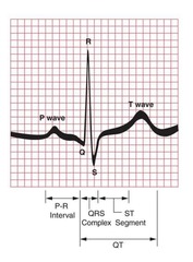
Components of cardiac cycle
P wave: electrical pulse starting in the SA node
P-R interval (Q): traveling to AV node
QRS: 3rd-5th steps are being fired up
T wave: ventricles relaxing, filling with blood
Information about Sinus Bradycardia
s/s
-usually none (fatigue,fainting, SOB)
treatment
-none if asymptomatic
-oxygen, atropine, dopamine or epinephrine
-pacing (pace maker)
-fix electrolyte imbalance
Information about Sinus Tachycardia
s/s
-none (angina or dyspnea, may be palpations)
-older adults are more sensitive
Interventions
-treat cause
-meds: adenosine, beta blockers, calcium channel blockers)
What is Atrial fibrillation
- Overloading of SA node, causes ventricles to respond quivering/twitch
Rhythm: irregular
HR: not measurable
P waves: non identifiable
Information about Atrial Fibrillation
s/s
-palpatations
-faint radial pulse
Purpose of interventions
-correct sinus rythm
-prevent blood clots
-control rate
Therapuetic interventions for Atrial Fibrillation
-synchronized cardioversion if unstable
-medications
-anticoagulation
-ablation (after)
-surgical maze procedure
What is asystole
Rhythm: none
Heart rate: none
P waves: none
PR interval: none
QRS: none
Signs and symptoms of Asystole
-unconscious
-no heart sounds, peripheral pulses, blood pressure
-respiratory arrest
-cyanosis
-pupil dilation
Asystole Therapuetic Interventions
-CPR
-ACLs protocols (intubation with oxygen)
-Treat cause
Information about Cardiac Pacemakers
Generate an electrical pulse
Traditional types:
-external and temporary
-internal and permanent
Leads placed in atria, ventricle or both
-spike on ECG begins paced beat
Leadless pacemaker
Pacemaker ECG Tracings
A. Atrial pacemaker (spike before P wave)
B. Ventricular only pacemaker (spike before QRS)
C. Dual chamber pacemaker (spike before P and QRS)

Cardiac pacemaker problems
-failure to sense
-failure to pace
-failure to capture
Nursing care for Pacemaker
-Monitor ECG
-monitor apical pulse
-monitor for symptoms
note: monitor enzymes for any cardiac procedure
Pacemaker education
-incision care
-activity restrictions
-symptoms of infection signs to report
(note: monitor for hiccups, could be problem with leads)
-ID card
-periodic pacemaker checks
Safety Considerations for Pacemakers
-microwaves are safe and can be used
-security devices safe but can trigger (avoid wands)
-Revo MRI Surecan Pacing System safe
-Move away if become symptomatic
Electromagnetic devices to avoid
-MRI
-welder
-radio tower
-touching running auto engine
What is defibrillation
Electrical shock to reset lethal ventricular arrhythmias
-pulseless VT
- V fib
Paddles placed on chest
-2nd intercostal space, right of sternum
-anterior axillary line, 5th intercostal space
Announce clear for safety, dont be touching patient
What is Automatic External Defibrillator (AED)
-automatically analyzes rhythms
-automatically delivers or prompts users to deliver electrical shock
(if shockable rhythm V fib or VT is detected)
Information about Implantable Cardioverter Fibrillator
-automatically defibrillates when lethal rhythm detected
-cardioverts when VT detected
-increased risk for falling (usually in person with pacemaker)
What is Synchronized Cardioversion
Allows R wave to be sensed for safe delivert
Used in arrhythmias if R wave present
-VT with pulse
- Afib, flutter, supraventricular tachycardia
Painful
-sedation used
Low energy
-25-50 joules
Definition of blood pressure
-pressure exerted by blood on walls of blood vessels
Determined by
-cardiac output
-peripheral vascular resistance
-vessel stretch
-blood viscosity
-blood volume
Blood pressure ranges
Normal: less then 120/80
Elevated: 120-129/80s
Hyper tension
Stage 1: 130-139/80-89
Stage 2: more then 140/90
Types of Hypertension
Primary hypertension
-unknown cause
Secondary hypertension
-known cause (medications, CKD, heart issues)
S/S of hypertension
None "silent killer"
Rare: headache, blood nose, aniexty, dyspnea
Diagnosis of Hypertension
-risk factor presence
-signs and symptoms
-kidney/heart failure
-BP readings
High potassium foods
-bananas
-orange juice
-dark leafy greens
-sweet potatoes
-chocolate
-milk
-french fries and potato chips
Complications of Hypertension
-atherosclerosis
-coronary artery disease
-eye damage
-kidney damage
-heart failure
-myocardial infarction
-stroke
What is hypertensive urgency
-severe BP elevation w/o target organ dysfunction progression
-severe headaches, nosebleeds, SOB and severe anxiety
-ambulatory treatment with oral medications
What does Hypertensive Emergency look like
-Systolic BP higher then 180
-Diastolic BP higher then 120
-risk for progression of target organ dysfunction
Treatment: IV nitroprusside
Patient Safety for Hypertension
- medication has possible side effects of hypotension
-dizziness may increase risk of falling
-rise slowly to decrease risk of orthostatic hypotension
-hydration balance is important
Information about Cardiovascular disease
-leading cause of death in US
-affects quality of life and ADLs
-unmodifiable/modifiable risk factors lead to CVD
-education is important
What is Coronary Artery Disease (CAD)
-obstruction of coronary artery blood flow
-usually from atherosclerosis
Blood flow reduction causes
-angina
-MIs
-sudden death
Prevention of CAD
-stop smoking
- low LDL
-lipid lowering agents
-Normal BP and blood sugars
-low dose aspirin as prescribed
Information about Angina Pectoris
-chest pain (CP)
-symptom of ischemia (inadequate blood flow)
Causes
-CAD
-vasospasm
-valvular heart disease
-hypertension
-heart failure
Angina Pectoris Pathophysiology
-narrowed blood vessels unable to dilate
-carry less blood/oxygen for heart muscle
-less oxygen causes myocardial ischemia which then causes pain
3 types of angina
-Stable angina
-Unstable angina
-Variant angina
Information of Stable Angina
-occurs with moderate exertion in familiar pattern
-usually stops with rest, Sl nitroglycerin
-pain is predictable
Information about Unstable Angina
-pain in unpredictable
-occurs with less exertion (at rest, during sleep)
-unrelieved with rest/ SL nitroglycerin
-can lead to MI
Information about Variant Angina (Prinzmetal)
-due to coronary artery spasm
-longer duration, pain is more severe
-can occur at rest
-often at same time each day (usually at night)
-serious condition
S/S of Angina
Pain
-heaviness,tightness,crushing pain in chest center
-pain may radiate to arms, shoulder,neck,jaw or back
Pale
Diaphoretic
Dyspnea
Angina Diagnostic Tests
-ECG, Stress test ECG
-echocardiography
-chemical stress testing
-CT,MRI
-Radioisotope imaging
-Coronary angiography
Anti-Anginal Medications and Therapies
-Vasodilators
-Beta blockers
-Calcium channel blockers
-Antiplatelets
-Statins
Nursing Care for CAD and Angina
-ensure IV access
-rate pain
-administer oxygen
-administer NTG, Aspirin, and analgesics
-remain with pt and provide emotional support
Acute Coronary Syndrome
Unstable angina
-can lead to MI
MI
-Non Stemi
-Stemi
Information about Silent Ischemia
MI without chest pain
-occurs in older patients, women
-often seen with hypertension and diabetes
Information about Myocardial Infarction
-Death of heart muscle
-Pathophysiology: coronary artery blockage reduces blood supply
Types
-Non stemi: partial blockage
-Stemi: complete blockage
S/S of Myocardial Infarction
-crushing, viselike pain (radiates)
-diaphoresis
-dizziness, fainting
-dyspnea
-nausea
-restlessness
Women and Myocardial Infarction
-CAD/MI leading cause of death in women
-African Americans more at risk
-Higher mortality rate and complications then men
-Prodromal symptoms the month before MI
Myocardial Infarction Atypical S/S for women and older adults
-absense of classic pain
-dyspnea
-fatigue
-anxiety, restlessness
-chest cramping
-epigastic or abdominal pain
-falling
Diagnostic testing for Myocardial Infarctions
-Serial ECG
- highly sensitive cardiac troponin (cTnT)
-myoglobin
-creatine kinase (CK)-MB
-CRP
-magnesium nd potassium
Myocardial Infarction Therapeutic Intervention Information
Goal: restore cardiac perfusion within 90 min
PCI
-ballon angioplasty, coronary artery stents
Thrombolytics
-time sensitive, administer 1-6 of symptom onset
Myocardial Revascularization
Coronary artery bypass graft (CABG)
-cornary artery occlusions bypassed with vein/artery graft
-increases blood flow/oxygen to myocardium
Myocardial Infarction Medications
-oxygen
-antiplatelets
-nitrates
-morphine sulfate
-vasodialators
-beta blockers
-antiarrthymics
Myocardial Infarction Therapuetic interventions
-bedrest/commode
-daily weight
-low sodium, LDL and fat diet
-weight loss
Nursing Care for Myocardial Infarction
-monitor vitals
-monitor for arrhythmias
-report symptoms
-provide incisional care
-monitor for depression
Patient education for MIs
-disease information
-medications
-diet
-activity and exercise
-stress reduction
-rehabilitation
Venous Thromboembolism Disease (VTE) information
DVT- blood clot develops in deep veins
PE- part of DVT clot breaks off and travels to lungs
Thrombophlebitis
-most common disorder
-clots forms and inflammation follows
-can be superficial or deep
VTE etiology
-made up of blood products, such as platelets, RBCs, WBCs and fibrin
-attaches to wall of blood vessel causing vessel to shrink in diameter
-three factors involved in formation
VTE Prevention
-identify risk factors
-maintain mobility
-antiembolism devices
-medications
-follow IV therapy protocol
VTE S/S
Superficial
-redness, warmth, swelling, tender, induration
-common sites are legs due to varicose veins or arms due to IV therapy
Deep
-swelling, pain, warmth, edema, venous distention
-cyanosis
-elevated temp
some have no S/S
VTE complications
-PE
-DVT
-Chronic venous insufficiency
VTE diagnostics
-D dimer (blood test for possible clots, doesn't give location)
-Ultrasound
-Venography
-CT/MRI
VTE Nursing Interventions
Superficial
-WMP
-analgesics, NSAIDS
-compression devices
-elevation
Deep
-WMP
-anticoagulants
-compression devices
-elevation
-early ambulation
-thrombolytic therapy/surgery if needed
Peripheral Arterial Disease Pathophysiology
-disorder of arterial circulation
-chronic, progressive arterial narrowing
-reduces blood supply
-ischemia develops
-affects lower extremities
Common cause of Peripheral Arterial disease
-Atherosclerosis (leading cause)
-Organic cause by structural changes from plaque and inflammation
-Functional cause from vasospasms as noted in Raynaud disease
Compensatory mechanisms for Peripheral Arterial Disease
-peripheral vasodilation
-anaerobic metabolism
-development of collateral circulation
-eventually lack blood supply produces ischemia
-results in ulceration, gangrene, necrosis of extremity
-amputation may be necessary
S/S of Peripheral Artery Disease
-pain in extremity
-muscle cramping
-hair loss on extremity
-dry, flaky scaly skin
-thickened toenails
-intermittent claudication (pain w/ activity)
-reddish purple when dependent, pale when elevated
-diminished or absent pulses
Diagnostic Testing for Peripheral Arterial Disease
-ankle, brachial index
-doppler ultrasound
-MRI and CT
-arteriography
-plethysmography
Therapuetic Interventions for Peripheral Arterial Disease
-low fat, cholesterol and calorie diet
-medications
Invasive therapies
-PTA
-Atherectomy
-Stents
-Aortofemor bypass
What is Heart Failure
-syndrome occurs from progressive inability of heart to pump enough blood to meet bodys oxygen/nutrient needs
Causes of Heart Failure
-CAD (most common)
-cardiomyopathy
-hypertension
-heart valve disorders
-MIs
Pathophysiology of Heart Failure
-each ventricle pumps an equal amount of of blood
-if it is more than either ventricle can handle, the heart is not an effective pump
-left ventricle typically weakens first
Left Sided Heart Failure
Afterload
-force generated by left ventricle to eject blood into aorta through aortic valve
Peripheral vascular resistance
-pressure within aorta/arteries
-influences afterload
Blood backs up from left ventricle into lungs
Alveolar Edema results
Right Sided Heart Failure
Major cause of L sided HF
-must continually pump blood against increased fluid in pulmonary artery and lungs
Cor Pulmonale
-right ventricle hypertrophies and fails due to increased pulmonary pressures
Backward build up of blood in systemic blood vessels
Peripheral edema results
Compensatory Mechanisms of Heart Failure
Maintain cardiac output when low
-sympathetic nervous system raises HR (⬆️ o2 needs)
-RAAS antidiuretic hormone save water
-chambers enlarge (dilation)
-muscle mass increase (⬆️ o2 needs)
What is Acute Heart Failure (Pulmonary edema)
-severe fluid congestion in alveoli
-life threatening
-feel like you can't breathe/drowning
S/S of Acute Heart failure
-pink,frothy sputum
-rapid resp with accessory muscles
- severe dyspnea, orthopnea
-crackles, wheezes
-anxiety, restlessness
-clammy, cold skin
Diagnostics for Acute Heart Failure
-arterial blood gases
-pulmonary pressure (will be increased)
-x ray
-clinical presentation
Therapuetic interventions for Acute Heart Failure
-reduce workload of left ventricle and treat underlying cause⭐️
-semi fowlers, provide oxygen
-IV: diuretics, inotropic agents and vasodilators
S/S of Chronic Heart Failure
Same as Acute Heart failure
-edema
-nocturia
-cyanosis
-malnutriton
-altered mental status
Complications of Chronic Heart Failure
-liver and spleen enlargement
-pleural effusion
-thrombosis and emboli
-cardiogenic shock
Diagnostic testing (LABS) for Chronic Heart Failure
-BNP ⭐️ (0-100)
-BUN, creatinine
-liver and thyroid function tests
-ferritin
Diagnostic Testing for Chronic Heart Failure
- X ray, MRI
-echo
-electrocardiogram
-stress test and sleep studies
-cardiac cath/ angiography
Therapeutic Interventions for Chronic Heart Failure
Purpose: improve hearts pumping ability and decrease hearts oxygen demands
-maintain optimum water and sodium balance
-identify and treat underlying cause
-medications
-activity
-pacemakers