Chapter 13
 Call Kai
 Learn
 Practice Test
 Spaced Repetition
 Match
 Flashcards
 Knowt Play
1/74

There's no tags or description
Looks like no tags are added yet.
75 Terms
neurons that are embedded in the heart that responsible for the heart’s conduction system
the ability of the heart to generate its own rhythm
autorhythmicity
autorhythmicity cells provide a starting point for
spreading excitation through the heart
pacemakers are what type of cells?
autorhythmic cells
neurons in our hearts that spontaneously depolarize their membranes → generating an action potential
coordinates and provides rhythm to heartbeat
pacemaker cells
rapidly conduct action potentials started by pacemaker cells to heart muscle/myocardium
conduction fibers
the conduction velocity of the conduction fibers is
4 meter per sec

autorhythmic neurons located on the upper right side of heart are
sinoatrial node

two sets of conducting fibers located in the medial region of the heart
atrioventricular node
have axons that go up and down through the interventricular septum
atrioventricular node
coming from the AV node, a bundle of axons is called
bundle of His aka atrioventricular bundle

conduction fibers of the myocardium
internodal pathways, bundle of His, purkinje fibers
the bundle of His separates into the left and right
av branches

Separates from the bundle of His into two branches
this one goes around towards the left ventricle at the apex of the heart
left bundle branch

Separates from the bundle of His into two branches
this one goes towards the right ventricular
right bundle branch

The smaller branches coming off of the left and right bundle branches are called the
purkinje fibers
the autorhythmic cells are found all around the different parts of the
nodes that have autorhythmic cells
SA node
AV node
Bundle of His
Purkinje fibers
conduction system
the node that does action potentials at rest the fastest is the
SA node
the cells within the conduction system of the heart are linked by
cardiac cells are linked by
gap junctions aka electrical synapses
no neurotransmitters needed between
cardiac muscle cells
fastest cells =
set the rate for rest of heart/ control other cells
pacemakers

interatrial pathway
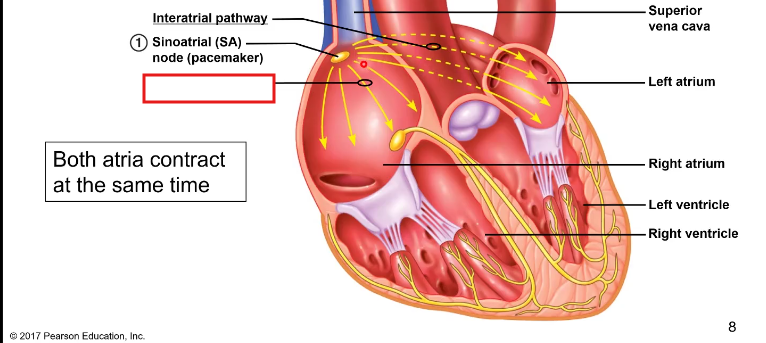
internodal pathway
both _____ contract at the same time
atria

conduction through the AV node is
slower

there is a delay with the AV node because it makes sure that both
atria contract before the ventricles
the conduction system of the heart
AP initiated in SA node, signals spread through atrial muscle via interatrial pathways
signal travels to AV node via internodal pathway—AV node delay
atrioventricular bundle (bundle of His)
splits into left and right bundle branches
purkinje fibers
Heartbeat is generated
within itself
Nodal cells are connected to the
muscle cells through connexions and gap junctions
autorhythmic cells have
pacemaker potentials
Pacemaker potentials
bring the membrane potential past AP threshold -50mV
Pacemaker potentials are
graded depolarizations caused by closing K channels and opening two types of voltage gated channels
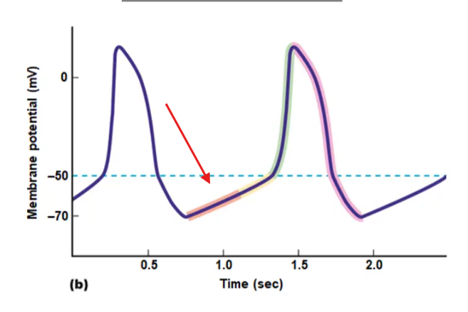
channels that are permeable to Na and K
as the membrane potential becomes hyperpolarized these channels open up
and once the open up they depolarize the cell
close at around -60 and -55mV
“initial subthreshold of cardiac neurons”
funny channels
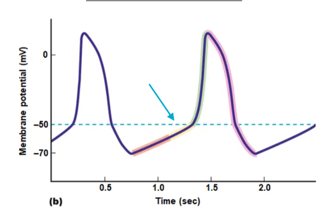
channel opens up at -60mV allowing Ca through
depolarizing AKA late subthreshold depolarization
after funny channels close
depolarizes potential until -50 mV (AP threshold)
brings membrane to AP threshold
T type calcium channels
Ca concentration is more; drives calcium
outside the cell; into the cell

once AP threshold is reached another Ca channel opens
allow Ca in faster → causing faster depolarization
generates calcium mediated AP
open longer to depolarize membrane
cells have delayed rectifier potassium channels (lazy)
L type calcium channels

after the delayed rectifier potassium channel opens up in L type calcium channels the membrane potential
repolarizes
After about -50 delayed rectifier potassium channels begin to; then _________ open
close; funny channels
action potentials of the cardio are from: ________ such as in the
autorhythmic neurons; SA node
excitable cells
driven by nodal neuronal AP
transferred to and between cardiac muscle fibers through gap junctions
cardiac muscle fiber cells
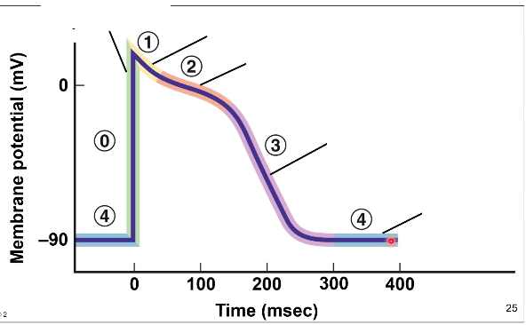
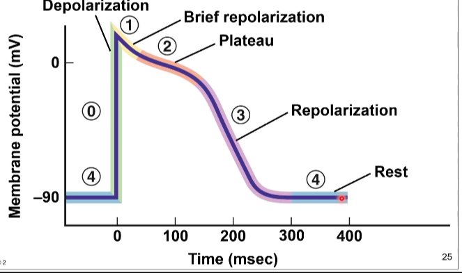
what are the phases from 0-1
depolarization, brief repolarization, plateau, repolarization, rest
in the cardiac muscle cells there are
rest periods
In the cardiac muscle there are
rest phases for the muscle to relax
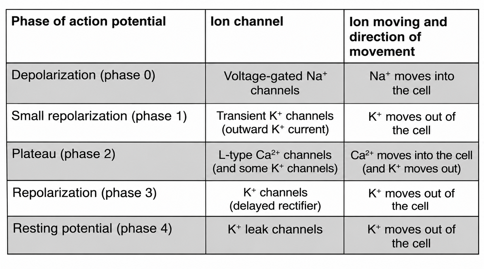
Five phases of electrical activity
depolarization of mV
very fast increased permeability to Na+
voltage gated Na+ channels open at a threshold
inactivation gate opens after short time
Phase 0
cardiac muscle AP are
longer
A inactivation gate opens up after a short while during phase 0 and causes the depolarization to
stop and creates the peak
Five phases of electrical activity
brief repolarization
inactivation of Na+ channels
leak channels that generate a resting mV start to repolarize
Phase 1
Five phases of electrical activity
slight plateau
increased permeability to Ca2+ (L type channels)
increasing permeability to inward rectifier K+ channels
Phase 2
what do inward rectifier K + channels do
open when cells is very depolarized and allow K to leave cell
Ca++ entering the cell while K+ leaves the cell causes a
balance with little to no change in mV
what phase extends the duration of the cardiac AP to keep cardiac muscle to keep contracted
phase 2
Five phases of electrical activity
L type Ca++ are starting to close
increased permeability to K +
delayed rectifier K channels open up
phase 3
Five phases of electrical activity
resting membrane potential
both K+ channels are open
phase 4
cardiac contractile cell AP have a
long duration of 250-300 msec


cardiac muscle fiber cells have a
straited appearance
Cardiac muscle fiber cells have properties that are similar to
skeletal muscles
cardiac muscle fiber cells have:
sarcomeres, t tubules, sarcoplasmic reticulum, troponin, tropomyosin cross bridge
properties similar to smooth muscles in the cardiac muscles are
gap junctions, extracellular Ca plays an important role in depolarization
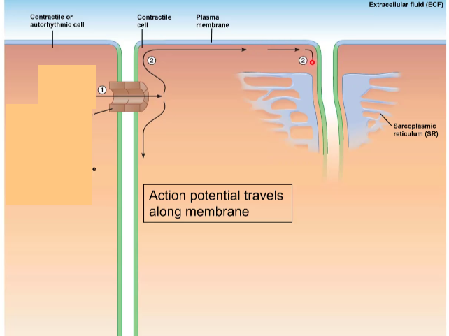
1
current spreads through gap junctions to contractile cell

2
action potentials travel along plasma membrane and T tubules

As ap goes by on membrane or sarcoplasmic reticulum
there are voltage calcium channels that open up
calcium voltage gated channels in the cardiac muscle are
L type ca channels
calcium voltage gated channels in the cardiac muscle are
attached to sarcoplasmic reticulum
AP triggers the release of Ca++ from the voltage gated channels, binding to _________ and pulls the ___________, exposing the binding sites on the actin
troponin; tropomyosin
After the binding sites are exposed with the help of Ca ________ binds to the sites on actin and this causes the cross bridge cycling
myosin
The cross bridge used for the actin uses ____, which allows the myosin heads to bind to actin
ATP
After the myosin heads does its power stroke the _____ falls off, disconnecting the myosin and actin
ADP
After Ca++ randomly unbinds from troponin it:
diffuses around and is bumped back into sarcoplasmic reticulum by an active transport pump that uses ATP
The pumps that pump Ca++ back into the sarcoplasmic reticulum can also be found on the ________ so it can be pumped out of the cell through _________
plasma membrane; ATP
Secondary transport in the cardiac muscle
calcium sodium cotransporter
The calcium sodium cotransporter uses the power of
sodium rushing in to pump calcium out