CHAPTER 3: ANOMALIES OF THE DEVELOPING DENTITION
 Call Kai
 Learn
Learn Practice Test
 Spaced Repetition
 Match
Match Flashcards
 Knowt Play
1/66

Earn XP
Description and Tags
sas
Name | Mastery | Learn | Test | Matching | Spaced | Call with Kai | Chat |
|---|
No analytics yet
Send a link to your students to track their progress
67 Terms
stage of tooth development
initiation → 8weeks → bud stage
proliferation → 9weeks → cap stage
differentiation (histology, morphology) → 11weeks → bell stage
calcification
attrition
initiation & proliferation
the stage of tooth development where anomalies on number of tooth usually occurs
conditions under histology defects
amelogenesis imprfecta
dentinogensis imperfecta
dentin dysplasia
anomalies on number of tooth
hyperdontia
hypodontia
under hyperdontia
mesiodens
distomolar
paramolars
hypodontia
occur more than hyperdontia
congenital absence of one or more teeth
occur more frequently in permanent
missing primary teeth are much less common than missing permanent teeth!
most common congenitally missing permanent teeth
lower 3rd molar
lower 2nd pm
upper lateral incisor
upper 2nd pm
hyperdontia
any number of extra (supernumerary) teeth
it may occur:
as part of a genetic syndrome
as an isolated developmental trait
mesiodens
an isolated developmental anomaly
not associated with syndromes & not hereditary, despite occasional increased familial occurrence
location:
located palatal to the permanent incisors
can be positioned to:
erupt normally & be inverted
clinical findings:
one or two extra teeth may be present
frequently conical or have an abnormal morphology
ectodermal dysplasia
a diverse group of genetic disorders
commonly affected:
teeth, hair, sweat glands
mammary glands, salivary glands
fingernails
oligodontia
missing more than six teeth, excluding third molars
anodontia
the complete absence of teeth
causes of hyper/hypodontia
genetic & molecular abnormalities affecting tooth development.
environmental stressors which may interrupt tooth bud development during later stages of development
2 chemotherapeutic agents commonly associated with dental anomalies are:
vincristine
cyclophosphamide (an alkylating agent)
—when these drugs are administered during tooth formation, pediatric patients are at risk of developing dental anomalies, including hypodontia
anomalies of tooth morphology
microdontia → more common
macrodontia
microdontia
refers to a reduction in tooth size.
it may result from:
decrease in the overall tooth size.
reduction in enamel thickness.
combination of both.
under microdontia
peg lateral
hutchinson’s incisors
mulberry molars
triad to confirm syphilis
hutchinson’s incisors
interstitial keratitis
deafness
under macrodontia
fusion
gemination
taurodontism
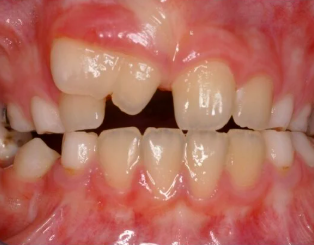
gemination
2 crowns, 1 root
most common cause of localized macrodontia
incomplete splitting (twinning) of a developing primary or permanent tooth
characteristics:
total number of teeth remains normal.
produces a tooth that may be almost twice the width of a normal tooth
etiology:
no known genetic predisposition
no known hereditary cause.
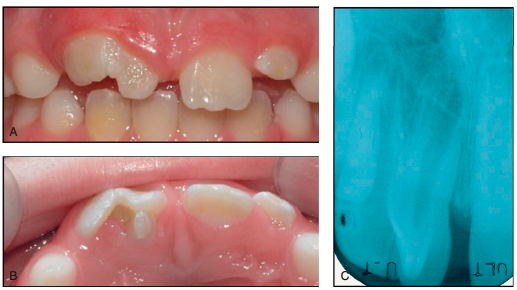
fusion
1 crown, 2 roots
occurs when two developing teeth join together
characteristics:
produces an enlarged tooth
more common in the primary dentition
results in one fewer tooth in the affected area.
taurodontism
enlarged pulp chamber, short roots
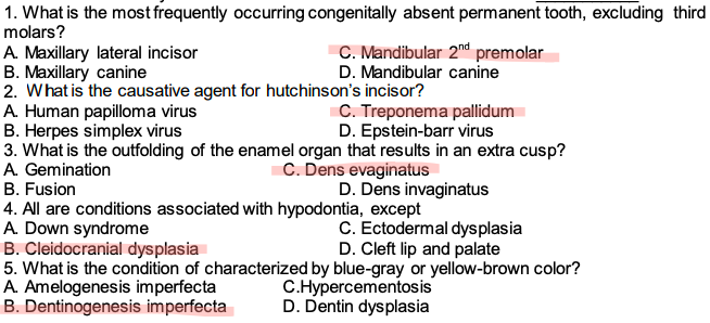
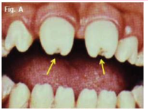
hutchinson’s incisor
tapered and notched incisal edges with screwdriver shapes
caused by congenital syphilis → treponema pallidum
tx: penicillin
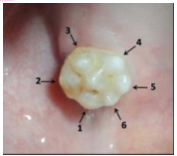
mulberrymolars
associated with congenital syphilis
irregular, accentuated occlusal patterns
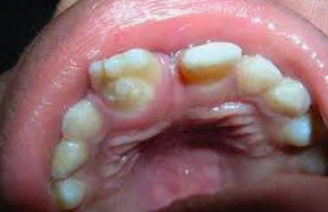
anomalies in shape
dens evaginatus
dens invaginatus (dens in dente)
dens evaginatus
approaching outward
an outfolding of the enamel organ that produces an extra cusp
pulp exposure can occur if excessive occlusal adjustment (equilibration) is performed bc pulp tissue extends into the cusp
location:
central groove or ridge of posterior teeth
cingulum area of anterior teeth → talon cusp
mx premolar → leon’s premolar
development:
results from evagination of the inner enamel epithelium (IEE), the cells that later become ameloblasts.
extra cusp contains:
enamel, dentin, pulp tissue
management:
careful enameloplasty.
preventive resin restorations (PRR)
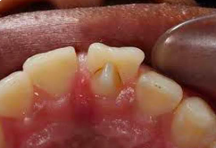
dens invaginatus / dens in dente
approaching inward
this creates the appearance of a tooth within a tooth
results from invagination of the inner enamel epithelium
most commonly affected tooth:
maxillary lateral incisors
clinical significance:
high risk of dental caries and pulpal involvement bc the invaginated portion on the lingual surface may communicate with the oral environment
management:
early diagnosis
prompt preventive treatment, including:
pit and fissure sealants
restorations if necessary
dilaceration vs flexium
dilaceration → >90 degrees
flexium → < 90 degrees
causes of root abnormalities
environmental
trauma
root dilacerations (abnormal bending or curvature of the roots
severe infections:
stevens-Johnson syndrome
meningococcemia
chemotherapy and radiation:
V-shaped roots
stunted (shortened) roots
genetic
tumoral calcinosis
singleton-merten syndrome
syndromes associated with short roots
sponastrime dysplasia
bardet-biedl syndrome 1
rothmund-rhomson syndrome
associated with short root anomaly specific gene mutations in this syndrome
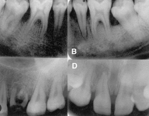
short root anomaly
frequently misdiagnosed as pathologic root resorption
these conditions are different because:
this anomaly is a developmental variation
root resorption is a pathologic process
oculo-facio-cardio-dental syndrome
an elongated root
cause:
mutations in the BCOR gene
mechanism:
uncontrolled root growth
increased proliferation of PDL cells
most commonly affected teeth:
permanent canines
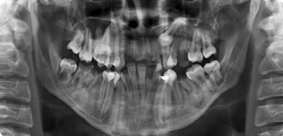
hypercementosis
an excessive deposition of cementum on the root surface
produces:
an abnormal root shape.
a bulbous enlargement at the root apex.
may result from:
trauma to the PDL
abnormal developmental processes
reactive response to periapical inflammation
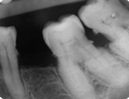
associated systemic conditions of hypercementosis
acromegaly
paget disease
atherosclerosis
taurodontism
enlargement of the body and pulp chamber of a multirooted tooth
characterized by:
an elongated pulp chamber.
apical displacement of the pulpal floor.
effects on root anatomy:
root furcation is displaced apically
individual roots become greatly shortened
overall root length may remain normal.
development:
root furcation forms through invagination of Hertwig's epithelial root sheath
bc of this developmental process, many ectodermal dysplasias exhibit this condition
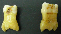
2 major types of enamel defects
enamel hypoplasia
enamel hypomineralization
hyperbilirubinemia
caused by biliary atresia
frequently produces green discoloration of teeth
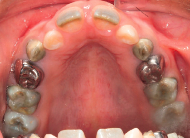
tetracycline staining
exposure to ultraviolet light oxidizes this complex, producing pigments that stain dental hard tissues
may cause severe intrinsic discoloration of:
primary teeth & permanent teeth
tooth discoloration may occur after treatment lasting as little as 3 days
among tetracycline antibiotics:
tetracycline hydrochloride → has the greatest staining potential
forms an orthocalcium phosphate complex with:
dentin & enamel
greatest risk occurs from:
intrauterine development
through 8 years of age
international association of dental traumatology (IADT)
recommends systemic doxycycline 2x daily for 7 days for avulsion injuries
dental fluorosis
hypomineralization of enamel due to excessive exposure to fluoride during enamel mineralization
severity ranges from:
mild, moderate, severe
degree of hypomineralization depends on the fluoride concentration in the serum during amelogenesis
individual susceptibility varies due to:
genetic makeup
overall health status
multiple genes contribute to differences in fluorosis susceptibility
mechanism:
fluoride contributes to fluorosis by affecting:
ameloblasts
developing enamel matrix
processing (maturation) of the enamel matrix
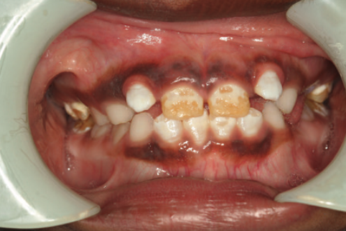
enamel hypoplasia (reduced enamel formation)
most common hereditary enamel phenotype
defect involving the amount (quantity) of enamel formed
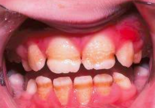
amelogenesis imperfecta (AI)
yellowish
characterized by defective or missing tooth enamel
refers to hereditary conditions that primarily affect enamel formation
classified according to:
mode of inheritance
mechanism causing the enamel defect
clinical phenotype (appearance of enamel)
secondary effects:
cracked tooth, early tooth decay
sensitivity to hot or cold exposures
susceptibility to multiple diseases of the tissues surrounding the teeth (periodontal tissues)
3 major types of amelogenesis imperfecta (AI)
hypoplastic amelogenesis imperfecta
hypocalcified amelogenesis imperfecta
hypomaturation amelogenesis imperfecta
hypoplastic amelogenesis imperfecta
quantity defect
results in reduced enamel thickness
caused by deficient enamel matrix formation
clinical appearance:
generalized thin enamel with pits/grooves
hypomaturation amelogenesis imperfecta
low value amount of ameloblast
produces hypomineralized enamel
caused by defective crystal growth and mineralization during the maturation stage of enamel development
clinical appearance:
mottled white-yellow-brown enamel that chips
soft
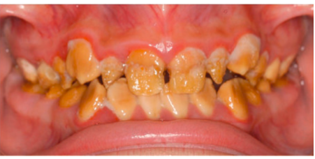
hypocalcified amelogenesis imperfecta
quality defect
also produces hypomineralized enamel
caused by abnormal initiation of enamel crystallites followed by defective mineralization
clinical appearance:
chalky yellow-brown enamel that wears rapidly
very soft
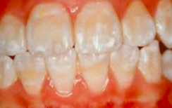
enamel hypomineralization
reduced mineral content
altered enamel translucency
higher protein content than normal enamel
weaker enamel, depending on the degree of mineral loss
defect involving the mineral content (quality) of enamel.
dentin
most abundant tissue in the tooth
poorly organized & less mineralized than normal
primarily responsible for determining crown-root morphology
as a result:
enamel frequently fractures away
it provides inadequate support for enamel
teeth become susceptible to rapid attrition bc hypomineralized dentin has poor wear resistance
vit. D deficiency during tooth development may result in:
large pulp chambers
reduced dentin production
reduced dentin mineral content
hereditary dentin defects
dentin dysplasia (DD)
dentinogenesis imperfecta (DGI)
are characterized by:
altered tooth morphology
abnormal dentin mineralization
dentinogenesis imperfecta (DGI)
discoloration results from abnormal dentin showing through translucent enamel
clinical features:
discolored teeth → blue-gray or yellow-brown
prone to rapid wear, breakage, loss
radiographic findings:
pronounced cervical constriction at the CEJ
small root structures that may appear:
sharp & tent peg-like
management:
once significant enamel loss and rapid attrition begin:
full-coverage crowns → treatment of choice
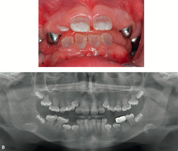
3 types of dentinogenesis imperfecta (DGI)
DGI type I
DGI type II
DGI type III
both type II and type II are:
autosomal dominant
highly penetrant
dentinogenesis imperfecta type I
bone involvement
blue-sclera on eyes, osteogensis imperfecta
occurs in people who have osteogenesis imperfecta
a genetic condition in which bones are brittle, causing them to break easily

dentinogenesis imperfecta type II
no bone involvement
not associated with a syndrome
most common type of dentinogenesis imperfecta
occurs in people without another inherited disorder
caused by mutations in the DSPP (dentin sialophosphoprotein) gene

dentinogenesis imperfecta type III
shell tooth
generally the same as dentinogenesis imperfecta type II except initially have large pulp chambers that obliterate with age results to multiple pulp exposure
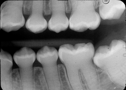
osteogenesis imperfecta
aka: brittle bone disease
caused by mutations affecting:
type I collagen
proteins involved in fibrillogenesis
dentin dysplasia (DD)
normal-looking crowns but abnormal roots and pulp
clinical features
short or no roots
pulp chamber obliteration
normal clinical crown morphology
radiographic features:
bowtie-shaped pulp chambers → molars
thistle tube-shaped pulp chambers → anterior
crescent-shaped root
sometimes absent root
2 types of dentin dysplasia (DD)
DD type I / radicular dentin dysplasia
DD type II / coronal dentin dysplasia
dentin dysplasia type I / radicular dentin dysplasia
affects the roots → rootless on xray
the exact cause remains unknown
tooth crowns appear clinically normal
dentin formation is markedly abnormal, characterized by:
pulp chamber obliteration
abnormal or nearly absent root development
a pathognomonic cascading waterfall histologic appearance
DD type II / coronal dentin dysplasia
affects the crown
pulp chamber has thistle tube appearance
2 stages of eruption of permanent dentition
preemergent eruption
postemergent eruption
eruption begins once root formation starts during the preemergent stage
normal eruption requires:
resorption of alveolar bone
resorption of primary tooth roots
propulsive movement of the tooth in an occlusal direction
mechanism of tooth eruption
one theory suggests that hard tissue proliferation at the root apex → the primary driving force for eruption
however, this does not explain all cases because:
intruded or luxated teeth may re-erupt without apical proliferation
tooth eruption is therefore considered a complex process involving multiple structures, including:
dental follicle
periodontal ligament (PDL)
ectopic eruption
one of the most common eruption anomalies
occurs when a tooth erupts along an abnormal eruption path
most commonly affected teeth:
permanent first molars
permanent lateral incisors
permanent canines
other eruption anomalies:
delayed exfoliation of primary teeth
delayed eruption of permanent teeth
environmental defects altering tooth color
excess fluoride exposure
fever, starvation, tetracycline
low birth weight, hyperbilirubinemia
trauma, hypoxia (e.g., severe cardiac defect)
infection (congenital syphilis, cytomegalovirus, congenital rubella)
practice question in sas