Molecular Biology - Exam II
 Call Kai
 Learn
Learn Practice Test
 Spaced Repetition
 Match
Match Flashcards
 Knowt Play
1/86

There's no tags or description
Looks like no tags are added yet.
Name | Mastery | Learn | Test | Matching | Spaced | Call with Kai | Chat |
|---|
No analytics yet
Send a link to your students to track their progress
87 Terms
In vivo
Study of tissue within a living organism (e.g. use of live model organisms)
Model organisms
Organisms used in In vivo studies that often…
Have well-characterized genetics
Grow and reproduce quickly (short life cycle)
Can be maintained in a laboratory setting
In vitro
Outside a living organism (e.g. in a test tube or petri dish)
In cell culture
In cells that are grown in a dish and passaged (e.g. used in study of cancer, viruses, proteins, and cellular differentiation)
Primary cell culture: cells that have been removed from an animal tissue and grown in culture
Cell line: cells that can be grown in cell culture and passaged multiple times
Passaging
Removal of calls growing in a dish filled with tissue culture media to another dish; enzymes involved in cell-cell-contact-protein digestion often used to remove cells
Cell culture media
An aqueous solution of salts, growth factors, and nutrients that will support cell growth
Ex vivo
In or on tissue that came recently from an organism
Organoids
Miniature organs grown in Ex vivo culture via self-organization of stem or progenitor cells
In silico
Computational simulations of biological processes
Light microscopy
Uses light to illuminate the sample
Up to 1000x magnification
0.2 mm - 200 nm diameter

Transmission electron microscopy (TEM)
Uses electrons to “illuminate” the sample
Up to 100,000x magnification
Can resolve structures smaller than 1 nm
Resolution higher than light’s

Scanning electron microscopy (SEM)
Generates microscopic images with 3D quality
Shows sample’s surface topography

Magnification
How big a microscopic sample appears
Resolution
Clarity and contrast of a microscopic sample
SDS-PAGE
Gel electrophoresis in which proteins are driven through a gel matrix and organized primarily by molecule size; progress is often tracked using dye

Western blots
Small scale study of proteins; allow detection of specific protein samples via…
Transfer of proteins from SDS-PAGE gel to piece of paper (nitrocellulose)
Paper is incubated with protein that will block non-specific binding of the antibody
Antibody, which is covalently attaches to an enzyme that produces a light reaction, recognizes specific protein on blot
Light reaction can be detected via camera of film to reveal protein band

Proteomics
Large scale study of proteins; provides a list of proteins within a sample by…
Digestion of proteins in a SDS-PAGE gel
Separation of peptides
Use of mass spectrometer to identify and gather data on proteins in sample

Non-ionic detergents
Triton X-100 and digitonin; permeabilize (make holes in) the plasma membrane or nuclear envelope to facilitate movement
Immunocytochemistry
Utilizes fluorophores attached to antibodies in order to recognize specific proteins in cells; done by…
Permeabilization of cell membrane via Triton X-100
Penetration of antibody through new holes in plasma membrane
Fluorescent tag (fluorophore) identifies antibody’s (therefore protein’s) location in the cell
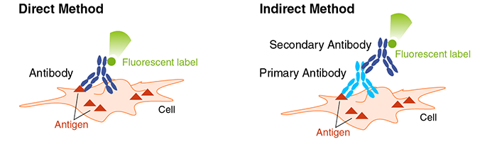
Immunohistochemistry
Utilizes stained antibodies to recognize specific proteins in tissues; done by…
Staining primary and secondary antibodies with DAB (brown dye)
Identifying the location of antibodies (therefore proteins) in tissue

Sanger Sequencing
DNA sequencing method used to determine the exact nucleotide sequence of a targeted segment by…
separation of DNA strands
introduction of a DNA primer
complementary strand synthesis via DNA polymerase w/ insertion of a fluorescent marker
disassociation of new strands, creating segments of different sizes
separation of segments via gel electrophoresis

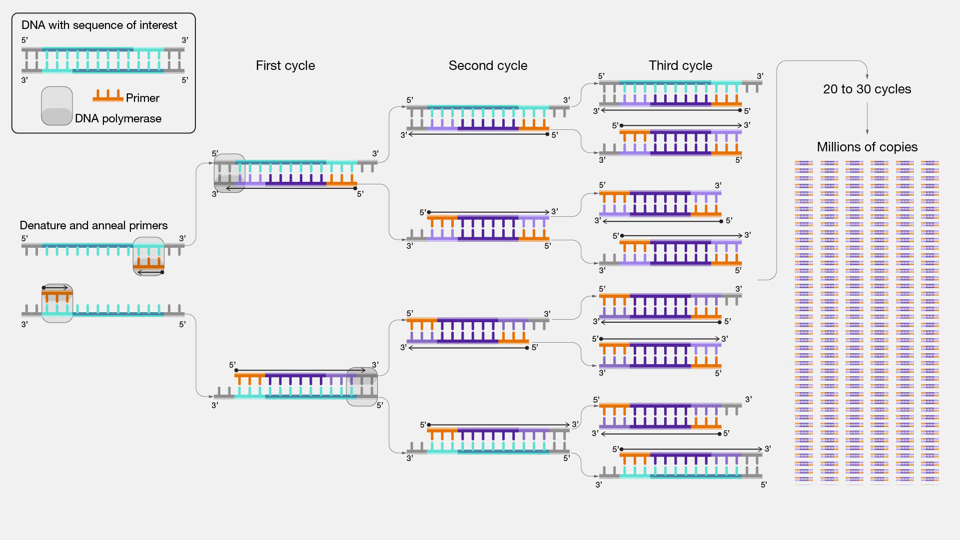
Polymerase chain reaction (PCR)
Creates exact copies of a target DNA sequence
Fluorescence In Situ Hybridization (FISH)
Analyzes and detects the location of a specific DNA or RNA sequence on a chromosome; labels complementary DNA fragments with fluorescence

RNA ISH
Localizes RNA by…
adding “probe” to specimen
denaturing probe and target
hybridization
binding of labeled antibodies
fluorescence => labeling/detection

RT-PCR
Converts RNA into DNA in order to replicate it for study

Single-cell RNA sequencing
Measures gene expression in individual cells using RNA by…
Collecting sample
Dissociating and isolating single cells (using oil)
Isolating RNA in each cell
Sequencing RNA

Bulk RNA sequencing
Measures gene expression in many cells using RNA; often used to compare genomes and find a mutation

Dissociation
Mechanical dissociation:
Mortar and Pestle
Scissors
Syringe
Enzyme dissociation:
Papain
Collagenase
Trypsin
Dnase
Dispase
Endogenous
Growing or originating from within an organism
Exogenous
Growing or originating from outside an organism
Recombinant DNA technology
Uses bacterial DNA to make a specific DNA segment by…
Linking a vector DNA (derived from bacterial genome) is linked to target DNA segment to create a plasmid
Bacterial cells are expanded and used to make many copies of plasmid
Plasmid DNA is isolated from bacteria for use in other cells

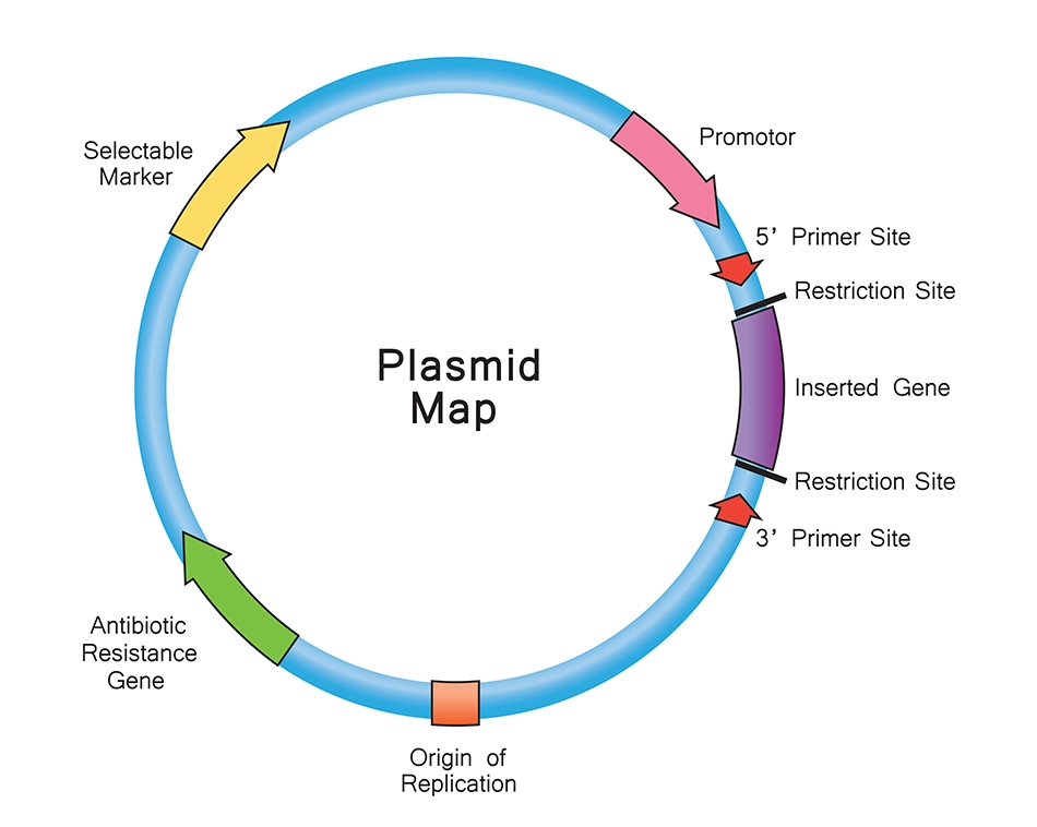
Recombinant plasmid
Requires…
Origin of replication (ORI)
Antibiotic resistance: allows positive selection in bacteria
Promoter
Target gene (YFG)
CRISPR-Cas9
DNA-dependent endonuclease that can cut DNA in a specific region and insert new DNA at cut site

GFP fusion proteins
Utilizes gene encoding green fluorescent protein (GFP) by…
Placing GFP gene in a plasmid vector with target gene
Transfecting plasmid into cell
Allowing plasmid to be transcribed and translated into fusion protein (containing target protein and GFP)

Transfection
Introduction of nucleic acids into eukaryotic cells

Lipid delivery agent
Utilizes membrane-enclosed vesicles containing target plasmid to induce membrane fusion and release of substance into cytoplasm

Electroportation
Cell is subject to short electric shock in order to temporarily permeabilize the membrane and allow target plasmid into cell

Transduction in plasmid delivery
Use of viral vectors to package exogenous nucleic acids in viral capsids, using cell surface proteins as receptors to enter cells

Adenoviral vector
Does not integrate with genome
Does not replicate during cell division
Quick, transient, high-level expression
Lentiviral vector
Integrates into genome
Transfects most cell types, but with varying efficiency
Signal transduction
The process by which an extracellular signal induces a cellular response
Cell surface receptors
Transmembrane receptors that bind to ligands on the outside of a cell’s plasma membrane and transduce the signal within the cell

Intracellular receptors
Cellular receptor within the target cell which requires the small, hydrophobic ligand to diffuse through the plasma membrane

Autocrine
The cell producing the ligand also accepts and responds to the message
Paracrine
Ligand must travel a short distance (i.e. to a neighboring cell) from the cell in which it was produced to the target cell
Endocrine
Ligand must travel a long distance (i.e. through the bloodstream) from the cell in which it was produced to the target cell
Hormones
Regulatory substances (steroids) produced in an organism and transported through the bloodstream (i.e. endocrine) to stimulate specific cells by…
Diffusing into the cell vis facilitated diffusion
Binding to intracellular receptor
Movement of signal-receptor complex into the nucleus
Binding of signal-receptor complex to promoter/enhancer region of DNA to activate gene transcription

Estrogen receptor signaling
Estradiol (E2) diffuses through membrane
E2 binds to estrogen receptor α (ERα)
E2 and ERα migrate into the nucleus to activate transcription

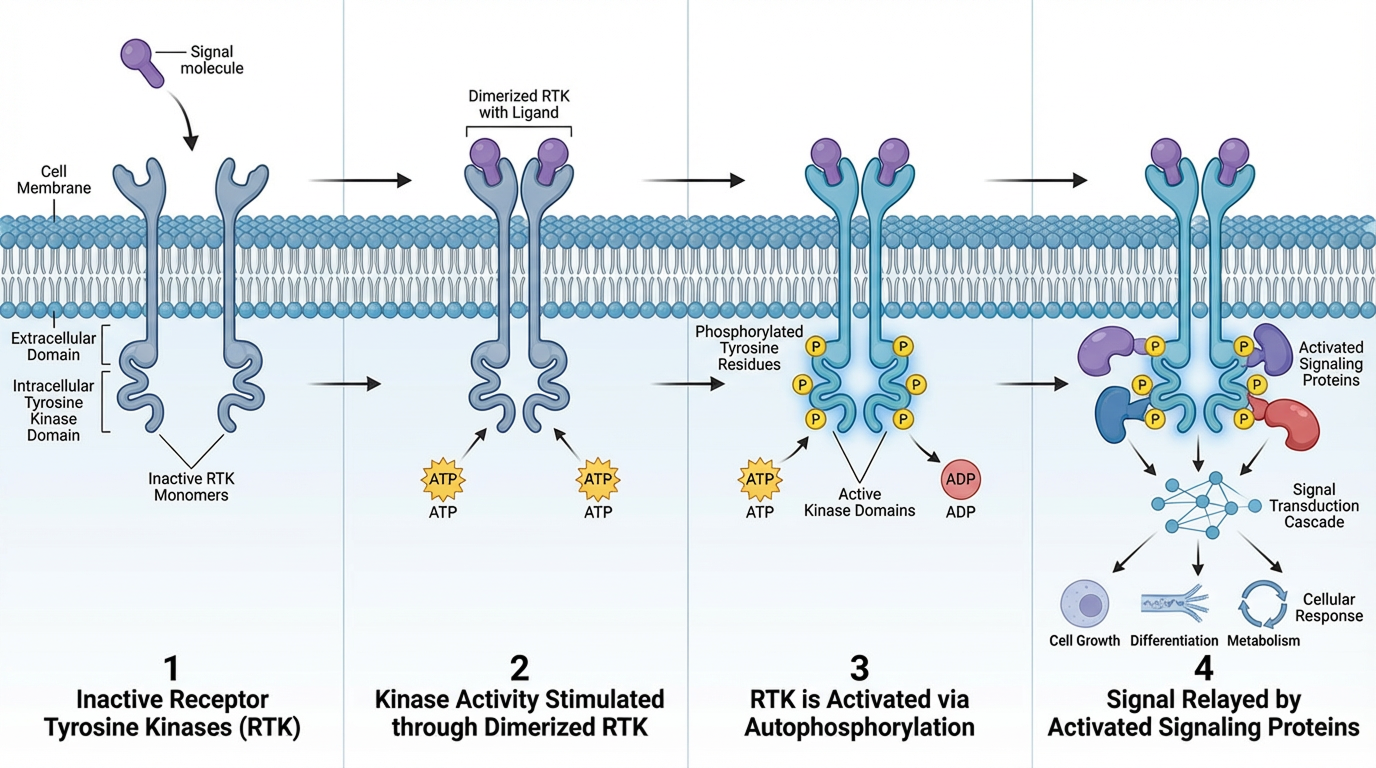
Receptor tyrosine kinases
Extracellular, transmembrane receptors that bind to growth factors and hormones; activate kinases (enzymes that phosphorylate proteins) by…
Awaiting signal molecule binding as monomeric, inactive receptors
Binding to signal molecule and stimulating kinase activity
Ligand-mediated dimerization: dimeric ligands induce dimerization and activation
Receptor-mediated dimerization: monomeric ligands are used; receptors must be dimerized in order to activate
Transphosphorylating tyrosines on partner receptor to activate it
Cytoplasmic kinase cascade
Kinases activate other kinases, which allow post-transcriptional modifications via phosphorylation of amino acids
Cytoplasmic kinases are generally Ser and Thr kinases

G-protein coupled receptor signaling
Transmembrane receptor couples with G-protein to induce a response by…
Binding of ligand to GPCR (receptor)
Association of G-protein complex with GPCR, activating it and forming GTP
Conformational change of G-protein, releasing α subunit
Binding of α subunit to target protein, activating it
Hydrolysis of GTP, inactivating the α subunit and causing dissociation from target protein
Re-association of α subunit with β and γ subunits for future G-protein use

Second messengers
Diffusible, non-protein molecules that relay signals from an effector molecule to activate target inside the cell; significantly amplify signals
Cyclic AMP (cAMP)
Second messenger produced by adenyl cyclase and derived from ATP; used to activate certain kinases

IP3
Small, water-soluble sugar phosphate molecule capable of rapid diffusion throughout the cell that bind to their specific receptors in the smooth ER

DAG
Lipid molecule that recruits and activates effector proteins across the plasma membrane bearing its specific binding domain

Ca2+
Ions that bind to various target molecules, triggering many specific cellular responses; associated with the smooth ER

Apoptosis
Cellular response to kill cells that are unnecessary, damaged, or dangerous in which…
Cell volume shrinks
Cells lose cell-cell contacts
Actin and chromatin are chopped into smaller pieces
Cell membrane forms blebs
Cell is phagocytosed

Extrinsic pathway (apoptosis)
Activation of apoptosis from outside the cell
Fas ligand of killer lymphocyte binds to Fas protein of target cell
Adaptor proteins (Apaf-1) and inactive casp-8 molecules associate with Fas protein
Aggregation of casp-8 molecules, therefore activating them and a caspase cascade
Dissociation of killer lymphocyte

Intrinsic pathway (apoptosis)
Activation of apoptosis from inside the cell
Cytochrome c releases from injured mitochondrion and binds to adaptor proteins (Apaf-1)
Aggregation of Apaf-1 and association with inactive casp-9
Activation of casp-9 and caspase cascade

Stages of the cell cycle
G1: organelles replicate, cell grows and carries out normal metabolic functions
S: DNA replication and chromosome duplication
G2: cell grows and prepares for division
M: mitosis, including prophase, prometaphase, metaphase, anaphase, telophase, and cytokinesis

Cell cycle checkpoints
G1: is environment favorable?
G2: is all DNA replicated properly? is environment favorable?
Metaphase: are all chromosomes attached to the spindle?

G0
Quiescence; state in which cells have exited the cell cycle, but can reenter when acted upon by a stimulus
Senescence
A permanent state of quiescence or G0
Cyclin-dependent kinase (Cdk)
Kinases that regulate cell cycle entry by direct phosphorylation of targets; activated by binding to cyclins
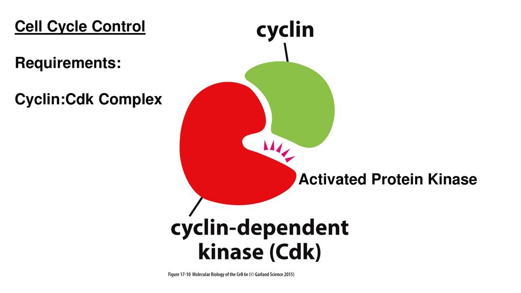
Cyclin
Protein partner for Cdks whose levels are regulated during the cell cycle

Ubiquitin-proteosome cycle
Cycle that degrades Cdk-cyclins throughout the cell cycle in order to regulate its progression

Activation of cdks
Inactive cdk must be phosphorylated once (by CAK) in order to be activated
Active cdk can be further phosphorylated (by Wee1 kinase) in order to be inactivated
Diphosphorylated (inactive) cdk can have a phosphate group removed (by Cdc25 phosphatase) in order to be activated
Active cdk/cyclin complex provides positive feedback to activate more complexes

p27
A cdk inhibitor that binds to the cdk/cyclin complex and inhibits its activity; must be removed for cell cycle to progress

p53
Tumor-suppressing transcription factor that directly impacts the binding of DNA or its conformation
Elephants have multiple copies of this gene, making cancer extremely rare

Gated protein transport
Proteins move from the nucleus to the cytoplasm via nuclear pore complex, which acts as a “selective gate”
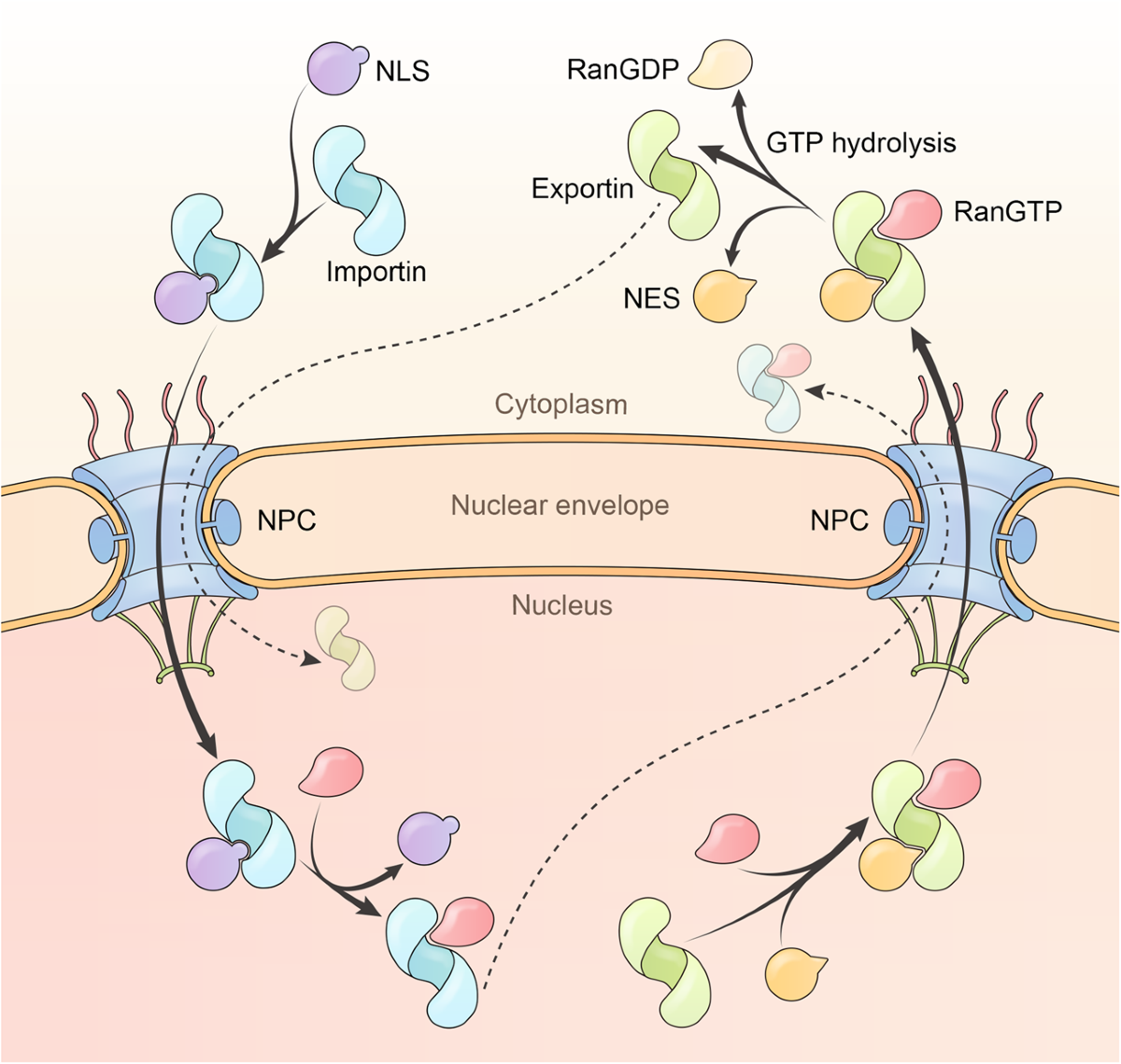
RanGTP
Regulates nucleocytoplasmic transport; interacts with RanGAP in the cytoplasm and the chromatin-associated RCC1 in the nucleus, creating a Ran-GTP gradient and concentrating Ran in the nucleus

Nuclear localization signals (NLS)
Target proteins for passage through the nuclear pore complexes
Transmembrane protein transport
Proteins move from the cytosol to an organelle across membranes via unfolding and “snaking” through a translocator

Co-translational import into ER
Ribosome translates mRNA
N-terminal signal peptide is recognized by signal recognition particle (SRP)
SRP is recognized by SRP receptor on ER surface
SRP receptor brings ribosome to translocator

Vesicular protein transport
Proteins move from the ER to the plasma membrane (or vice versa) via membrane-bound vesicles formed to “transport cargo”

Rough ER
Organelle containing ribosomes for protein synthesis; serve to…
synthesize integral membrane proteins, secretory proteins, and soluble proteins
fold and oligomerize proteins (with the help of chaperones) into their tertiary and quaternary structures and run a “quality control check” (ERAD pathway)
initiate glycosylation or proteins and lipids
transport proteins to Golgi complex for further processing
N-linked glycosylation
Linking of a sugar to the N-terminus of a protein, specifically the amino acid asparagine; starts in the ER and is modified in the Golgi
O-linked glycosylation
Linking of a sugar to the O-terminus of a protein, specifically the amino acids serine and threonine; occurs entirely in the Golgi
Chaperones
Proteins that undergo a change in conformation (typically requiring ATP) to facilitate protein folding; occurs during co-translational import into the ER
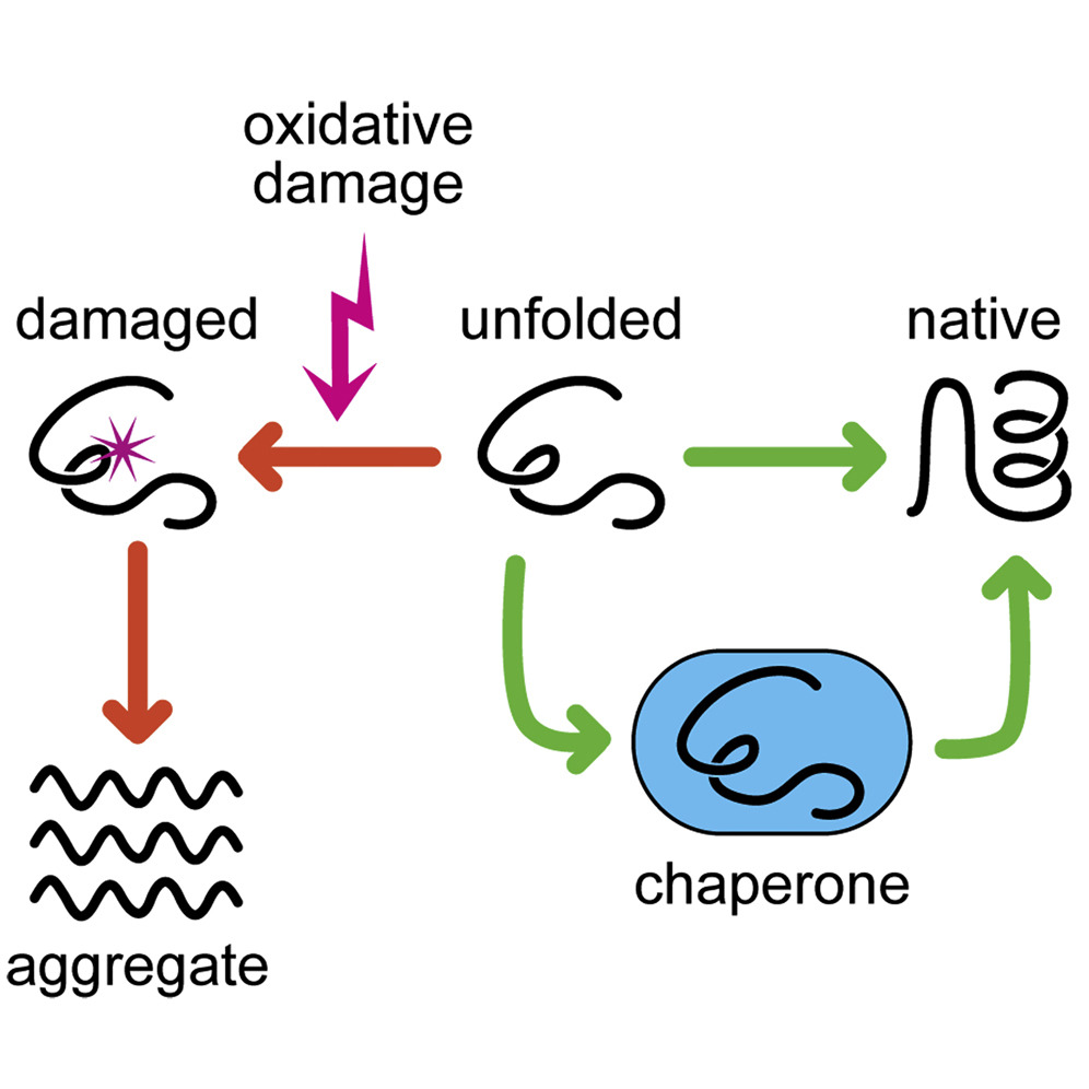
ERAD pathway
Checks the quality of proteins to ensure they’re normally folded before leaving the ER; if released into the cytoplasm misfolded, proteosomes (organelles containing digestive enzymes) will disassociate them back into ammino acids

KDEL and KKXX
Amino acid sequences that signify a protein’s return to the ER; are recognized by their own specific receptor complex
ER entry signaling
Proteins require a signal sequence and ER retention signal
Golgi entry signaling
Proteins require a signal sequence and a Golgi retention signal
Lysosomes entry signaling
Proteins require a signal sequence and a Mannose 6-phosphate
Nucleus entry signaling
Proteins require an NLS
Mitochondria entry signaling
Proteins require an MTS
Peroxisomes entry signaling
Proteins require a PTS