AE 1.10 - Systolic & Diastolic Function, Heart Failure/Transplant

0.0(0)
Studied by 2 people
call kaiCall Kai
learnLearn
examPractice Test
spaced repetitionSpaced Repetition
heart puzzleMatch
flashcardsFlashcards
GameKnowt Play
Card Sorting

1/32

encourage image

There's no tags or description

Looks like no tags are added yet.

33 Terms

1
New cards

Basic pathophysiology of heart failure

  • any structural or functional cardiac disorder that impairs the ability of the ventricles to fill correctly or eject blood completely

  • leading to congestion & reduced systemic perfusion

  • characterized by fatigue, breathlessness, and edema


  • Valve disease – stenosis/regurgitation

  • Coronary disease – segmental wall dysfunction

  • Congenital disease – abnormal anatomy

  • Pericardial disease – effusion/constriction

  • Hypertension – LVH

  • Cardiomyopathy – composite (cavity size, wall thickness, contraction)


<ul><li><p><span style="background-color: transparent;">any structural or functional cardiac disorder that impairs the ability of the ventricles to fill correctly or eject blood completely</span></p></li><li><p><span style="background-color: transparent;">leading to congestion &amp; reduced systemic perfusion</span></p></li><li><p><span style="background-color: transparent;">characterized by fatigue, breathlessness, and edema</span></p></li></ul><p></p><ul><li><p><span style="background-color: transparent;">Valve disease – stenosis/regurgitation</span></p></li><li><p><span style="background-color: transparent;">Coronary disease – segmental wall dysfunction</span></p></li><li><p><span style="background-color: transparent;">Congenital disease – abnormal anatomy</span></p></li><li><p><span style="background-color: transparent;">Pericardial disease – effusion/constriction</span></p></li><li><p><span style="background-color: transparent;">Hypertension – LVH</span></p></li><li><p><span style="background-color: transparent;">Cardiomyopathy – composite (cavity size, wall thickness, contraction)</span></p></li></ul><p></p>
2
New cards

2 main HF types

  1. Heart failure with reduced ejection fraction (HFrEF) “heff-reff”

    • EF < 40%

    • “systolic HF”


  1. Heart failure with preserved LV ejection fraction (HFpEF) “heff-peff”

    • EF > 40%

    • “diastolic HF”

    • approximately 50% of patients in contemporary registries and studies of HF have preserved EF

    • more than 80% of patients have diastolic dysfunction

    • preserved LV systolic function


<ol><li><p><span style="background-color: transparent;">Heart failure with <em><u>reduced ejection fraction</u></em> <strong>(HFrEF) </strong>“heff-reff”</span></p><ul><li><p><span style="background-color: transparent;">EF &lt; 40%</span></p></li><li><p>“systolic HF”</p></li></ul></li></ol><p></p><ol start="2"><li><p><span style="background-color: transparent;">Heart failure with <em><u>preserved LV ejection fraction</u></em> <strong>(HFpEF) </strong>“heff-peff”</span></p><ul><li><p><span style="background-color: transparent;">EF &gt; 40%</span></p></li><li><p><span style="background-color: transparent;">“diastolic HF”</span></p></li><li><p><span style="background-color: transparent;">approximately 50% of patients in contemporary registries and studies of HF have preserved EF</span></p></li><li><p><span style="background-color: transparent;">more than 80% of patients have <em>diastolic dysfunction</em></span></p></li><li><p><span style="background-color: transparent;">preserved LV systolic function</span></p></li></ul></li></ol><p></p>
3
New cards

4 sub types of HF

  1. Acute (pulmonary edema)

  1. Chronic stable

  1. Right – sided failure

  1. Left – sided failure


<ol><li><p><span style="background-color: transparent;">Acute (pulmonary edema)</span></p></li></ol><ol start="2"><li><p><span style="background-color: transparent;">Chronic stable</span></p></li></ol><ol start="3"><li><p><span style="background-color: transparent;">Right – sided failure</span></p></li></ol><ol start="4"><li><p><span style="background-color: transparent;">Left – sided failure</span></p></li></ol><p></p>
4
New cards

Acute vs Chronic HF

  1. Acute (pulmonary edema)

    • Symptoms appear suddenly but go away quickly, caused by a sudden change in structure or function

    • Typically a medical emergency and requires immediate medical intervention

    • Causes fluid buildup:

      • lungs – pulmonary edema

      • abdominal cavity – ascites

    • Worsening kidney function

    • Worsening liver function


  1. Chronic stable

    • Systolic

      • Symptoms are continuous and don’t improve over time

      • Most heart failure cases are chronic

      • Heart doesn't pump well 

    • Diastolic

      • Occurs when the heart muscle becomes stiffer than normal and your heart doesn’t fill with blood easily

      • It leads to a lack of blood flow to the rest of the organs in your body


5
New cards

Right vs Left sided HF

  1. Right – sided failure

    • Usually triggered by left-sided heart failure

    • The accumulation of blood in the lungs caused by left-sided heart failure 

      • makes the right ventricle work harder

    • This can stress the right side of the heart & cause it to fail

    • Etiology:

      • Lung disease

      • Valve disease


  1. Left – sided failure

    • Most common type

    • LV doesn’t pump efficiently

    • This prevents your body from getting enough oxygen-rich blood

    • The blood backs up into your lungs instead, which causes shortness of breath & a buildup of fluid


6
New cards

Clinical signs & symptoms of left-sided heart failure

  • SOB (dyspnea)

  • fatigue

  • reduced exercise

    tolerance

  • orthopnea (SOB lying flat)

  • paroxysmal nocturnal dyspnea

  • edema

  • weight loss >4.5 lb in response to treatment

  • elevated jugular venous pressure

  • third heart sound (gallop)

  • rales on auscultation

  • cardiomegaly on x-ray

  • hepatojugular reflux

  • hepatomegaly

  • pleural effusion

  • tachycardia


<ul><li><p><span style="background-color: transparent;">SOB (dyspnea)</span></p></li><li><p><span style="background-color: transparent;">fatigue</span></p></li><li><p><span style="background-color: transparent;">reduced exercise</span></p><p><span style="background-color: transparent;">tolerance</span></p></li><li><p><span style="background-color: transparent;">orthopnea (SOB lying flat)</span></p></li><li><p><span style="background-color: transparent;">paroxysmal nocturnal dyspnea</span></p></li></ul><ul><li><p><span style="background-color: transparent;">edema</span></p></li><li><p><span style="background-color: transparent;">weight loss &gt;4.5 lb in response to treatment</span></p></li></ul><ul><li><p><span style="background-color: transparent;">elevated jugular venous pressure </span></p></li><li><p><span style="background-color: transparent;">third heart sound (gallop) </span></p></li><li><p><span style="background-color: transparent;">rales on auscultation</span></p></li><li><p><span style="background-color: transparent;">cardiomegaly on x-ray</span></p></li><li><p><span style="background-color: transparent;">hepatojugular reflux</span></p></li><li><p><span style="background-color: transparent;">hepatomegaly</span></p></li><li><p><span style="background-color: transparent;">pleural effusion</span></p></li><li><p><span style="background-color: transparent;">tachycardia</span></p></li></ul><p></p>
7
New cards

Effects of heart rate on ventricular filling (HF)

  • Diastole is the longest cycle

    • normal diastolic function allows for adequate filling of the ventricles during rest to ensure there is an adequate stroke volume (SV) during systole


  • higher HR = less time for filling = lower EDV

  • lower HR = more time for filling = higher EDV


8
New cards

Relationship between heart failure & cardiac output

  • CO ↓ with both systolic & diastolic HF


CO = SV x HR

SV = LVEDV – LVESV


<ul><li><p>CO <span style="background-color: transparent;">↓ </span>with both systolic &amp; diastolic HF </p></li></ul><p></p><p><span style="background-color: transparent;">CO = SV x HR</span></p><p>SV = LVEDV – LVESV</p><p></p>
9
New cards

Phases of the cardiac cycle

  • Diastole

    • Phase 1: Isovolumic Relaxation

    • Phase 2a: Rapid Filling

    • Phase 2b: Slow Filling (Diastasis)

    • Phase 3: Atrial Systole


  • Systole

    • Phase 4: Isovolumic Contraction

    • Phase 5a: Rapid Ejection

    • Phase 5b: Slow Ejection


<ul><li><p><span style="background-color: transparent;">Diastole</span></p><ul><li><p><span style="background-color: transparent;">Phase 1: Isovolumic Relaxation</span></p></li><li><p><span style="background-color: transparent;">Phase 2a: Rapid Filling</span></p></li><li><p><span style="background-color: transparent;">Phase 2b: Slow Filling (Diastasis)</span></p></li><li><p><span style="background-color: transparent;">Phase 3: Atrial Systole</span></p></li></ul></li></ul><p></p><ul><li><p><span style="background-color: transparent;">Systole</span></p><ul><li><p><span style="background-color: transparent;">Phase 4: Isovolumic Contraction</span></p></li><li><p><span style="background-color: transparent;">Phase 5a: Rapid Ejection</span></p></li><li><p><span style="background-color: transparent;">Phase 5b: Slow Ejection</span></p></li></ul></li></ul><p></p>
10
New cards

Timing of mitral inflow (E-wave & A-wave)

E = rapid filling / early diastole

  • after T wave


A = atrial systole/kick

  • after P wave


<p>E = rapid filling / early diastole </p><ul><li><p>after T wave </p></li></ul><p></p><p>A = atrial systole/kick</p><ul><li><p>after P wave </p></li></ul><p></p>
11
New cards

Rapid filling, diastasis, & atrial systole

Rapid filling

  • 70% filling

  • ventricles are relaxing, pressures ↓

  • ventricular pressures drop low enough that the pressure in the atria becomes higher than ventricles → causes MV to open

  • Atria act as conduit 


Diastasis

  • brief period when minimal amount of blood continues to flow from the atria just before atrial contraction


Atrial systole

  • 30% filling

  • mechanical contraction of the atria due to rise in pressure in the atria

  • atria are acting as a pump


12
New cards

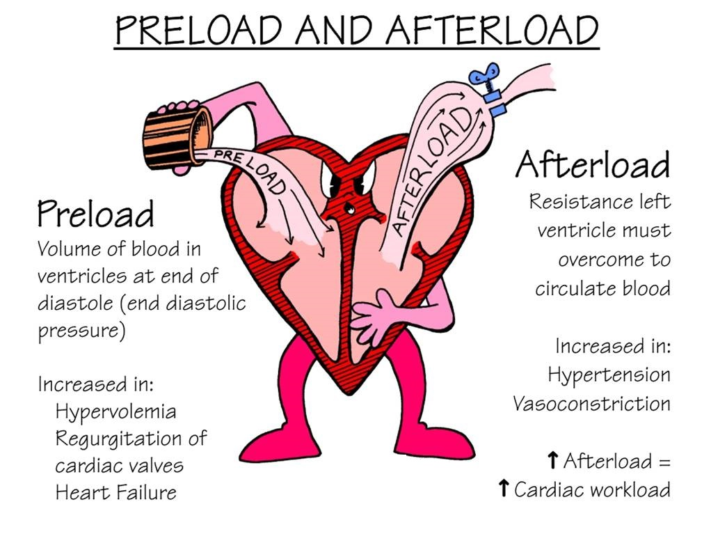
Preload

  • initial ventricular volume / the initial stretching (filling) of the chamber prior to contracting

  • leads to increased LV pressure, power, and work (Starling Law)

  • is quantified by end-diastolic volume

  • can also be affected by the ability of the myocardium to relax

<ul><li><p><span style="background-color: transparent;">initial ventricular volume / the initial stretching (filling) of the chamber prior to contracting</span></p></li><li><p><span style="background-color: transparent;">leads to increased LV pressure, power, and work (Starling Law)</span></p></li><li><p><span style="background-color: transparent;"><em>is quantified by end-diastolic volume</em></span></p></li><li><p><span style="background-color: transparent;">can also be affected by the ability of the myocardium to relax</span></p></li></ul><p></p>
13
New cards

Afterload

  • resistance heart encounters when emptying / the amount of resistance that the heart encounters as it ejects

  • under normal condition is “good”

    • most of the vital organs need some level of blood pressure so they can operate their blood supply systems

  • too little afterload (hypotension) = shock

  • too much afterload = ↓ SV


<ul><li><p><span style="background-color: transparent;">resistance heart encounters when emptying / the amount of resistance that the heart encounters as it ejects</span></p></li><li><p><span style="background-color: transparent;">under normal condition is “good”</span></p><ul><li><p><span style="background-color: transparent;">most of the vital organs need some level of blood pressure so they can operate their blood supply systems</span></p></li></ul></li><li><p><span style="background-color: transparent;">too little afterload (hypotension) = shock</span></p></li><li><p><span style="background-color: transparent;">too much afterload = ↓ SV</span></p></li></ul><p></p>
14
New cards

Stroke Volume

  • reflects the pump performance of the heart

  • amount of blood pumped with each contraction

  • SV = LVEDV – LVESV

  • can be calculated using volumes or by formula using LVOT diameter & LVOT VTI

  • affected by both your diastolic filling (preload) & systolic emptying (afterload)


15
New cards

Factors that influence stroke volume

  • affected by both your diastolic filling and systolic emptying

  • Contractility (ability of myocardium to contract)

  • Preload (initial ventricular volume)

  • Afterload (resistance heart encounters when emptying)

  • Ventricular geometry (walls, cavities) 


16
New cards

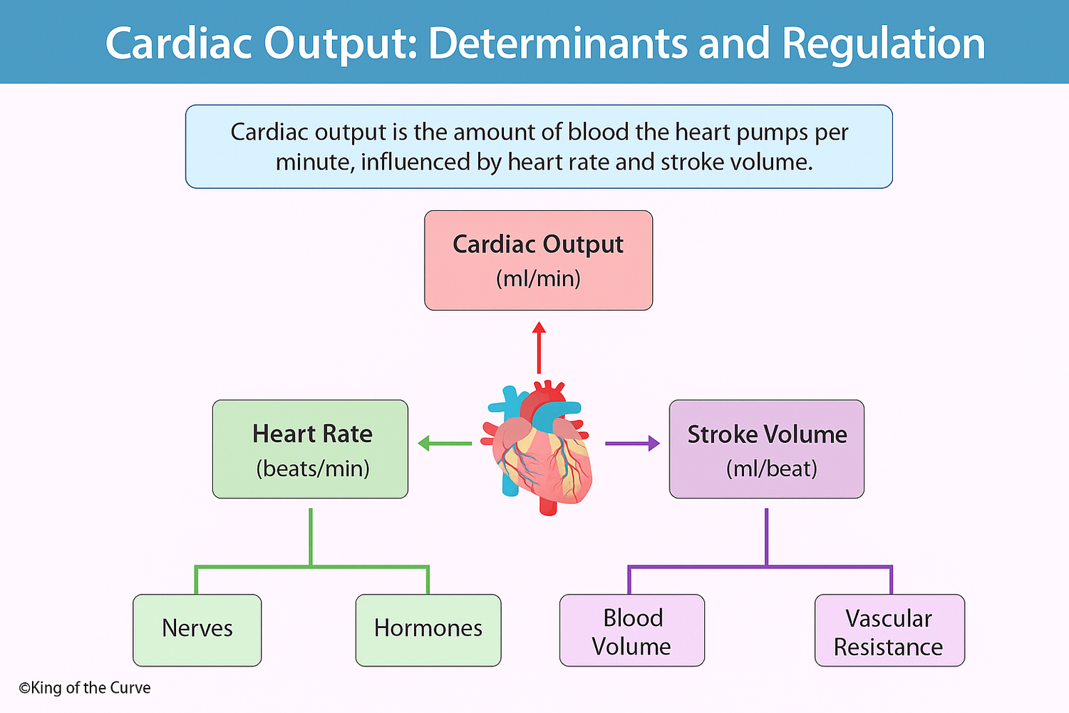
Cardiac Output

  • normal = 4-6 L/min

  • the amount of blood ejected by the ventricle in one minute

  • CO = SV x HR

  • CO systemic = CO pulmonic

  • Cardiac Index (CI): CO / BSA

    • relates heart performance to the size of the individual

<ul><li><p><span style="background-color: transparent;">normal = 4-6 L/min</span></p></li><li><p><span style="background-color: transparent;"><em>the amount of blood ejected by the ventricle in one minute</em></span></p></li><li><p><span style="background-color: transparent;">CO = SV x HR</span></p></li><li><p><span style="background-color: transparent;">CO systemic = CO pulmonic</span></p></li><li><p><span style="background-color: transparent;">Cardiac Index (CI): CO / BSA</span></p><ul><li><p><span style="background-color: transparent;">relates heart performance to the size of the individual</span></p></li></ul></li></ul><p></p>
17
New cards

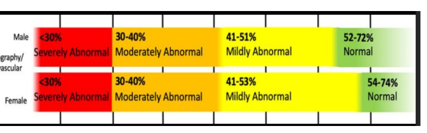
Ejection Fraction

  • reflects overall ventricular function

  • the % of the LV diastolic volume that is ejected with systole

    • a normal, healthy heart doesn’t eject all the blood (ejects 50-55%)

  • (EDV – ESV) / EDV x 100

  • highly dependent on loading conditions

    • EF is derived from SV, which is determined by preload & afterload

      • (ie – aortic stenosis, mitral regurgitation, etc)

<ul><li><p><span style="background-color: transparent;">reflects <strong><em>overall ventricular function</em></strong></span></p></li><li><p><span style="background-color: transparent;">the % of the LV diastolic volume that is ejected with systole</span></p><ul><li><p><span style="background-color: transparent;">a normal, healthy heart doesn’t eject all the blood (ejects 50-55%)</span></p></li></ul></li><li><p><span style="background-color: transparent;">(EDV – ESV) / EDV x 100</span></p></li><li><p><span style="background-color: transparent;">highly dependent on loading conditions</span></p><ul><li><p><span style="background-color: transparent;">EF is derived from SV, which is determined by preload &amp; afterload</span></p><ul><li><p><span style="background-color: transparent;">(ie – aortic stenosis, mitral regurgitation, etc)</span></p></li></ul></li></ul></li></ul><p></p>
18
New cards

Contractility

ability of myocardium to contract

19
New cards

Effects of changes in contractility on stroke volume & end-systolic volume

SV

  • ↑ contractility = ↑ SV

  • ↓ contractility = ↓ SV


ESV

  • ↑ contractility = ↓ ESV

  • ↓ contractility = ↑ ESV


<p><strong>SV </strong></p><ul><li><p><span style="background-color: transparent;">↑ contractility = ↑ SV </span></p></li><li><p><span style="background-color: transparent;">↓ contractility = ↓ SV</span></p></li></ul><p></p><p><strong>ESV</strong></p><ul><li><p><span style="background-color: transparent;">↑ contractility = ↓ ESV</span></p></li><li><p><span style="background-color: transparent;">↓ contractility = ↑ ESV</span></p></li></ul><p></p>
20
New cards

Relationship between preload, afterload, contractility, & ventricular performance

  • ventricular function & pump performance depend on all of these (loading conditions + function)


Ventricular performance (stroke volume & cardiac output) is governed by 3 primary mechanical determinants: preload, afterload, & contractility

  • these factors control how much blood fills the ventricles & how forcefully & completely they eject it
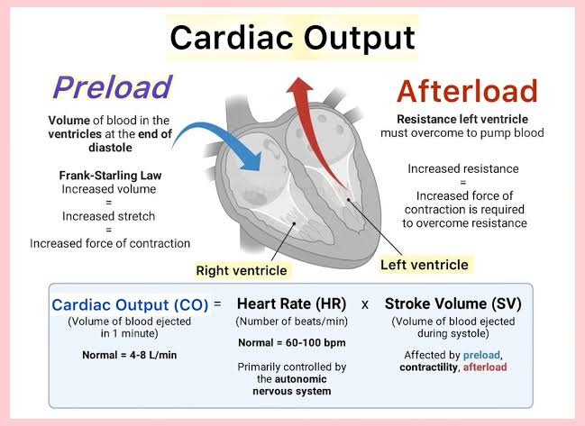

<ul><li><p><span style="background-color: transparent;">ventricular function &amp; pump performance depend on all of these (loading conditions + function)</span></p></li></ul><p></p><p>Ventricular performance (stroke volume &amp; cardiac output) is governed by 3 primary mechanical determinants: preload, afterload, &amp; contractility</p><ul><li><p>these factors control how much blood fills the ventricles &amp; how forcefully &amp; completely they eject it</p></li></ul><p></p>
21
New cards

Simpson's Biplane Method of Disks

  • divides the LV cavity into multiple discs of equal height

  • the length determines the # of disks

  • the volume of each disk will be calculated and formulated into the calculation package within our ultrasound machines to provide us with LVEDV & LVESV

  • preferred method for EF 


  • limitations

    • need to visualize all walls

    • foreshortened apicals = inaccurate measurements

    • using 2D to measure something that is 3D


<ul><li><p><span style="background-color: transparent;">divides the LV cavity into multiple discs of equal height</span></p></li><li><p><span style="background-color: transparent;">the length determines the # of disks</span></p></li><li><p><span style="background-color: transparent;">the volume of each disk will be calculated and formulated into the calculation package within our ultrasound machines to provide us with LVEDV &amp; LVESV</span></p></li><li><p><span style="background-color: transparent;">preferred method for EF&nbsp;</span></p></li></ul><p></p><ul><li><p><span style="background-color: transparent;">limitations </span></p><ul><li><p>need to visualize all walls </p></li><li><p>foreshortened apicals = inaccurate measurements </p></li><li><p>using 2D to measure something that is 3D</p></li></ul></li></ul><p></p>
22
New cards

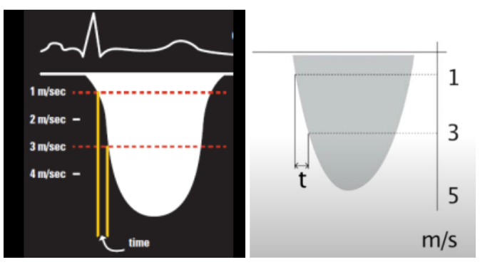
dP/dt

  • Not influenced by loading conditions

  • dP/dt is the rate of rise of LV pressure

  • ∆ pressure/ ∆ time

  • Taken from MR Doppler jet (machine package)

    • Draw a vertical line from intercept of mitral regurgitation jet at 1 m/s and 3 m/s

    • Measure time (msec)

  • Normal value > 1200 mm Hg/sec


  • limitations

    • need good MR jet to measure


<ul><li><p><span style="background-color: transparent;"><em>Not influenced by loading conditions</em></span></p></li><li><p><span style="background-color: transparent;">dP/dt is the <em>rate </em>of rise of LV pressure</span></p></li><li><p><span style="background-color: transparent;">∆ pressure/ ∆ time</span></p></li><li><p><span style="background-color: transparent;"><u>Taken from MR Doppler jet</u> (machine package)</span></p><ul><li><p><span style="background-color: transparent;">Draw a vertical line from intercept of mitral regurgitation jet at 1 m/s and 3 m/s</span></p></li><li><p><span style="background-color: transparent;">Measure time (msec)</span></p></li></ul></li><li><p><span style="background-color: transparent;">Normal value &gt; 1200 mm Hg/sec</span></p></li></ul><p></p><ul><li><p><span style="background-color: transparent;">limitations </span></p><ul><li><p>need good MR jet to measure </p></li></ul></li></ul><p></p>
23
New cards

Tissue Doppler e′ velocity

  • apical 4-chamber

  • PW Doppler at MV septal and lateral annulus

  • SV size 5-6 mm

  • sweep speed = 100 mm/s

  • Measure: e’ wave, also gives E/e’ ratio

  • E/e’ ratio used to estimate LAP

  • normal E/e’ ratio < 14

  • technical Tips:

    • Optimal spectral waveforms should be sharp

    • Scale set to 20 cm/sec


  • limitations


<ul><li><p><span style="background-color: transparent;">apical 4-chamber</span></p></li><li><p><span style="background-color: transparent;">PW Doppler at MV septal and lateral annulus</span></p></li><li><p><span style="background-color: transparent;">SV size 5-6 mm</span></p></li><li><p><span style="background-color: transparent;">sweep speed = 100 mm/s</span></p></li><li><p><span style="background-color: transparent;"><u>Measure</u>: e’ wave, also gives E/e’ ratio</span></p></li><li><p><span style="background-color: transparent;">E/e’ ratio used to estimate LAP</span></p></li><li><p><span style="background-color: transparent;">normal E/e’ ratio &lt; 14</span></p></li><li><p><span style="background-color: transparent;">technical Tips:</span></p><ul><li><p><span style="background-color: transparent;">Optimal spectral waveforms should be sharp</span></p></li><li><p><span style="background-color: transparent;">Scale set to 20 cm/sec</span></p></li></ul></li></ul><p></p><ul><li><p>limitations </p><ul><li><p></p></li></ul></li></ul><p></p>
24
New cards

Appropriate Doppler sample volume locations

TDI Mitral Annulus e’

  • Apical 4-chamber

  • PW Doppler at MV septal & lateral annulus


PW Doppler: Mitral Inflow (E, A, E/e’ ratio)

  • Apical 4-chamber

  • PW Doppler at MV leaflet tips


Pulm Vein S/D ratio

  • A4C view

  • PW placed 5-10 mm into the pulmonary veins


IVRT

  • A5C

  • CW in between MV & LVOT


25
New cards

Normal left ventricular twist (torsion)

26
New cards

Direction of apical & basal rotation during systole (LV mechanics)

27
New cards

Clinical significance of ventricular torsion (LV mechanics)

28
New cards

Expected echocardiographic findings following heart transplantation

29
New cards

Common surveillance testing following transplantation

30
New cards

General indications for stress echocardiography in transplant patients

31
New cards

General principles of diastolic function assessment

32
New cards

Situations in which ASE diastolic grading guidelines should not be applied

33
New cards

Clinical conditions that may alter interpretation (Diastolic Function)