ua week 7
 Call Kai
 Learn
 Practice Test
 Spaced Repetition
 Match
 Flashcards
 Knowt Play
1/83

There's no tags or description
Looks like no tags are added yet.
84 Terms
CSF fluid
subarachnoid CNS space, formed by selective filtration of plasma and active transport secretion by epi membranes
csf physiology
floes through subarachnoid space between arachnoid and pia mater, reabsorbed into blood, formed by selective filtration
csf total vol
adult 80-150, baby 10-60
meninges
lines brain and spinal cord, dura mater (outer), arachnoid (middle), pia mater (surfaces of brain and spinal cord)
csf functions
provide nutrients to nervous tissue, remove metabolic wastes, maintain intracranial pressure, mechanical barrier to cushion brain and spinal cord
csf fluid production
choroid plexuses of the 4 ventricles, 20 mL/min produced in adults
csf collection
3-5 lumbar puncture, aliquoted into 3+ sterile tubes- 1 chem freeze, 2 micro rt count in 1hr, 3 hem cell count 4 hr fridge, 4 other, if only 1 tube do micro first
CSF milky vs cloudy/turbid
protein, lipid, vs wbc (meningitis) infec, poteins lipids
csf pink-red
intracranial bleed (consistent color/cell count, no clot), traumatic tap (gradual decrease color/count from 1st to last tubes, clots- web like= TB)
csf xanthrochromia
yellow/amber/orange, hgb degradation products due to older intracranial bleed, elevated serum bili and protein, if bloody centrifuge & observe supernatent, jaundice
csf cell count range
0-5 wbc adults, <30 newborn, 0 rbc, manual hemocytometer (neubauer counting chamber), hematology analyzer body fluid mode
granulocytes
lyse within 1 hr stat count
csf diff
cytospin concentrated slide, adult primary lymphs:monos 70:30, kids 30:70
neutrophils
bacterial meningitis, phagocytize bacteria, increase early virla, fungal, tubercular, parasitic, may have vacuoles, bm contamination, can pyknotic
csf diff abnormals
high neutrophil- bacterial meningitis, high lymph/monos- viral meningitis, macrophages- previous hemorrhage, malignant cells- hematologic origin, metastatic carcinomas, CNS tumors
abnorm csf values due to
alteration in permeability of blood-brain barrier, increased metabolism by neural cells in response to pathological condition
multiple sclerosis
<50 lymphs norm and reactive, no bands in serum only in csf, compare to IgG index
eosinophils
coccidioides immitis, med and shunts into CNS
csf protein
15-45 method dependent, increased in infants and over 40, low= leak, high= meningitis or hemorrhage damaging blood brain barrier
IgG index
csf IgG / serum IgG / csf alb / serum alb, >0.7 = IgG production within CNS
detection of oligoclonal bands
cns inflammation, located in the gamma region or protein electrophoresis, simultaneous serum electrophoresis must be done
csf glucose
60-80% plasma glucose if low sig, compared to blood (drawn 2 hrs before spinal tap), selective transport across blood brain barrier
low csf glucose
with high wbc neutrophils= bacterial meningitis, high lymphs= TB meningitis, w/o high wbc= alteration of transport across blood brain barrier or increased glu metabolis by brain cells
high csf glucose
due to increase plasma glucose
additional csf testing
lactate, GS and culture, molecular, serology
csf lactate
dx and manage meningitis (>25= bact TB fungal, <25= viral), elevated until effective treatment falling rapidly, due to low O2 flow to tissue condition (severe head injury)
csf glutamine
by brain cells from ammonia and a-ketoglutarate to remove toxic ammonia, Indirect detects ammonia in csf, 8-18, better than direct csf
elevated csf glutamine
liver disease, kids with reye syndrome, disturbance of consciousness when glutamine >35
CSF glutamine staining
acid fast or fluor ab only it Tb meningitis, india ink for cryptococcus neoformans, starburst pattern= cryptococcus
immunologic assays- latec agglu
more sensitive for C. neoformans than india ink, confirm with culture dye to false pos frequency (rheumatoid factor), can also use enzyme linked immunosorbent
GS csf
on sediment of centrif at 1500g for 15 min, cytocentrif helps, draw blood culture, few orgs and debris = difficult, best for detection
csf GS and immunologic assays for
S. pneumo, H. influ, E. coli, N. meningitis, L. monocytogenes, S. agalactiae, (MTB, C immitis immuno only)
naegleria fowleri
in ponds/lakes/pools, enters nasal passage migrated to brain, motile amoeba in wet prep, nonmotile in cytospin prep, elongated with tapers posterior
PCR
nuc acid amp detect cause of meningitis with pathogen dna, high sens and spec, rna detects and diff path of meningitis
serologic testing
for neurpsyphilis, uncommon due to early penicillin treatment, detection of AB is dx, VDRL specific with pos FTA-ABS
serous fluid
ultrafiltrate of plasma constantly produced and reabsorbed, pleural, pericardial, peritoneal, lubricant between membranes surrounding organs and organ cavities
effusion
excess fluid between membranes, transudate (CHF or hypoproteinemia) or exudate (direct damage)
transudate effusion congestive heart failure
imbalance of filtration & reabsorption, bad heart pumping> excess blood in bv= increased hydrostatic pressure> push fluid into serous space)
transudate effusion hypoproteinemia
imbalance of filtration & reabsorption, low protein in plasma due to nephrotic syndrome> osmotic pressure change> fluid flow into serous spaces to even out concentrations
exudate effusion
direct damage to membranes by infection or malignancy
transudate lab values
protein <0.5 fluid:serum, LDH < 0.6 fluid:serum, low wbc and rbc
exudate lab values
protein >0.5 fluid:serum, LDH >0.6 fluid:serum, high wbc and rbc
collection of serous fluids
pleural- thoracentesis, pericardial- pericardiocentesis, peritoneal- paracentesis, large vol, sterile for micro/cytology, EDTA for cell count/diff, ice for pH
hematology serous fluid tests
cell count- wbc, total nucleated cells, rbc; nucleated cell diff- wbc, mesothelial, malignant
chemistry serous fluid tests
pH, protein, LDH, amylase, tumor markers
pleural
transudate, exudate (pneumonia, malignancy, PE), low pH= pneumonia not responding to abx, pH6= esophageal rupture

mesothelial cells
line serous membranes, norm or reactive
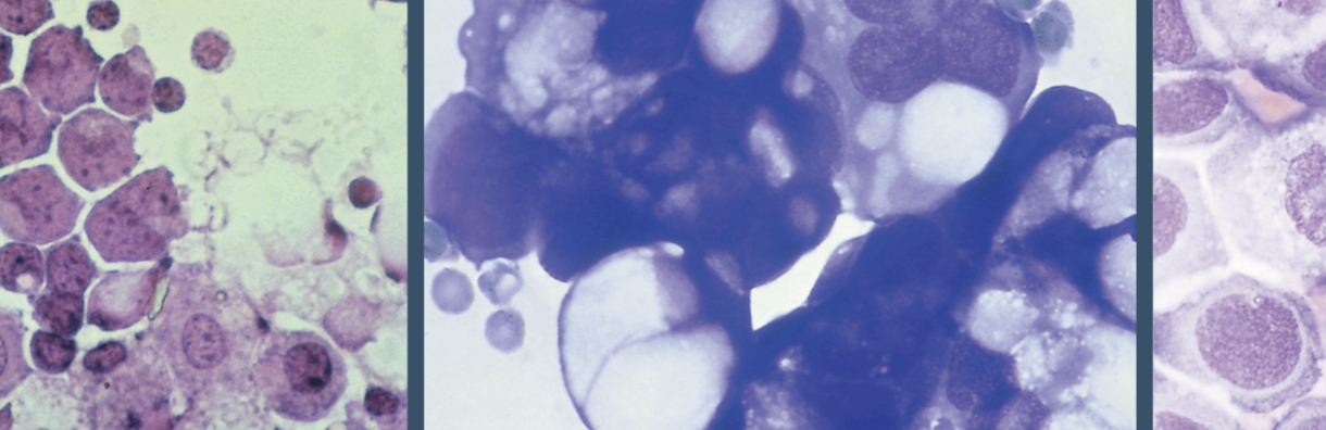
malignant cells
high nucleus:cytoplasm, irregular chromatin, nuclear and cytoplasmic molding (no windows between cells)
pericardial
cardiac tampnade by effusions, transudate, exudate (malignancy, trauma, pericarditis or endocarditis), high neutrophils & >1000 wbc= bacterial endocarditis
peritoneal
ascites, transudative (hepatic disorder- cirrhosis), exudate (bac infec= peritonitis, appendicitis, intestinal perforaton; malignancy)
peritoneal green
bile, high amylase= pancreatitis and GI perforation, high CEA= GI malignancy, high CA125= ovarian malignancy
synovial fluid
lubricate joints (diarthroses), shock absorbing in activity, nutrients for cartilage, ultrafiltrate of plasma containing hyaluronic acid
joint disorders
noninflam- degenerative (osteoarthritis), septic- infect, inflam- immunologic (RA) or crystal inducted (gout, pseudogout), hemorrhagic- traumatic injury/ tumor
synovial collection
arthrocentesis, EDTA (anticoag if disease fluid has fibrinogen)- cell count/ diff/ crystal ID, hyaluronidase- decreases viscosity
synovial fluid appearance
norm= clear-pale yellow, turbid= cells crystals fat droplets fibrin, deep yellow= effusion, bloody, green= bacterial infec
synovial cell count
norm= 200 wbc, abnormal highest in inflam and septic
synovial diff
norm= mono/macro/lining cells, septic= high neutrophils, inflam= high lymphs

synovial crystal analysis
monosodium urate MSU= uric acid gout, calcium pyrophosphate dihydrate CPPD= pseudogout birefringence, polarizing microscope
synovial fluid other tests
GS and culture, glu, protein, uric acid, lactate, enzymes
ammniotic fluid collection
amniocentesis, genetic disease- neural tube disorder, fetis distress- bili for HDFN, fetal maturity- resp distress (common in preme) and lung maturity by surfactant level
fetal lung maturity- surfactant level
phospholipid lowers surface tension in alveoli (stays open), analyizes L/S ratio, phosphatidyl glycerol, foam stability, lamellar bodies
L/S ratio
TLC, labor intensive
phosphatidyl glycerol
immunologic agglutination test
foam test
amniotic fluid added to 95% EtOH (antifoam), shake for 15 sec, stable bubble ring after 15 min= adequate surfactant

lamellar body count
phospholipid storage granule, same size as plt, measured on hem analyzer using plt channel
fecal occult blood
colorectal cancer screening, anemia investigation, guaiac method, immunochemical method
fecal leukocytes
ulcerative colitis, infec diarrhea, wright stain
fecal fats and elastase
pancreatic insufficiency, quantitative
fecal occult blood- guaiac method vs immunochemical method
pseudoperoxidase activity of hgb= color change vs ab specific for human hgb
fecal occult blood false neg
testing before stool has dried on card, collected in closed container can heat and break down hgb, too much hgb= prozone
vaginal secretion wet prep
wbc, yeast, clue cells, trich
vag secretions rupture membranes testing
fetal fibronectin- preterm labor, AmniSure, actin PROM, ROM plus, fern test

semen analysis
infertility, assisted reproductive tech, postvasectomy, forensic analyses, sperm count/motility/morph
neubauer calc formula
cells counted x dilution/ cells counted x col of 1 square
cytocentrifugation
makes monolayer, 30% alb increases yield and decreases distortion, positive slide attracts cells
leukemias
lymphoblasts, myeloblasts, monoblasts, nucleoli more prominent than in blood, bands in serum and csf
lymphomas
dissemination from lymph organs, cleaved nucleii and prominent nucleoli, bands in serum and csf
HIV bands
in serum and csf
electrophoresis and immunophoretic- elevated CSF total protein but a normal cell count and a clear, colorless specimen
encephalitis, neurosyphilis, guillain-barre, and neoplasms, ref symptoms
myelin basic protein
presence in csf indicated demyelination of myelin sheath around axons of neurons, monitors MS treatment, immunoassay
A fluid specimen submitted as "CSF" is of questionable origin. Which finding would help confirm that the fluid is truly CSF?
Presence of the tau transferrin (carbohydrate-deficient transferrin) fraction, which is unique to CSF
csf/serum alb
>9 blood brain barrier damage
A control slide for the detection of monosodium urate crystals is prepared with which material?
Betamethasone acetate
A MLS counts 63 cells in all nine large squares of a Neubauer chamber using a 1:2 dilution with saline. What is the WBC count (cells/uL)?
140