quiz 5 (E2)
 Call Kai
 Learn
Learn Practice Test
 Spaced Repetition
 Match
Match Flashcards
 Knowt Play
1/104

Earn XP
Description and Tags
tissues
Name | Mastery | Learn | Test | Matching | Spaced | Call with Kai | Chat |
|---|
No analytics yet
Send a link to your students to track their progress
105 Terms
Connective Tissue General Features
most abundant tissue in body
major functions: strength and support, protection, insulation, transport substances around body

Substantial Matrix: basis for classification
predominant type of matrix, composition of each type of matrix

Substantial Matrix: protein fibers
collagen, elastin (elastic fibers), reticular fibers

Collagen
most common protein in body, “rope” of three fibers wound around each other, flexible and strong, but not elastic

Elastin (elastic fibers)
long, thin proteins that form branching networks, elastic

Reticular Fibers
thin, short collagen fibers, generally continuous with collagen, form “nets”

Blasts
cells that create the matrix
Cytes
cells that maintain the matrix
Mesenchyme
embryonic connective tissue: all connective tissue arises from mesenchyme, some remains as source of stem cells in adult
structure: star-shaped mesenchymal cells, fluid ground substance

Areolar Tissue (loose connective tissue)
structure: gel like matrix, loosely-arranged protein fibers, hyaluronic acid

Areolar Tissue (loose connective tissue): cells
has fibroblasts
other cells: macrophages, mast cells, fat cells
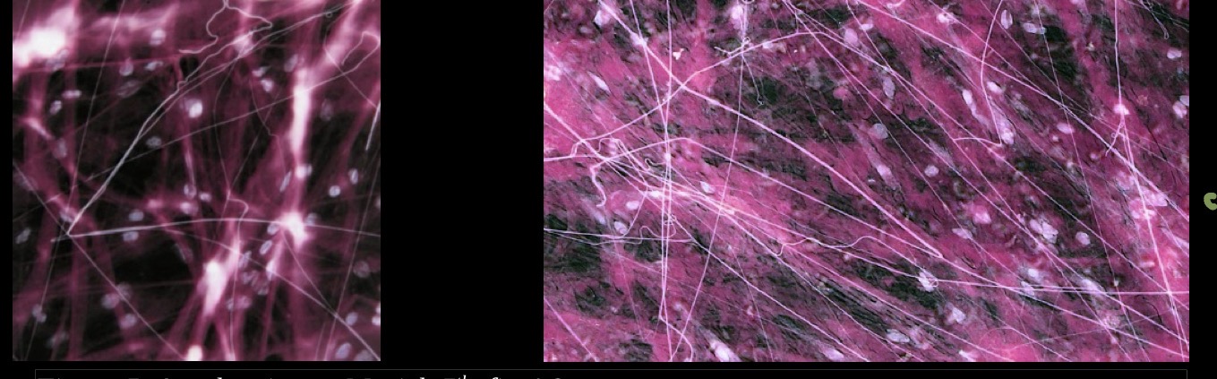
Areolar Tissue (loose connective tissue): function
“universal packing material”, allows diffusion for nutrition and wastes, reservoir for water and salts

Areolar Tissue (loose connective tissue): location
most widely distributed connective tissue, surrounds organs and capillaries, attaches skin to underlying tissues

Adipose Tissue: structure
closely packed cells (adipocytes), each cell contains so much lipid that nucleus & cytoplasm pushed to side of cell, just under plasma membrane, low levels of mitosis

Adipose Tissue: function
energy storage, insulation, supports and protects organs (padding)

Adipose Tissue: location
subcutaneous, around kidneys, mammary glands

Reticular Connective Tissue (reticular tissue): structure
reticular cells, reticular fibers

Reticular Connective Tissue (reticular tissue): function
support soft tissue of organs and small blood vessels

Reticular Connective Tissue (reticular tissue): location
lymph nodes, spleen, bone marrow

Dense Regular Connective Tissue: structure
matrix of parallel fibers, cell type is primarily fibroblast

Dense Regular Connective Tissue: function
tensile strength and stretch resistance in ONE directio

Dense Regular Connective Tissue: location
ligaments, tendons

Dense Irregular Connective Tissue: appearance
interwoven fibers, cell type is primarily fibroblast
Dense Irregular Connective Tissue: function
tensile strength and stretch resistance in MULTIPLE directions
Dense Irregular Connective Tissue: location
dermis of skin, fibrous joint capsule
Elastic Connective Tissue: appearance
coiled appearance of elastin, elastin fibers parallel
Elastic Connective Tissue: function
elastic: returns to original shape, recoil of tissue following stretching
Elastic Connective Tissue: location
walls of large arteries, walls of bronchial tubes
Cartilage: general characteristics
avascular: used in compression areas, slow healing
chondrocytes widely dispersed in lacuna
matrix: firm; fairly solid but not completely rigid, primarily collagen
fibers associated with bone
Hyaline Cartilage: structure
matrix appears transparent, chondrocytes found in spaces (lacunae) within the matrix
Hyaline Cartilage: function
forms the embryonic skeleton, allows growth of long bones, rigid and smooth, yet slightly flexible
Hyaline Cartilage: location
articular cartilage, epiphyseal plate, costal cartilage of ribs, nose, trachea
Elastic cartilage: structure
elastin fibers, lacuna

Elastic Cartilage: function
flexibility, shape retention

Elastic Cartilage: location
external ear, epiglottis

Fibrocartilage: structure
bundles of collagen fibers, lacuna

Fibrocartilage: function
absorbs compression, tensile strength

Fibrocartilage: location
intervertebral disks, articular disks, pubic symphysis

Bone: general characteristics
most of skeleton, solid matrix, excellent blood supply, osteocytes found in lacunae in the matrix
Blood: structure
40-45% of volume is cells, primarily fluid: plasma, fibrous proteins in the plasma help with clotting
Blood: function
transportation of gasses, nutrients, wastes, hormones
Blood: location
cardiovascular system
Serous Membrane characteristics
made of thin layers of epithelial and connective tissues, “fist pushed into balloon”
Serous Membrane parts
Visceral Serous Membrane, Parietal Serous Membrane, Serous Fluid
Serosal membranes associated with ventral cavities
pleura - lungs, pericardium - heart, peritoneum - organs in abdominal cavityFunctions of Skin
Functions of Skin
Protection, body temperature regulation, cutaneous sensation, Vitamin D synthesis, blood reservoir, excretion
Skin Protection
physical barrier, chemical barrier, biological barrier
physical barrier of the skin
hard and thick, oil and glycolipid secretion (from glands waterproofs skin), melanin, hair
Chemical barrier of the skin
pH, RNAse
biological barrier of the skin
Langerhans cells within epidermis, macrophages within dermis
body temperature regulation of the skin
autonomic nervous system: heat loss required, sweating, vasodilation and greater opening of skin capillary beds
heat retention required: decreased sweating, vasoconstriction and decreased opening of skin capillary beds
Cutaneous sensation of the skin
touch in epidermis and dermis, temperature, pressure, pain receptors in dermis, hair follicles well innervated
Vitamin D synthesis of the skin
produced in skin with UV light exposure, required for absorption of calcium from gut
blood reservoir of the skin
highly vascularized, when blood needed elsewhere, skin capillary beds close
excretion of the skin
nitrogen wastes, water and NaCl loss
Skin General Structure
Epidermis, Dermis, Hypodermis
Epidermis
thick stratified squamous keratinized epithelium
Epidermis: Keratinocytes
produce keratin (soft form), Desmosomes, developmental progression, turnover time ≈ 52-75 days
Epidermis: Melanocytes
produce melanin: pigment, provides protection from UV light
number of melanocytes is similar for people with different skin tones
people with darker skin have more active melanocytes that produce more pigment
how melanocytes create skin tone
melanin stored in membrane granules (packages), granules move to tips of processes, uptake by keratinocytes by phagocytosis
in response to UV light, melanin production increases
Epidermis: Langerhans cells
phagocytic scavengers, immune protection
Epidermis: Merkel Cells
detect touch, located at the junction between the epidermis and dermis
Epidermis: Strata
epidermis has several megalayers of cells
bottom layer contains
1) stem cells for keratinocytes
2) melanocytes
3) a few Merkel cells
as cells mature, they move to the surface
1) fill with keratin
2) die
surface strata contains multiple layers of dead cells
1) 20-30 cell layers
a) in thick skin can be up to 50 layers
b) completely keratinized cells: “cornified”
2) dead cells can “flake” of
Dermis
connective tissue; fibers primarily collagen, but also elastin and reticular fibers, cells primarily fibroblasts, ground substance is gel-like
Dermis: structural support for skin
the “hide” of animals, rich supply of blood and nerves, relationship to accessory structures
Dermis: Papillary Layer
areolar connective tissue, papillae project into epidermal layer: capillary loops, nerve endings
Dermis: dermal ridges
large mounds, force overlying epidermal layer to form ridges assist with grip, friction
Dermis: Reticular Layer
dense, irregular connective tissue, thick bundles of collagen
Hypodermis (subcutaneous tissue)
Loose connective tissue, Adipose tissue
Dermis: Thick Skin
in palms, fingertips, soles of feet
more layers of strata than thin skin
more papilla of dermal papillary layer than thin skin
has epidermal ridges
can have calluses
Dermis: Thin Skin
everywhere else that thick skin isnt
less strata than thick skin
less papilla of dermal papillary layer than thick skin
hair follicles found here
Hair characteristics
shaft formed by dead, keratinized cells, shaft is visible hair, cells added at base of hair root, “hard” keratin, rather than “soft” keratin of skin
Hair texture
determined by shape of shaft in cross section: flat/ribbon-like tight curls oval wavy round straight, usually coarse
Hair Genetics
straight hair: KSKS
curly hair: KCKC
wavy hair: KSKC
Hair bulb
epidermal growth zone, epithelial tissue, single cell layer, actively dividing, producing hair cells, melanocytes add pigment to hair cells
Vellus Hair
short, fine hair; usually unpigmented, puberty over most of body
Terminal hair
coarse, longer and pigmented, tends to be localized in body
Hair growth cycle
1) hair growth
2) resting phase
3) hair shed, new growth begins
male pattern baldness
hair follicles lost, remaining follicles revert to producing vellus hair
Nails
modified epithtelial tissue
Nail components
nail root, nail body, free edge of nail
Nail matrix
by nail root, part of nail bed, responsible for most of nail growth
Nail bed
underneath nail body and root, contributes to nail thickness
Sebaceous Gland: produce sebum
oily, rich in lipids, released in holocrine manner, testosterone can increase release
Sebaceous Gland: actions
waterproofs, softens and lubricates skin, antibacterial
ducts usually terminate in hair follicles
Sebaceous Glands: distribution
cover body, except palms and soles, large on face, neck, upper chest
Eccrine Sweat Gland
most common, watery secretion, secretory portion located in dermis, duct opens to surface, not associated with hair follicles
Eccrine Sweat Glands: watery secretion
99% water, salts, trace metabolic wastes, released in a merocrine fashion, temperature regulation
Eccrine Glands: distribution
over the entire body surface, highest concentration in palms, soles, forehead
Apocrine Sweat Gland
merocrine secretion of sweat, proteins and lipids, larger than eccrine sweat glands, ducts terminate in hair follicles
~ ceruminous gland
~ mammary gland
Apocrine Sweat Gland: merocrine secretion of sweat, proteins and lipids
released in a merocrine fashion, protection from bacteria, odor results from bacterial degradation
Apocrine Sweat Gland: distribution
axillae, anogenital region
Skin Cancer
the most common type of cancer, correlates with sun damage
Basal Cell Carinoma
most common, originates in stratum basale, least malignant, 99% cure rate
Squamous Cell Carcinoma
stratum spinosum keratinocytes, rapid growth, spread, good cure rate if caught early
Melanoma (malignant melanoma)
5% of skin cancers
2) develops from melanocytes, usually pre-existing mole
3) unless caught early, generally fatal
4) ABCDE Rule
ABCDE Rule Melanoma
Asymmetry
Border irregularity
Color
Diameter
Evolving
Burns: process
denaturing of cell proteins, cell death, causes
Burns: 1st Degree
epidermal damage, redness, swelling and pain, generally heal in a few days
Bunrs: 2nd Degree
epidermis and upper dermis/all of dermis, results in blisters (in addition to previous symptoms), healing time depends on depth of damage in dermis