403 Unit 3: CRR
 Call Kai
 Learn
 Practice Test
 Spaced Repetition
 Match
 Flashcards
 Knowt Play
1/267

There's no tags or description
Looks like no tags are added yet.
268 Terms
Vasculature: 2-85
Introduction to Cardiovascular: 86-195
Renal/Urinary: 195-268
Artery Layers
Tunica Interna
Tunica Media
Tunica Externa
Artery Tunica Interna Components
Endothelium and an Elastic lamina
Artery Tunica Media Components
Smooth muscle (ANS) and an Elastic lamina
Artery Tunica Externa Components
Elastin and Collagen
Elastic vs. Muscular Arteries
Elastic: “Pressure reservoir” arteries where walls are about 10% of the vessel’s diameter
Muscular: “Vascular tone” arteries where walls are about 25% of the vessel diameter (thick muscular layer of tunica media)
Arterioles are _ vessels with a thick _
Resistance, Tunica media
Capillaries are _ vessels composed only of _
Exchange, A tunica interna with endothelium and a basement membrane
Capillary diameter is _ compared to the size of an erythrocyte
About the same
Vein Layers
Tunica Externa
Tunica Media
Tunica Interna
Vein Tunica Externa Components
Elastin and Collagen
Vein Tunica Media Components
Smooth Muscle
Vein Tunica Interna Components
Endothelium/basement membrane and Valves
3 methods of venous return
Muscle milking
Respiratory milking
Venae comitantes
3 Portal Systems and Venous/Arterial Classification
Hepatic - Venous (GI/liver)
Hypophyseal - Venous (hypothalamus/anterior pituitary)
Renal - Arterial (glomerulus/peritubular capillaries)
Which layer of a blood vessel is most affected by autonomic stimulus?
Tunica Media
Aorta “Parts”
Ascending Aorta
Arch of Aorta
Descending Aorta - Thoracic
Descending Aorta - Abdominal
The aorta bifurcates at _ into _
L4, Common iliac arteries which supply the abdominopelvic cavity and lower limb
The coronary arteries originate from _
The ascending aorta
The aortic arch is at _ spinal level
T4
The aortic arch splits into which four arteries?
Right common carotid artery (via brachiocephalic trunk)
Right subclavian artery (via brachiocephalic trunk)
Left common carotid artery
Left subclavian artery

1
Brachiocephalic Artery/Trunk
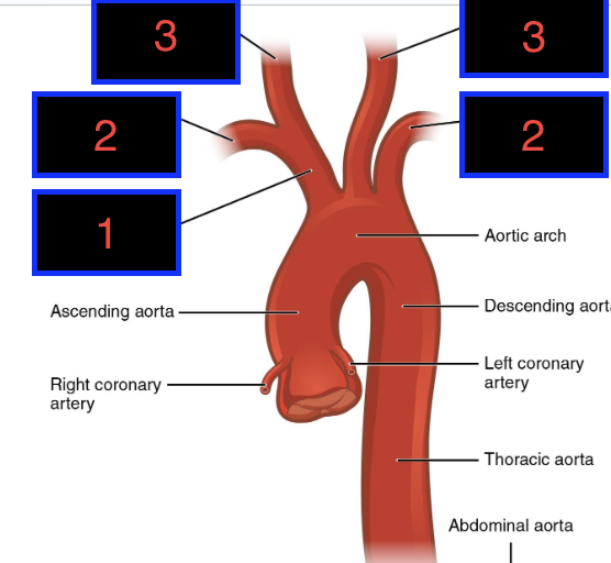
2
Right and Left Subclavian Arteries

3
Right and Left Common Carotid Arteries
The descending aorta spans from _ to _ spinal level
T5, T12
The visceral branches of the thoracic aorta supply _
The parietal branches of the thoracic aorta supply _
Thorax contents
Thoracic wall
The aortic hiatus is located _
At T12, within the diaphragm
Abdominal Aorta Anterior Visceral Branches (3)
Celiac Trunk
Superior Mesenteric Artery
Inferior Mesenteric Artery
The celiac trunk is located at _ spinal level and supplies _
T12, Foregut
The superior mesenteric artery is located at _ spinal level and supplies _
L1, Midgut
The inferior mesenteric artery is located at _ and supplies _
L3, Hindgut
Abdominal Aorta Lateral Visceral Branches (2)
Renal Arteries
Gonadal Arteries
The renal arteries are located at spinal level _ and supply _
L1, Kidneys
The gonadal arteries are located at _ spinal level and supply _
L2, Ovaries/testes
Abdominal Aorta Parietal Branches supply _
The diaphragm, lumbar wall, etc.
The brachiocephalic trunk predominantly supplies which areas of the body with blood?
Right head and neck, upper limb

1
Celiac Trunk

2
Gonadal Arteries

3
Inferior Mesenteric Artery

4
Renal Artery/Vein

5
Superior Mesenteric Artery

6
Common Iliac Vein and Artery
Carotid Sheath
Neck structure containing the Common carotid artery, Internal jugular vein, and Vagus nerve
Internal carotid artery supplies _
Cranial cavity/brain, Orbit, and Forehead
External carotid artery supplies _
Superior neck, Face, and Scalp
External Carotid Artery Branches (3)
Facial Artery (superficial face)
Maxillary Artery (deep face)
Superficial Temporal Artery (external cranium, scalp)
Internal Carotid Artery supplies which components of the Circle of Willis?
Anterior cerebral a.
Anterior communicating a.
Middle cerebral a.
Posterior communicating a.
Which areas external to the cranial cavity are supplied by the internal carotid aa.?
Orbit and Forhead
Branches of Common Iliac Artery
Internal Iliac Artery
External Iliac Artery
Internal iliac artery divisions and regions each one supplies
Anterior: Pelvic viscera, perineum, gluteal region
Posterior: Posterior pelvic wall, gluteal region
External Iliac Artery Pathway
Common Iliac Artery
External Iliac Artery
Femoral Artery
Popliteal Artery
Posterior Tibial Artery
The femoral artery appears at _ and supplies _
The inguinal ligament, Anterior/posterior thigh
The popliteal artery appears at _ and supplies _
Adductor hiatus (knee), Anterior leg/dorsal foot
The posterior tibial artery is located _ and supplies _
On the posterolateral leg, Plantar surface of foot
Femoral Triangle
Femoral Vein, Femoral Artery, Femoral Nerve
Which landmark indicates the transition of the external iliac a. to the femoral a.?
Inguinal Ligament
The axillary artery becomes brachial at _
Teres major m.
Upper Limb/Arm Blood Supply Pathway
Ascending Aorta → Subclavian Artery → Axillary Artery → Brachial Artery → Radial and Ulnar Arteries
The subclavian artery becomes axillary at _
Rib 1 (lateral margin)
Subclavian Artery 1st Part supplies _
Vertebral artery (brain) and Internal thoracic artery (thorax/abdominal walls)
The subclavian artery transitions from 1st-2nd and 2nd-3rd part based on which landmark?
Scalenus Anterior m.
The axillary artery transitions from 1st-2nd and 2nd-3rd part based on which landmark?
Pectoralis Minor m.
The brachial artery divides into radial/ulnar arteries at _
The cubital fossa
The radial artery becomes the _ in the hand
Deep palmar arch
The ulnar artery becomes the _ in the hand
Superficial palmar arch
Allen Test
A way of measuring if the radial and ulnar arteries have an anastomose based on whether blood can return to the hand upon occlusion of one of the arteries
If present, an anastomosis with which artery allows for safe cannulation of the radial artery?
Ulnar Artery
There is/are _ brachiocephalic arteries and _ brachiocephalic veins
1, 2
The superior vena cava is a union of _
Right and Left Brachiocephalic veins
The brachiocephalic veins combine _ and _ at a _
Internal Jugular Vein, Subclavian vein, Venous angle
Lymph returns to venous circulation at approximately _
The venous angles
The internal jugular vein drains _
Head and neck
The subclavian vein drains _
Upper limb, head, neck, and thorax
The cephalic vein drains into _
The axillary vein
The basilic vein drains into _
The axillary vein (and becomes it)
Vein used for venipuncture
Median Cubital Vein
The venous angle is the union of the subclavian v. with which other vein?
Internal Jugular Vein
IVC Tributaries
(Common) Iliac Veins, Lumbar Veins, Testicular/Gonadal Vein, Renal Veins, Suprarenal Veins, Hepatic Veins
“I Like To Rise So High”
Superficial Venous Return from Lower Limb
Small Saphenous Vein → Popliteal Vein (+ Great Saphenous Vein) → Femoral Vein → External Iliac Vein (+ Internal Iliac Vein) → Common Iliac Vein
Deep Venous Return from Lower Limb
Via venae comitantes
Which structures’ venous pathway includes the left renal vein?
Left gonad, inferior surface of diaphragm, kidney, and suprarenal gland
Left Renal Vein Tributaries (4)
Kidney (ipsilateral)
Hemi-diaphragm (ipsilateral)
Suprarenal Gland (ipsilateral)
Gonad (ipsilateral)
Hepatic Portal System Flow
Inferior Mesenteric Vein → Splenic Vein (+ Superior Mesenteric Vein) → Hepatic Portal Vein → Liver → Hepatic Veins→ IVC
Portal Hypertension
Increased portal pressure due to numerous etiologies, leading to esophageal, peri-umbilical, or anorectal submucosal varices
The hepatic portal system delivers material from which source tissues to the sinusoids of the liver?
GI Tract
Blood Broad Components (2) and Functions (3)
Components: Plasma and Formed Elements
Functions: Transportation, Regulation, Protection
Thoracic Cavity Boundaries - Superior, Inferior, Anterior, Posterior, Lateral
Superior: First Ribs
Inferior: Diaphragm
Anterior: Sternum
Posterior: Vertebral Column
Lateral: Intercostal Spaces/Lateral Ribs
Thoracic Cavity Spaces
Pulmonary Cavities and Mediastinum
The pulmonary cavities contain _ and are lined by _
Lungs, Pleurae
The mediastinum contains _
Heart, viscera, great vessels/nerves
Mediastinal Parts
Superior
Anterior
Middle
Posterior

Blue Highlight
Superior Mediastinum

Green Highlight
Middle Mediastinum

Red Highlight
Posterior Mediastinum
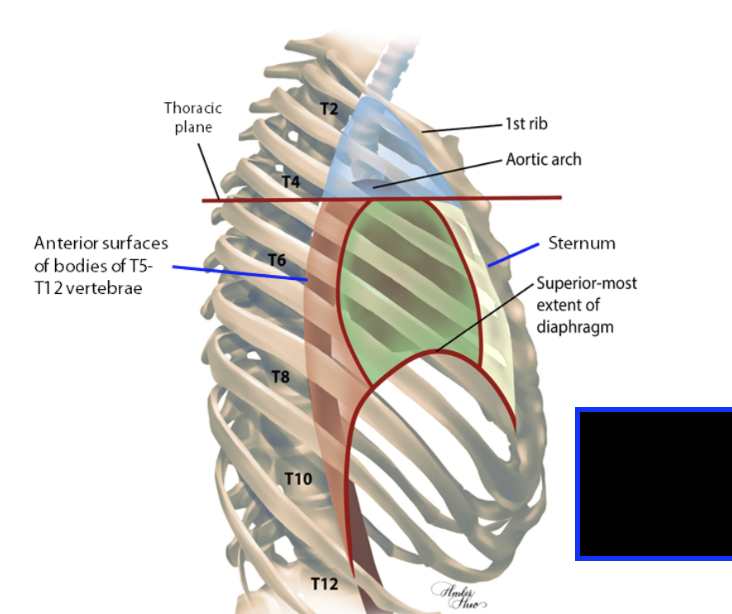
Yellow Highlight
Anterior Mediastinum
The superior mediastinum spans _
T2-T4, ending at the thoracic plane
Thoracic Plane
T4/5 intervertebral disc to sternal angle
The anterior mediastinum spans _
T5-T9
The middle mediastinum spans _
T5-T8
The posterior mediastinum spans _
T5-T12