Ortho Path Learning Objectives - Exam 1
 Call Kai
 Learn
Learn Practice Test
 Spaced Repetition
 Match
Match Flashcards
 Knowt Play
1/125

There's no tags or description
Looks like no tags are added yet.
Name | Mastery | Learn | Test | Matching | Spaced | Call with Kai | Chat |
|---|
No analytics yet
Send a link to your students to track their progress
126 Terms
Name the 3 phases of healing
inflammation, proliferation, remodeling
Describe the function of hemostasis and a blood clot after injury
body's response to stop bleeding and start healing after a blood vessel innjured
- Blood clotting- release of pro-inflammatory molecules such as cytokines and growth factors
Identify the time frame for each phase of tissue healing
Inflammatory phase - occurs immediately after
Proliferation phase - few weeks
Remodeling phase - months up to a year
Compare and contrast the difference betweeen intraarticular and extraarticular ligament healing and how this guides clinical decision making
Intra-articular - environment without supporting soft tissue vascular supply to allow healing
- ACL - within joint
Extra-articular - environment with ample surrounding microvasculature to facilitate healing
- MCL - around joint capsule
Describe the role of stress on healing tissue and a positive effect from early stress
Stress will promote adaptation and improved function
Contrast intramembranous bone formation from endochondral bone growth
Intramembranous - osteoblast differentiate from mesenchyme and form bone directly. There is no cartilaginous stage.
Endochondral - during 6th gestational week mesenchyme turn into chondrocytes. Then the chondrocytes form preliminary skeleton. Then they maturen and calcify to turn to bone.
Name a common connective tissue disorder
Ehlers Danlos Syndrome (EDS) - happens when someone has faulty collagen which leads to fragile skin and unstable joints
List 2 risk factors that lead to impaired wound and injury healing
- Age
- nutrition
- comorbidities (diabetes, cardiovascular disease, autoimmune)
- medications (anti-inflammatory and immune medications)
- lifestyle (smoking, diet, alcohol consumption)
Describe the role of proteoglycans in cartilage formation
intracellular matrix of hyaline cartilage
Proteins attach to glycosaminoglycans = proteoglycans.
combine with hyaluronic acid and water = elastic
Describe the role and importance of articular cartilage
Smooth, specialized CT that covers the end of bones in moveable joints. It provides low-friction movement and acts as a shock absorber
Name the 3 types of embryonic tissue
Ectoderm, mesoderm, endoderm
State the healing time under normal circumstances for bone healing from injury to fully remodeled
Proliferation/Repair (2-3 Weeks)
Remodeling (4-12 Weeks)
Contrast healing by primary vs secondary intention
Primary intention: Wound edges joined by fibrin plug, regrowth of basel layer of epidermins, lysis of fibrin and re-epithelialisation, restoration to intact skin
Secondary intention: Large defect filled by fibrin clot, new blood vessels and fibroblasts grow from the dermis into fibrin, collagen laid down by granulation tissue fibroblasts to restore integrity, maturation of collagen achieves structural integrity and allows regrowth of epidermis
Joints that make up the shoulder girdle
Glenohumeral joint
Acromionclavicular joint
Sternoclavicular joint
Scapulothoracic joint
Neonatal/congenital pathologies - adult and adolescent
- Shoulder dislocation
- Recurrent instability/bone loss
- Hill-Sachs Lesion
- Bankart tear
- SLAP tear
- Multi-directional instability (MDI)
- Acromioclavicular joint injury
- Clavicle fracture
- Proximal humerus fractures
- transient brachial plexus neuropraxia
- Supracondylar humerus fracture
- Neonatal brachial plexus palsy
- Symbrachydactyly
- Amniotic band syndrome
- Osteochondritis dissecans
- Parsonage-Turner Syndrome
- Sternoclavicular joint dislocations/physeal fractures
- Sternoclavicular joint injury
Describe simple elbow dislocation criteria
no fracture
- Nonoperative treatment when stable through passive range of motion after closed reduction
Describe complex elbow dislocation criteria
dislocation and associated fracture
ex: radial head, coronoid, or medial epicondyle fracture
Name the three joints that make up the elbow joint complex
Ulnohumeral, radiocapitellar, and proximal radioulnar
Name the injuries that make up the elbow "Terrible Triad"
Elbow dislocation + coronoid fracture + radial head fracture
Describe the importance of the medial epicondyle and UCL in throwing athletes
The UCL has three bands, and the anterior band is the most important for resistance to valgus movement. UCL injury is seen primarily in overhead throwing athletes and is usually chronic/attritional. Athletes may have prodromal elbow pain during activity followed by a "pop" and inability to pitch. The UCL is essential for pitching well and without pain.
Be able to name the order of elbow apoptosis ossification
Capitellum → Radial head → Internal/medial condyle → Trochlea → Olecranon → External/lateral condyle. o After age 1, a new ossification center appears every 2 years.
Describe the importance of the lateral ulnar collateral ligament in elbow stability
The LUCL =varus ulnohumeral stability
without= posterolateral rotary instability
first structure to fail in elbow dislocation.
Describe the mechanism of compression for carpal tunnel and cubical tunnel syndrome
- Carpal tunnel: compression of the median nerve
roof = transverse carpal ligamen
floor =proximal carpal row
radial boundaries= scaphoid tubercle/trapezium
ulnar boundaries = hook of hamate/pisiform.
Night splinting prevents passive wrist flexion and compression.
- Cubital tunnel: compression of the ulnar nerve
between the two heads of FCU distally,
within the arcade of Struthers proximally, or between the UCL and Osborne's fascia.
Describe the location of scaphoid fractures and associated risks of missed or undertreated injuries
proximal pole, waist, and distal pole
blood enters distally.
Nondisplaced distal-pole fractures =good healing
waist and proximal-pole fractures= operative
Missed or nonhealing fractures = malunion/nonunion, avascular necrosis
Describe treatment options for scaphoid fracture based on location
Nondisplaced distal-pole fractures have a good chance of healing. Nondisplaced waist fractures can be treated nonoperatively in young patients. The slides list casting options including long-arm thumb spica, short-arm thumb spica, and short-arm cast. Waist and proximal-pole fractures, especially when displaced, are considered operative. Vascularized bone grafting is identified as a treatment for complex nonunion/AVN problems.
Describe the mechanism for "Gamekeepers thumb"
Gamekeeper's (Skier's) thumb is a first MCP medial collateral ligament sprain caused by acute traumatic abduction/valgus of the first MCP joint. The slides also describe chronic attritional injury
Describe an associated injury that requires operative intervention in treating thumb UCL injuries
A Stener lesion: the thumb UCL avulses and displaces above the aponeurosis and will not heal without surgery. The slides also state operative treatment when valgus motion is >20° compared with the other side.
Describe normal developmental milestones
- head control — 3 months
- sit — 6 months
- stand with support — 12 months
- walk unsupported — 15 months
- Infant gait is less stable and efficient, with a wide base, irregular cadence, instability, and poor energy efficiency due to a high center of gravity, low muscle-to-body-weight ratio, and immature nervous/postural control.
Define metatarsus adductus
- A medial deviation of the forefoot
- It is secondary to intrauterine positioning and/or genetics, is noted at birth, has a convex lateral border and a normal-to-high arch, and the majority resolve with growth.
Describe normal patterns of growth in the tibia
- Torsional
o internal tibial torsion- by the foot-thigh angle.
corrects over time age 3, by up to 10-15°
- Varus/Valgus
10-15° of varus at birth
maximum valgus of 10-15° at 3-4 years.
Describe normal femoral growth patterns
- The slides state that upper-limb growth occurs earlier than lower-limb growth
- the foot grows earlier than the rest of the lower limb
- during childhood the trunk grows most rapidly
- during adolescence the lower limbs grow the fastest
- For the femur specifically, the slides discuss femoral anteversion: newborn femoral anteversion is 45-50° and normal adult anteversion is 15°
Describe components of a rotational profile
- determine the foot progression angle during gait
- assess hip rotation
- assess tibial rotation
- determine the alignment of the foot
- hip rotation is measured with the child prone and includes internal and external rotation
Define anteversion and retroversion
- Femoral anteversion = forward angulation of the femoral neck
o It is usually noted after age 3
o the slides list easy W-sitting and kissing patellas as observations.
- Femoral retroversion = backward angulation of the femoral neck
Gait pattern of anteversion and retroversion
anteversion - feet inward during walking
retroversion - feet point outward during walking
Define vara and valgum
- Vara - bowed leg alignment
o measured by intercondylar distance or the femoral-tibial angle
- Valgum - knock-knee alignment
o measured by intermalleolar distance
Describe the changes in genu varus/valgum as a child ages
Normal alignment progresses from 10-15° of varus at birth to a maximum of 10-15° of valgus at 3-4 years
What are the possible contributors of in/out toeing?
Femoral anteversion, internal tibial torsion and metatarsus adductus can all contribute to in/out toeing
Describe diseases that can lead to pathologic bowing/knockknees
- Rickets - radiologic growth-plate changes, vitamin D problem, include inherited rickets
- blount disease - pathologic abnormallity of the proximal medial metaphysis, the medial growth plate slows or stops while the lateral growth plate continues
Joints of the ankle
- subtalar
- talocrural
- talocalcaneonavicular (midtarsal)
- calcaneocuboid (midtarsal)
How they correspond to motion and function
Subtalar - allows pronation, supination, rotation
o diagonal axis makes pronation/supinaton triplanar
Midtarsal
o During pronation, their axes are parallel, unlocking the foot and making it hypermobile for shock absorption.
o During supination, the axes are nonparallel and the joint becomes locked for efficient force transmission.
Normal development of the foot in terms of arch development
Infant feet = flat - of joint laxity/ abundant subcutaneous fat Adult foot length = earlier than the rest 1/2 by 12-18 months.
Be familiar with deformities of the toes and forefoot
- Hallux valgus
- Hallux varus
- Cleft foot
- Polydactyly
- microdactyly
- turf toe
- mallet toe
- hammer toe
- claw toe
Hallux valgus
- bunions
- proximal joint creates deformity
Hallux varus
- medial deviation of the big toe
- daylight sign
cleft foot
Lobster claw deformity
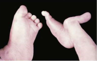
Polydactyly
extra digits

microdactyly
overly small digits

Turf toe
hyperextension of first MTP joint
Mallet toe
the part of the toe that should be in contact with the ground isn't
- distal phalanx bent
Hammer toe
- middle joint of a smaller toe bends upward and curls downward
Claw toe
mtp bends upward pip and dip joints curl downward
Vertical talus
- congenital deformity and the most severe/serious pathologic flatfoot, with flattening and convexity of the sole.
- It is normally associated with myelodysplasia or arthrogryposis.
- Surgical treatment includes anterior release to reduce the contracted ankle capsule/talus, posterior release to allow the foot to dorsiflex to neutral, and immobilization with casting.
Clubfoot
- congenital deformity occurring in about 1 in 1,000 births.
- Components are equinus, cavus, adductus, varus, and internal rotation.
- Clinical position: hindfoot equinus/varus, forefoot adductus/supination, and cavus.
- The foot may be small with a skinny calf and short leg ("clubleg").
- Types include postural, congenital idiopathic, and syndromic/teratologic.
- The standard approach is the Ponseti method
Ponseti method
Correction in order:
- cavus
- rotate foot from under talus
- then equinus
- serial casting is followed by bracing/stretching
- with a foot-abduction brace worn until 3-5 years.
Cavus
- increased height of the longitudinal arch, often associated with clawing of the toes and heel varus.
- physiologic cavus representing the extreme end of normal variability of longitudinal arch shape
joints that make of the shoulder girdle
glenohumeral, acromioclavicular, sternoclavicular, and scapulothoracic joints
Describe the shoulder and upper extremity anatomy and pathology
- The glenoid is the articular surface on the lateral scapula
- the labrum deepens the socket and increases stability
- the humeral head is covered with articular cartilage. - The GHJ is described as a "golf ball on a tee."
Describe Neonatal/congenital pathologies
neonatal brachial plexus palsy
adolescent and adult pathologies
shoulder dislocation
bankart tear
hill-sachs lesion
recurrent shoulder instability
SLAP tear
Multidirectional Instability (MDI)
AC joint injury
clavicle fracture
Parsonage-Turner syndrome
Sternoclavicular joint injury
Shoulder dislocation
anteroinferior dislocation caused by= forced abduction and external rotation
injure the labrum, ligaments, and bone/cartilage.
Bankart tear
anterior band of the IGHL pulls anteroinferior labrum away from the glenoid
instability
Hill-Sachs lesion
An injury to the articular surface of the humeral head that occurs during shoulder dislocation
Recurrent Instability
Repeated shoulder instability can cause labral injury and loss of glenoid bone, sometimes requiring a Latarjet procedure to increase articular surface.
SLAP tear
A tear of the superior labrum
anterior to posterior
athletes and involves long head of the biceps attachment
MDI
Shoulder instability in multiple directions is distinguished from simple hyperlaxity by the presence of pain, with the sulcus sign being a classic finding.
AC joint injury
Usually occurs from a fall onto the lateral shoulder with the arm adducted and can range from no deformity to obvious deformity.
Clavicle fracture
The most commonly fractured bone, which generally has strong healing potential and can often tolerate some deformity without functional loss.
Parsonage-Turner syndrome
An acute brachial plexus neuropathy causing sudden shoulder/upper-extremity pain followed by weakness, numbness, and eventual muscle wasting.
Sternoclavicular joint injury
anterior or posterior displacement
posterior = subclavian artery.
Neonatal brachial plexus palsy
injury to brachial plexus in delivery= weakness
improves by 3 months
Horner syndrome can occur
Compare and contrast of soft tissue injury vs bone and/or cartilage injury
- Soft tissue injuries involve labrum and ligament
o Bankart tear - where the anterior band of the IGHL tears the anteroinferior labrum from the glenoid
- Bone and/or cartilage injuries involve glenoid, humeral head, or articular cartilage
o Hill-Sachs lesion - which is an injury to the articular surface of the humeral head that occurs during dislocation
Describe the proximal humeral physis on growth and development
- The proximal humeral growth plate provides approximately 90% of humeral growth
- Proximal humerus fractures frequently involve the growth plate in pediatric patients and have tremendous healing and remodeling potential.
Define the Function of the AC joint
The AC joint allows motion in three planes:
- anterior-to-posterior gliding of the clavicle
- rotation of the clavicle
- tilting of the acromion during GHJ abduction/adduction
AC ligament and coracoclavicular (CC) ligaments
- AC joint's stabilizing structures
CC ligaments as the coracoclavicular stabilizers
Describe Mechanism of injuries for brachial plexus
Brachial plexus injuries occur when the nerves of the brachial plexus are damaged by traction, stretching, or compression, causing weakness, numbness, or loss of movement in the upper limb.
Neonatal vs trauma
Neonatal - Usually occurs from traction on the brachial plexus during delivery, especially with high birth weight, shoulder dystocia, prolonged labor, or maternal obesity.
o occurs during delivery
Trauma - Can occur from traction or compression of the brachial plexus, such as during a traumatic event or collision.
o traction/compression from an injury or accident
Describe Upper limb deficiency causes and types
- Causes - Vascular ingrowth and timing of vascular injury/disruption dictates the type and severity of deficiency.
- types: Symrachdyactyly, amniotic bands, trauma, and resection from malignant processes
Describe Mechanism of amniotic band syndrome
- The amnion is the lining inside the uterus during fetal development
- Injury to the amnion can cause a sheet or band to form, which can entangle parts of the fetus and disrupt development
- Severity is determined by blood flow distal to the amniotic band.
Describe of osteochondritis dissecans
occur in any joint - vascular phenomenon
- Diseased subchondral bone loses blood supply and turns into fibrous scar tissue;
adjacent articular cartilage cracks and fails- leaving an osteochondral defect / early arthritis / dysfunction.
- Articular cartilage does not have a direct blood supply
it receives nutrition from synovial fluid and joint compression.
Describe treatment options of osteochondritis dissecans of capitellum of humerus
- Osteochondral allograft - replaces bone and articular cartilage - donor tissue
Define the cause and symptoms of Parsonage-Turner syndrome
- an acute brachial plexus neuropathy
- sudden acute shoulder and upper-extremity pain, weakness, numbness, and wasting
- Supraspinatus and infraspinatus wasting , with suprascapular nerve involvement in 97% of cases
- Onset can coincide with recent viral illness; the slides describe an autoimmune component, possible recurrence, spontaneous resolution, and a duration of 18-24 months
Describe clavicle fracture management principle
- Clavicle fractures have tremendous healing potential, and deformity can often be tolerated without functional loss
- Surgical indications listed are greater than 100% displacement, shortening greater than 2 cm, open fractures or fractures threatening the skin, and comminution.
Describe a systematic approach to understanding evaluation of the shoulder
The physical exam begins with inspection for swelling, deformity, discoloration, and wounds, followed by range of motion.
- Special tests include:
o apprehension/relocation for anteroinferior GHJ instability
o O'Brien's for SLAP
o clunk for labral tear/crepitance
o load-and-shift for degree of instability
o sulcus sign for inferior GHJ instability
o cross-arm testing for ACJ pain.
- A neurovascular exam is critical:
o distal motor testing includes radial/PIN (thumbs up)
o median/AIN ("OK" sign)
o ulnar nerve finger abduction/adduction.
Describe knee anatomy and functional anatomy
- The knee is primarily made up of the femur, tibia, and patella.
- The knee includes the tibiofemoral joint and patellofemoral joint.
- The knee primarily allows flexion and extension, with a small amount of rotation when the knee is flexed.
- The menisci, ligaments, muscles, and articular cartilage provide stability and help distribute forces through the knee.
Ligaments - location and function
- ACL (anterior cruciate ligament): prevents excessive anterior translation of the tibia and helps control rotational movement.
- PCL (posterior cruciate ligament): prevents excessive posterior translation of the tibia.
- MCL (medial collateral ligament): resists valgus forces at the knee.
- LCL (lateral collateral ligament): resists varus forces at the knee.
meniscus - function
- Help distribute load across the knee.
- Increase joint congruency.
- Provide shock absorption.
- Contribute to joint stability.
- Assist with lubrication and nutrition of the joint.
Articular cartilage
- Articular cartilage covers the ends of the bones within the knee joint.
- It provides a smooth, low-friction surface for movement.
- It helps distribute forces and absorb some of the stress placed on the joint.
- It has a limited ability to heal because it has poor blood supply.
Compartments of the knee
- Medial compartment - medial femoral condyle and medial tibial plateau.
- Lateral compartment - lateral femoral condyle and lateral tibial plateau.
- Patellofemoral compartment - patella and trochlea of the femur.
Quadriceps
Extends the knee.
Helps stabilize the patella and knee.
Hamstrings
Flex the knee.
Can assist with rotation of the tibia.
Gastrocnemius
Assists with knee flexion.
Also contributes to plantarflexion of the ankle.
Popliteus
Helps initiate knee flexion.
Assists with rotation of the tibia/femur.
Sartorius
Assists with knee flexion and rotation.
ACL injury
- Sudden deceleration.
- Cutting or changing direction.
- Landing from a jump.
- Knee moving into valgus with rotation.
- Hyperextension.
MCL injury
- valgus force applied to the knee.
- lateral side of the knee hit
PCL injury
- Often caused by a force pushing the tibia posteriorly.
- Example: dashboard injury in a motor-vehicle accident.
- Can also occur from a fall onto a flexed knee.
LCL injury
- Usually caused by a varus force applied to the knee.
- Often involves a force directed toward the medial side of the knee.
Meniscus injury
- Twisting or rotation of the knee.
- A planted foot combined with knee rotation.
- Acute trauma or degenerative changes.
Discoid meniscus
- A discoid meniscus is an abnormally shaped meniscus, most commonly involving the lateral meniscus.
- It is thicker and more disk-shaped than a normal meniscus.
- It may become symptomatic because of tearing, instability, or mechanical symptoms.
ACL treatment - graft type
- Patellar tendon autograft
- Hamstring tendon autograft
- Quadriceps tendon autograft
- Allograft