Lecture 9: O2 transport, Hb saturation curves, Co2 transport, Co2 relationship with pH, control of ventilation, peripheral chemoreceptors, central chemoreceptors)
 Call Kai
 Learn
Learn Practice Test
 Spaced Repetition
 Match
Match Flashcards
 Knowt Play
1/33

There's no tags or description
Looks like no tags are added yet.
Name | Mastery | Learn | Test | Matching | Spaced | Call with Kai | Chat |
|---|
No analytics yet
Send a link to your students to track their progress
34 Terms
most of O2 in the blood is….
98.5% of O2 in the blood is bound to hemoglobin (Hb) in red blood cells
what is the major determinant of Hb saturation
Po2
Hb saturation
% of heme sites that have O2 bound (if Hb is 100% saturated, every single heme binding site has O2 bound)
O2 binds where on hemoglobin
binds to heme. there are 4 hemes per hemoglobin.
what happens during oxygen diffusion in lungs
alveolar Po2 > cap Po2
O2 diffuses into blood and binds Hb
what happens during oxygen diffusion in tissues
cap Po2 > ISF Po2
Hb releases O2 and O2 diffuses into ISF and cells
right shift factors in Hb saturation curve
increase Pco2
increase temperature
increase 2,3 DPG
increase H+ (decrease pH)
difference between blood Po2, Hb saturation, total blood oxygen content
blood Po2: amount of O2 dissolved in blood
Hb saturation: percentage of hemoglobin binding sites occupied by oxygen.
total blood oxygen content: total amount of oxygen carried in the blood.
write out the relationship between Co2, H+, HCo3-. what enzymes catalyzes the reaction

how is Co2 transported in the tissues
PCo2 in tissues > PCo2 in capillaries , so Co2 diffuses into capillaries
Co2 + H2O → H2Co3 by carbonic anhydrase
H2CO3 → HCO3- + H+
what is the effect of CO2 in blood on O2
increase Co2 in blood will cause more O2 to be unloaded
how is Co2 transported in the lungs
PCo2 in lung capillaries > PCo2 alveolus → co2 diffuses and is exhaled
why is arterial vs. venous pH values different
increased PCo2 in venous blood → increase H+ (decreased pH) in venous blood, so venous pH (7.36) < arterial pH (7.4)
dead space value
150 mL
breathing rate (frequency) value
12 breaths/min
TV value
500 ml/breath
arterial Po2 value
100 mmHg
arterial PCo2 value
40 mmHG
arterial pH value
pH = 7.4
venous Po2 value
40 mmHg
venous PCo2 value
45-46 mmHg
venous pH value
pH = 7.36
pre-botzinger complex
central pacemaker neurons
explain how AP in motor neurons controls inspiration and expiration
inspiration is initiated by burst of APs in the motor neurons to inspiratory muscles
expiration occurs when the APs cease
increasing the # of AP bursts/min will
increase frequency of breaths
increasing the # of APs within a burst will
increase depth of breaths
where are peripheral chemoreceptors located and what changes in variables do they detect
located in aortic arch and each of the carotid arteries
detect changes in Po2 (direct), pH (direct), and PCo2 (indirect through H+ change)
where are central chemoreceptors located and what changes in variables do they detect
located in medulla oblongata
detect changes in Pco2 (indirectly)
explain mechanistic steps of how elevated Co2 in blood is detected by central chemoreceptors
elevated Co2 in blood → elevated Co2 in brain → elevated H+ in brain (cannot cross blood-brain barrier) → central chemoreceptors sense the increase brain H+ from CO2
effect of H+ on ventilation
increase in H+ → increase in minute ventilation
response mediated by peripheral chemoreceptors

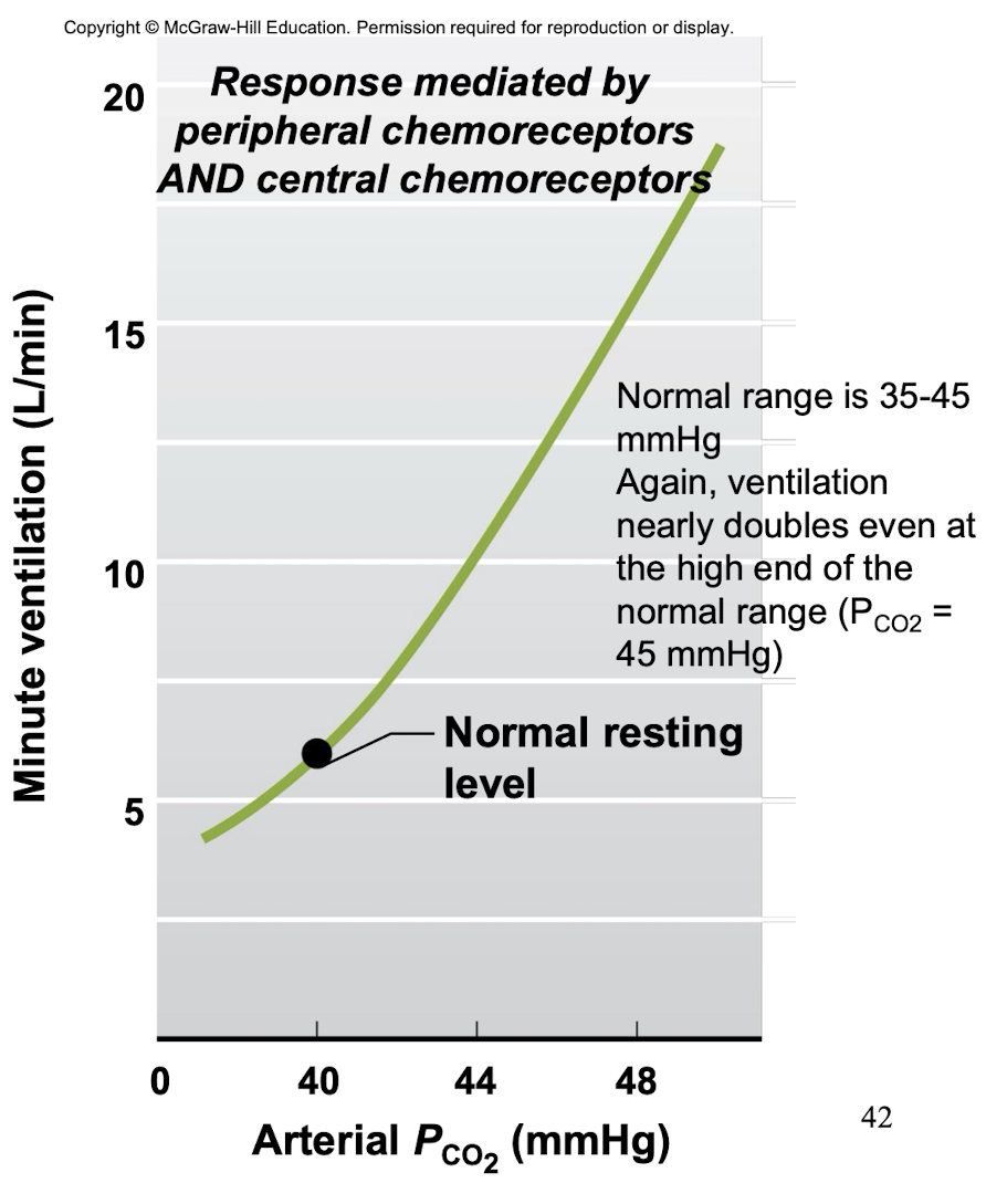
effect of PCo2 on ventilation
increase PCo2 → increase ventilation
response mediated by peripheral AND central chemoreceptors
explain what happens when you breathing normally and then hold your breath
Po2
PCo2
chemoreceptors
breaking point
Po2 decreases
PCo2 increases
peripheral and central chemoreceptors both respond
breaking point will be caused by Co2
if you hyperventilate first, then hold your breath…
after hyperventilating, Po2 + Pco2
during breathing holding, Po2 + Pco2
chemoreceptors
breaking point
after hyperventilating, Po2 increases and Pco2 decreases a lot
during breathing holding, Po2 decreases and Pco2 increases
peripheral chemoreceptor respond
breaking point will be caused by o2
effect of Po2 on ventilation
