BIOL324 Exam 4
 Call Kai
 Learn
 Practice Test
 Spaced Repetition
 Match
 Flashcards
 Knowt Play
1/55

There's no tags or description
Looks like no tags are added yet.
56 Terms
By what week in development can you morphologically tell what sex an embryo is
Week 7 the gonads differentiate, which marks the end of chromosomal control, hormones control the rest
What are the two cells in an embryo's testes that control/help development
1. Sertoli cells (aka nurse cells)- in the seminiferous tubules, their functions are on another slide
2. Leydig cells (aka interstitial cells)- in conn tissue outside of the seminiferous tubules, use cholesterol to produce androgens like testosterone, DHT, and estradiol
Sertoli cells (5 functions)
- Produces anti-mullerian hormone
- Their junctional complexes make the blood-testis barrier
- Provide physical support to sperm cells in tubule walls
- Physiological support: contains androgen binding protein which collects androgens to supply to the developing germ cells
- Controls cell division via 2 hormones: activin (promotes mitosis/meiosis) and inhibin (represses mitosis/meiosis)
Two possible ducts in the developing embryo's gonads
1. Mullerian duct (develops into female reprod tract)
2. Wolffian duct (develops into male reprod tract)
In a male embryo what do Testosterone and DHT do together
- Both hormones
- Stimulates the growth of the external male genitalia
In a male embryo what do Testosterone and Estradiol do together
- Both hormones
- Programs the part of the hypothalamus that controls the gonads
At which point in male embryonic development is the embryo finished making the gonads and programing the brain? Affect on Leydig cells?
4.5 months, the leydig cells disappear until puberty starts
What are do the primary sex organs of a male secrete
Germ line cells aka gametes aka spermatozoa
There were 4 main secondary (accsesory) sex organs in males that we talked about, what are they? What do three of them secrete? (4 secretions total)
- Epididymis: maturing/storing station for immature spermatozoa
- Seminal vesicles: store/produce thick fluid rich in fructose which is used by spermatozoa for motility
- Prostate: produces prostate-specific antigen (PSA, protein), serine protease
- Bulbourethral gland: creates a lubricant for the urethra (not part of the semen)
- Other: penis, scrotum, vas deferens, etc.
Semen
70% liquid from seminal vesicles, 30% spermatozoa
Serine protease (function)
Enzyme made by prostate gland to liquify the liquid produced by the seminal vesicles
Seminiferous tubules
- Tunica albuginia is the conn tissue outer covering
- Seminiferous tubules exist inside lobules (the noodle lookin things)
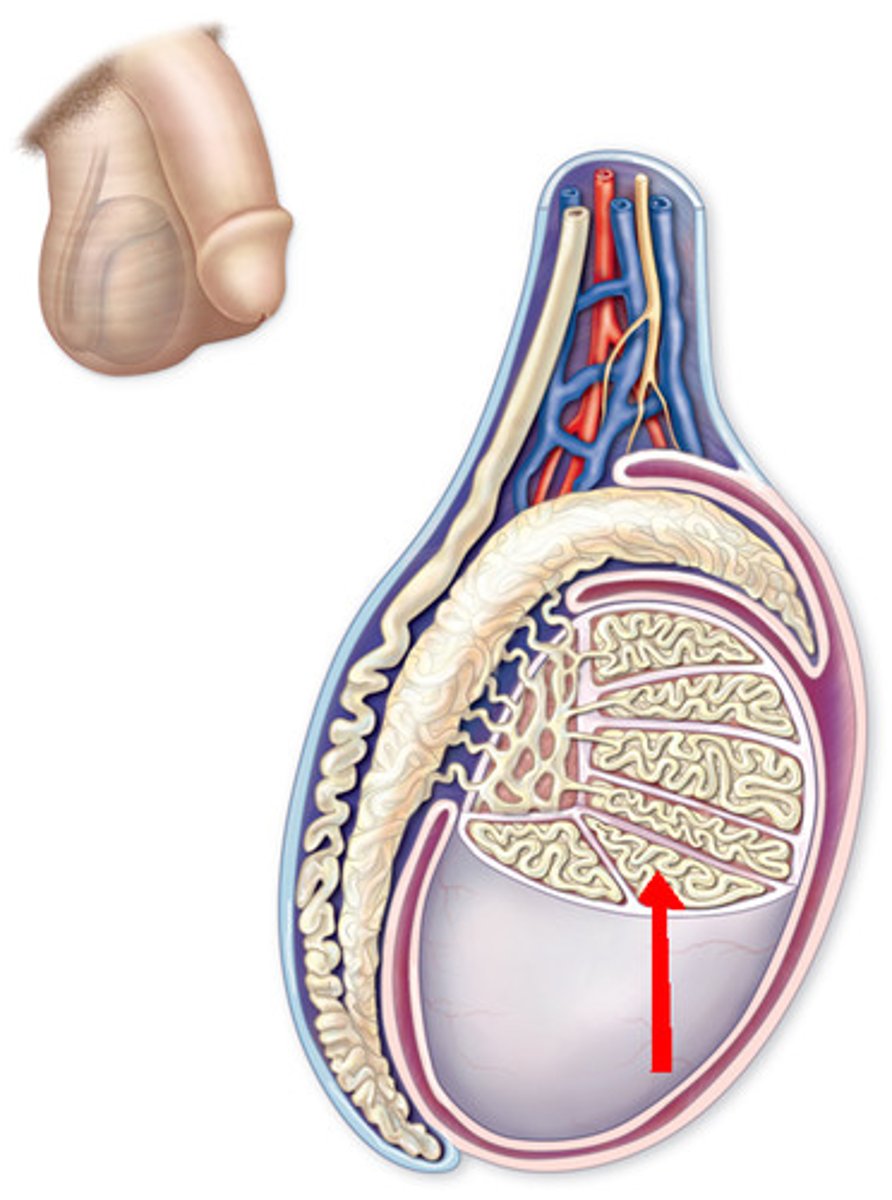
Seminiferous tubules (function, include ducts and epididymis)
- Inside the lobules
- The walls produce spermatozoa, the spermatozoa is swept away through convoluted (folded, twisted) tubules
- Swept towards Rete testis by Straight tubules
- Efferent ductules take the spermatozoa to the "head" of the epididymis
Head to tail of epididymis, how long
After entering from efferent ductules, it takes spermatozoa 2-4 weeks to go from head to tail of the epididymis, and spermatozoa is then considered mature, this is where the spermatozoa is stored until ejaculation
Pathway of sperm from testes to urethra
Seminiferous tubules, epididymis, vas deferens passes through inguinal canal into abdominal cavity and collects liquid from the seminal vesicle, prostate, bulbourethral gland, and then exits penis through the urethra
Is there epithelium in seminiferous tubules
- Yes, technically there are two epitheliums
- Basement membrane separating the two
- One epithelium is from the basement membrane => lumen, containing spermatid-making machinery, Sertoli cells, etc.
- The other is from the basement membrane => out, containing lamina propria with 3-5 layers of myofibroblasts (smooth muscle myoid cells), these cells contract to aid in release of spermatids from the wall, into the lumen
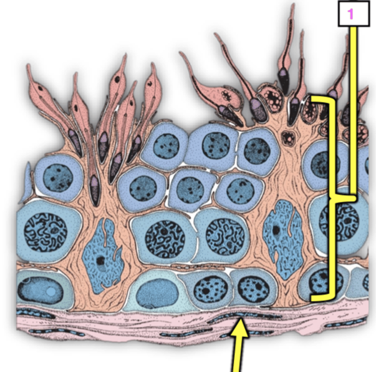
Cells found in the seminiferous tubule epithelium
- Referring to the inner epithelium (B.M. => lumen)
- Sertoli cells (sustentacular cell): long columnar cells that extend from BM to lumen
- Germ cells (spermatogenic): cytomorphosis (lots of variability and differentiation) of germ cells from spermatogonia to spermatocyte to immature spermatids. This is why this epithelium is considered stratified
Spermatogonia (3 different types based on nucleus structure, which is the "reserve stem cell"?)
1. Dark type A spermatogonia- intensely basophilic chromatin that has fine granules, homogenous, considered the reserve stem cell for all germ cells
2. Pale type A spermatogonia- rises from the above cell, still has fine granular chromatin but it stains lighter and less basophilic
3. Pale type B spermatogonia- rises from the above cell, has clumped darker heterochromatin against the edge of the nucleus and lighter euchromatin on the inside
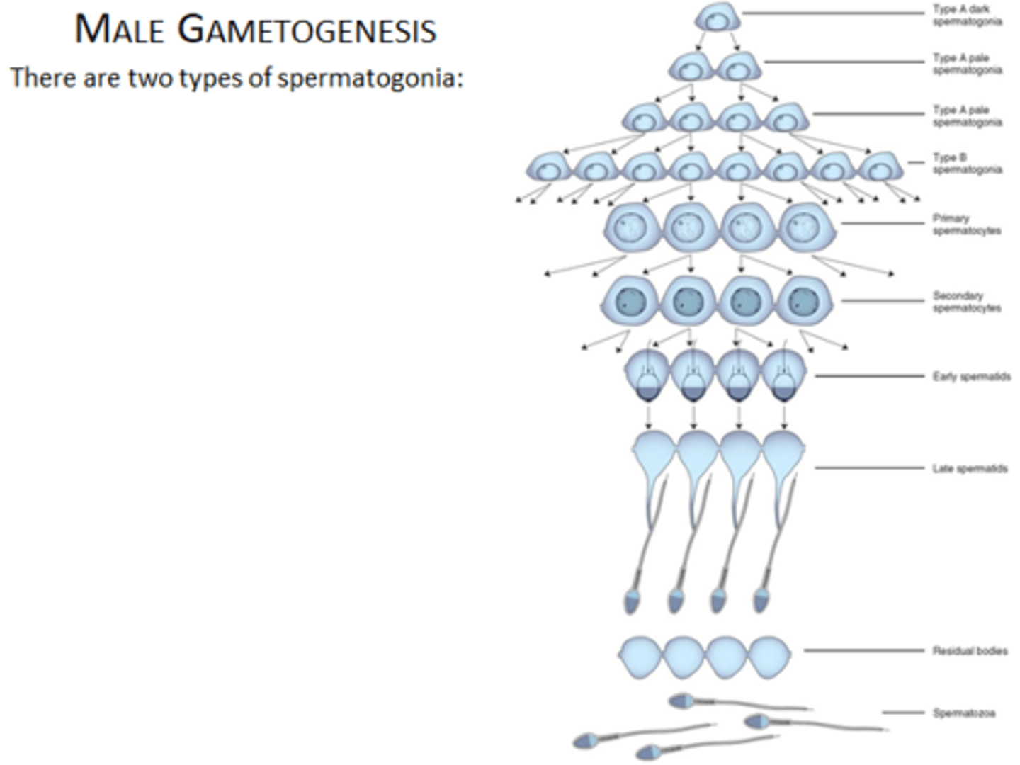
How do spermatogonia divide
- Mitotically
- Dark type A spermatogonia splits into another dark type A cell and a pale type A cell
- Pale type A spermatogonia mainly divides into more of itself, but eventually, a pale type A cell divides into two pale type B spermatogonia
- Pale type b spermatogonia divides into two primary spermatocytes (above spermatogonia on the basement membrane)
How do male gametes move in the seminiferous tubules
- Overall "centripetally" meaning from the outside towards the center where the lumen is
- But in spermatogonia, the cells are connected to the basement membrane meaning that when they divide they move laterally until pale type b spermatogonia create spermatocytes
Basal compartment
- Part of seminiferous tubules containing the BM and the spermatogonia, technically sealed off from the spermatocytes above (some spermatocytes are within the compartment as well)
- The inner edge of the compartment is the meeting lateral processes of the Sertoli cells (called junctional complexes)
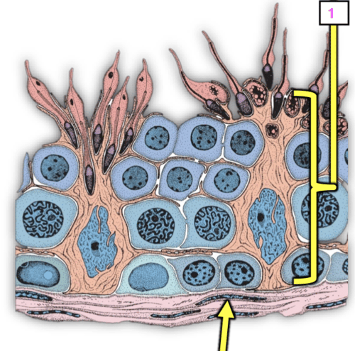
Junctional complex (anatomy) (include zona occludens, lines of fusion, actin, ER, etc)
- In the seminiferous tubules
- Contains unique tight junctions that have zonula occludens which are scaffolding protein complexes that help hold tight junctions together
- 50+ lines of fusion
- The lateral processes have stacks of endoplasmic reticulum inside and hexagonally arranged actin filaments under the cell membrane
Junctional complex (function)
- Adhesion device
- Physiological barrier called blood-testis barrier: keeps stuff out of the lumen of the seminiferous tubules like: intercellular fluid, blood from capillaries in testes, antibodies and other immune system material (too large to get through)
- Allows nutrients and waste products to move too and from
Spermatozoa vs spermatid
Spermatids are still attached to the wall of the lumen in the seminiferous tubules, spermatozoa are the free-floating, still immature sperm cells that move to the epididymis
Why does the junctional complex of seminiferous tubules keep immune system material out?
- Immune system recognizes self vs non self
- Pretty much everything after this junctional complex barrier (spermatocytes) divides through meiosis instead of mitosis, meaning that the cells produced are haploid instead of diploid
- If not for this barrier, immune cells would kill gametes bc they wouldn't recognize them as "self"
Name and give the difference between the two compartments of the seminiferous tubules
- Basal compartment: under junctional complexes, contains spermatogonia which divide via mitosis and are diploid (2n)
- Adluminal compartment: above junctional complexes, contains spermatocytes which divide via meiosis and are haploid (n)
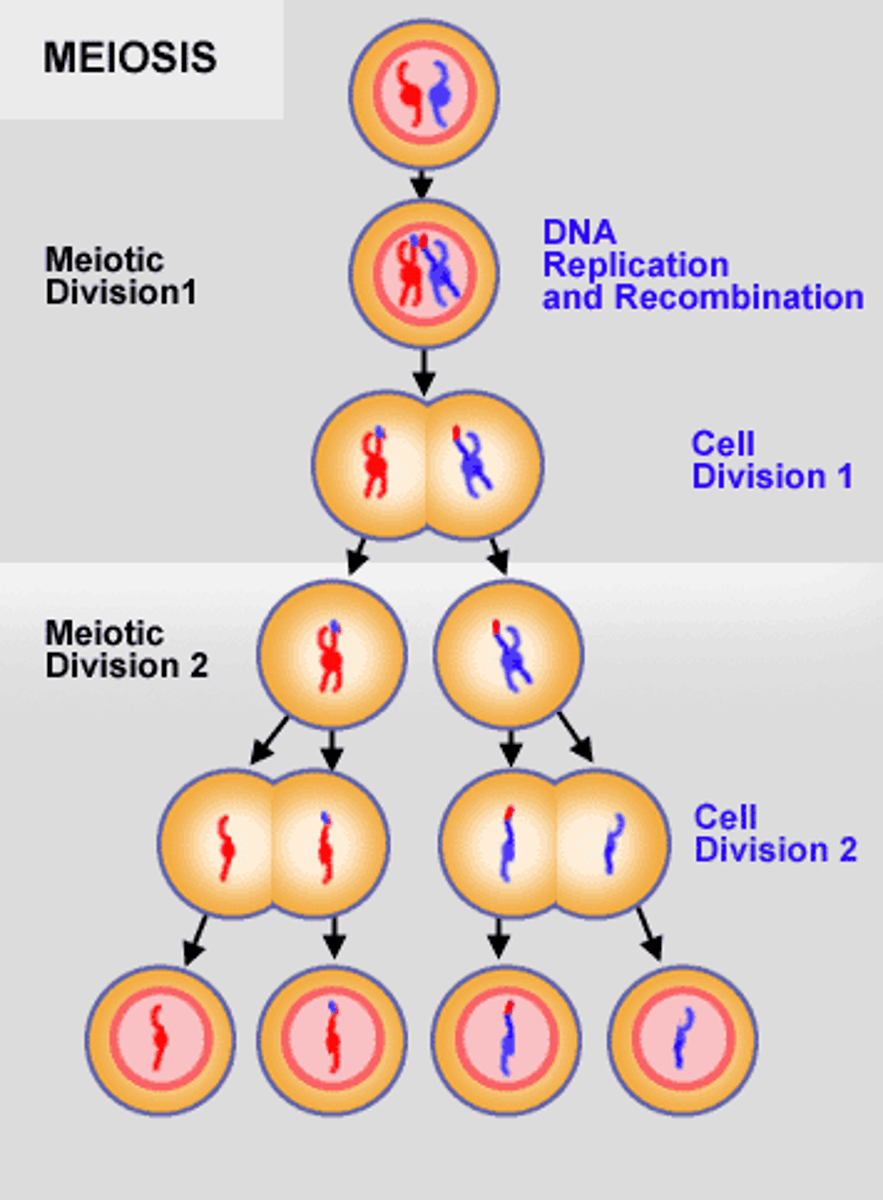
Steps in germ cell meiosis
1. Replication of DNA- Happens before meiosis begins, the genetic material doubles so before there are 2 chromatids/chromosome and after there are 4 chromatids/chromosome
2. Meiosis 1- Primary spermatocytes (diploid) => Secondary spermatocytes (haploid # of chromosomes, diploid DNA)
3. Meiosis 2- Secondary spermatocytes (haploid # of chromosomes, diploid DNA) => Spermatids (truly haploid)
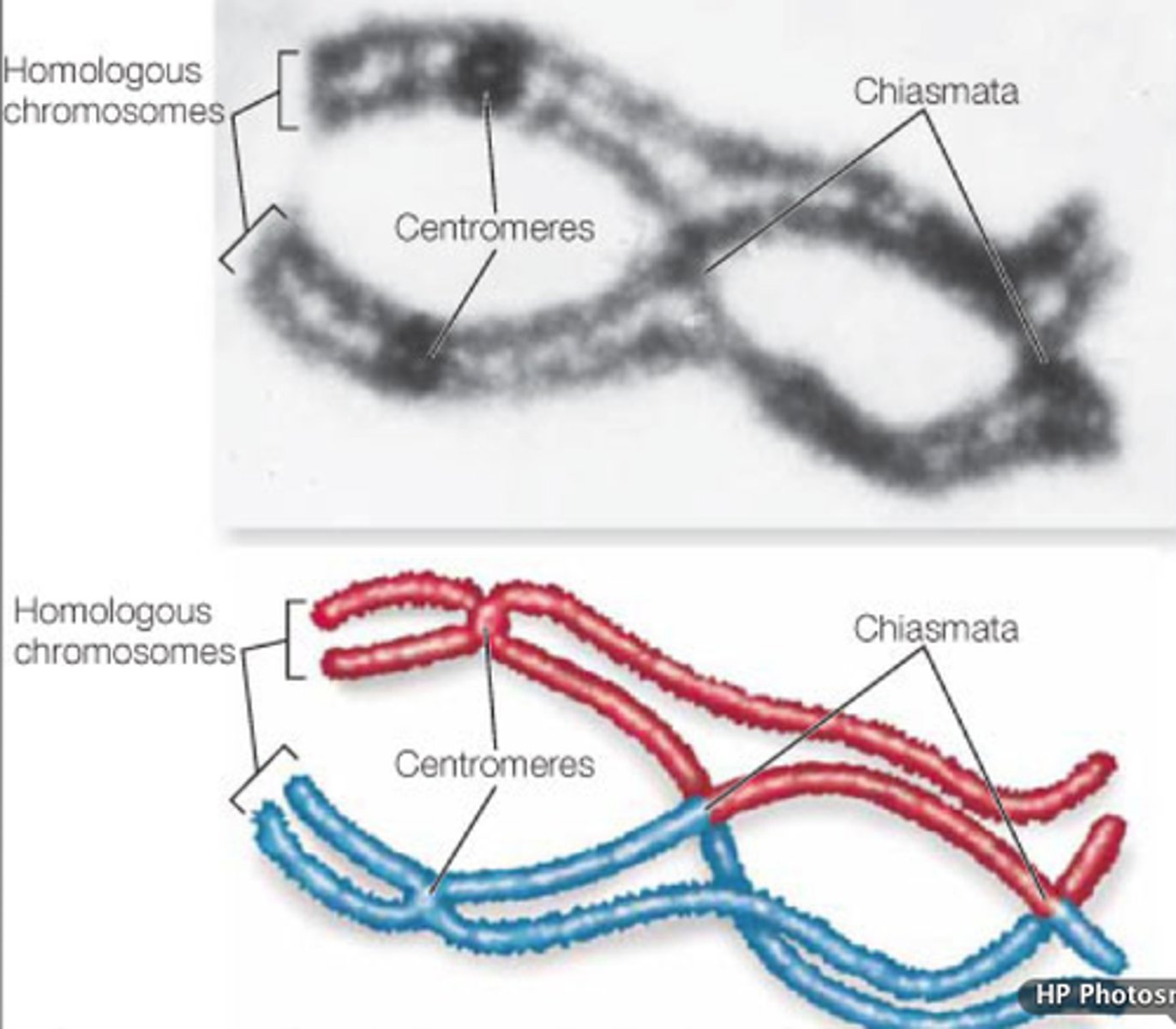
Chiasmata
Point of contact btwn two chromosomes which facilitates crossing over
Spermiogenesis vs spermatogenesis
- Spermiogenesis: spermatids => spermatozoa
- Spermatogenesis: spermatogonia => spermatozoa
Spermiogenesis (develop 5 things)
- Spermatids develop a flagellum (tail), acrosome (head), condensed chromatin (also part of the head), and a midpiece (contains mitochondria that use fructose to make ATP)
- When spermatids come off wall, the cytoplasm that they got rid of also gets propelled out into lumen, which are called "residual bodies", this extra cytoplasm is resorbed by Sertoli cells and used again
Spermiation
Release of mature spermatozoa into lumen of seminiferous tubule
Function of endocrine system
Produces various secretions (hormones) that regulate activities of various cells, tissues, organs, etc
Communication network = ?
Endocrine system + Nervous system
How does NS communicate
- Action potentials
- Quick stimulation, quick/fleeting response
Three types of hormone control
1. Endocrine control mechanism- hormones moved by circulatory system to affect far tissues (slower and more long-term than NS)
2. Paracrine control- endocrine glands secrete hormones into surrounding conn tissue to affect nearby tissue (localized)
3. Autocrine control- endocrine glands secrete hormones into surrounding tissue, which bind to receptors on that gland's surface to affect that gland only
Diffuse neuroendocrine system (DNES)
- Cells/glands in other systems of the body which secrete hormones, even though the larger purpose of the organ they inhabit isn't to secrete hormones (ex. small granule cells in the lungs)
- These cells incorporate nerve cells and hormone-producing characteristics
Multi-function organ
- Lungs, "gut", testes/ovary (primary function is to produce gametes, but also make steroid hormones)
- Play multiple roles
- Any organs with DNES cells are multi-function
Discrete endocrine glands (list 5)
- Endocrine organs whose primary function is the production and secretion of hormones
- There are only 5: pituitary gland, thyroid, parathyroid, adrenal gland, pineal gland
Pituitary gland
- Discrete endocrine gland
- Balls-lookin gland at the base/center of the brain
- Aka hypophysis
- Neurohypophysis (posterior) is nervous tissue and the adenohypophysis (anterior) is glandular tissue
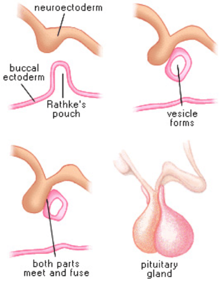
How is the pituitary gland formed? (mention diencephalon, Rathke's pouch, what tissue is each made of?)
- In embryological development there is the base of the brain (diencephalon) that forms a "stalk" coming down, that becomes the neurohypophysis (posterior pituitary), and is made of nervous tissue
- The stalk mentioned above keeps growing down until it touches the roof of the mouth, which stimulates that tissue to "mound up" through the mechanism of "contact induction"
- The mound is called "Rathke's pouch" which becomes the adenohypophysis (anterior pituitary), and is made of glandular tissue
- These come tg to form the hypophysis (pituitary)
3 parts of the neurohypophysis (posterior pituitary)
- Median eminence
- Infundibulum
- Pars nervosa (lobe of the posterior pituitary)
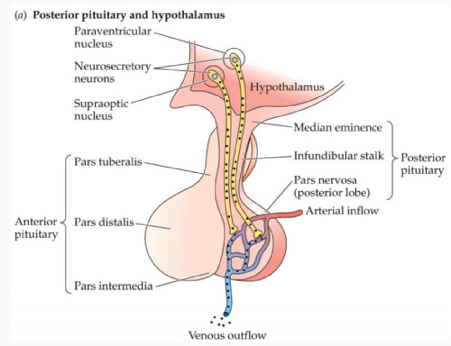
3 parts of the adenohypophysis (anterior pituitary)
- Pars tuberalis
- Pars intermedia
- Pars distalis (lobe of the anterior pituitary)
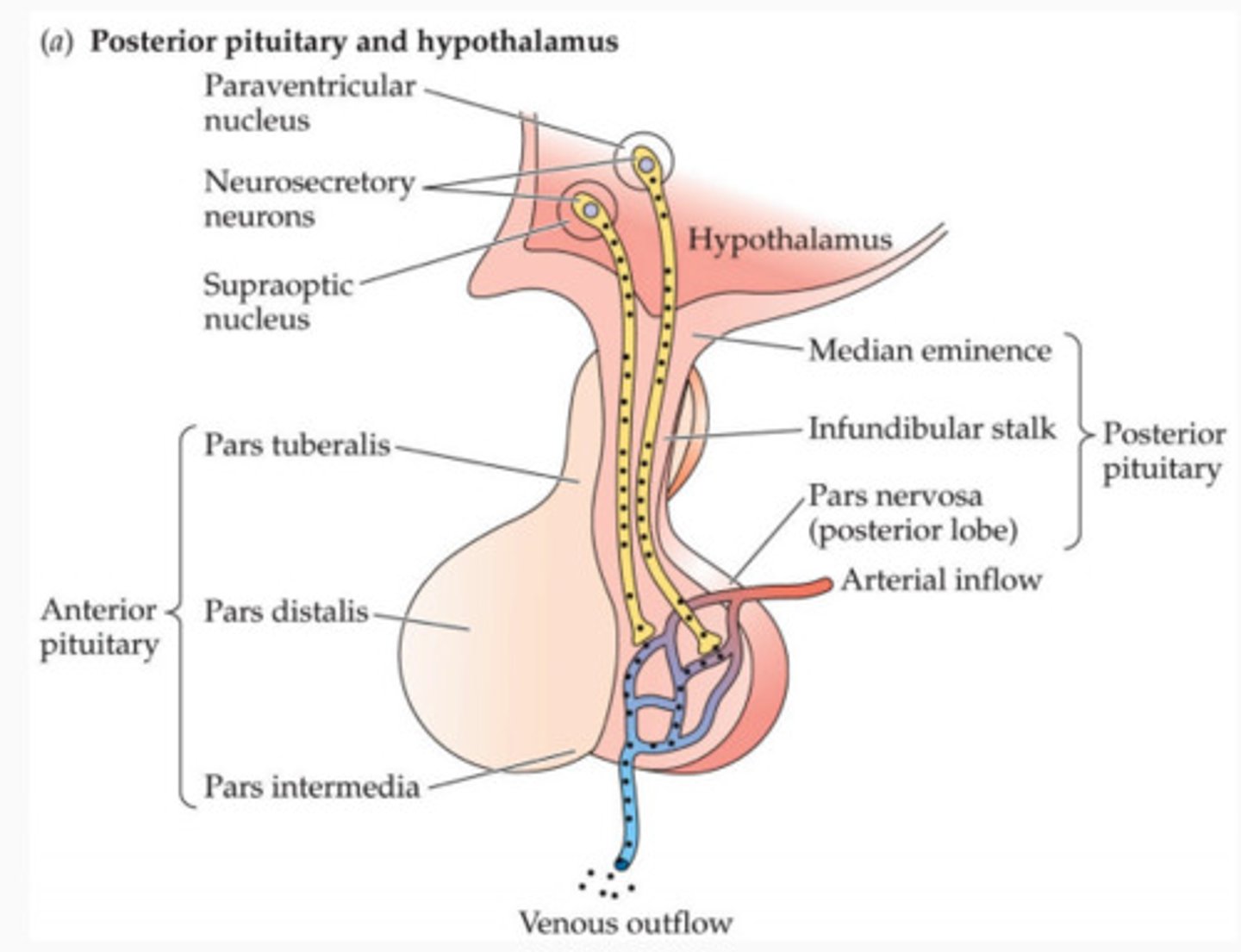
Neurons of the pituitary (explain, 2 types)
- Axons start in the hypothalamus, come down and enter the pars nervosa
- Split into 2 branches: supraoptic and paraventricular nuclei, together, they are called the hypothalamo-hypophyseal tract
- These neurons specialize in secreting hormones, not neurotransmitters
- These hormones are specialized to the pituitary gland, even though they are connected to the hypothalamus
Supraoptic nuclei vs paraventicular nuclei: which secretes what hormone
- Supraoptic: ADH
- Paraventricular: Oxytocin
What are two hormones secreted by the neurohypophysis? Give their function
1. Antidiuretic hormone (ADH)- Conserves water by taking it out of the urine filtrate in the collecting ducts of the kidneys and restoring into the blood. This makes urine more concentrated.
2. Oxytocin- when stimulated, enables the breast to release milk
Why is ADH also called vasopressin?
Another affect of ADH is vasoconstriction (raising BP)
Diabetes insipidus
- Unable to secrete ADH, meaning the kidneys can't retain water back into bloodstream
- Constantly drinking water and peeing very dilute urine (polyuria)
Neurohypophysis hormones: tell story of hormones from production to secretion (mention neurophysin and herring bodies)
- Two hormones referenced are ADH and oxytocin
- Made by cell bodies in the hypothalamus
- Hormone molecules are bound to transport protein called neurophysin
- Taken down their respective axons (supraoptic/paravent.) to the neurohypophysis
- Stored in axon terminals
- When the axon terminals become full with hormone and swell, they are called Herring bodies
Herring bodies: baso/acidophilic?
Acidophilic
Main type of cell in the neurohypophysis (posterior pituitary)
Pituicytes- specialized glial (neural support) cells containing GFAP (glial fibrillary acidic protein) and pigment similar to astrocyte
What type of hormones does the adenohypophysis secrete
Tropic hormones, which are hormones "that have other endocrine glands as their target" and increase the activity of the target gland (stimulates gland to release its hormones)
How are the hypothalamus and pituitary connected?
- Physically connected by the supraoptic and paraventricular neurons and make the hormones that use these neurons
- Hypothalamus can control the release of hormones from the adenohypophysis through the use of hypothalamic regulating factors
Hypothalamic regulating factors (function, 2 types)
- Controls the release of hormones from the adenohypophysis
1. Releasing factors- stimulate secretion
2. Inhibiting factors- inhibit secretion
- Uses receptors to detect levels of tropic hormones, will release the releasing factors accordingly
5 cell types of cells in the Pars Distalis, explain their functions (2 acidophilic, 3 basophilic)
- "Lobe" of the adenohypophysis (anterior pituitary)
Acidophilic:
- Somatotropic cells: secrete growth hormone which stimulates long bone growth
- Lactotropic cells: secretes prolactin which stimulates Leydig cells to secrete testosterone (males) and stimulates lactation (females)
Basophilic:
- Gonadotropic cells: secrete LH (luteinizing hormone) and FSH (follicle stimulating hormone) which stimulate Leydig cells (males) and ovulation (females)
- Thyrotropic cells: secretes thyroid stimulating hormone (T3 and T4)
- Adrenocorticotropic cells: adrenal corticotrophin (ACTH) which influences cells in the zona fasciculata (in adrenal gland)
Function of pars intermedia
- Part of the adenohypophysis (anterior pituitary)
- Produces MSH (melanocyte stimulating hormone)
Main type of cell in the pars tuberalis
- Part of the adenohypophysis (anterior pituitary)
- Gonadotropic cells