Anatomy Lab Quiz 3
 Call Kai
 Learn
Learn Practice Test
 Spaced Repetition
 Match
Match Flashcards
 Knowt Play
1/52

There's no tags or description
Looks like no tags are added yet.
Name | Mastery | Learn | Test | Matching | Spaced | Call with Kai | Chat |
|---|
No analytics yet
Send a link to your students to track their progress
53 Terms
Cranial bones (how many?)
8
frontal (1)
sphenoid (1)
Ethmoid(1)
Parietal (2)
Temporal (2)
Occipital (1)

Frontal bone: Supraorbital notch
tiny blue part, artery, vein & nerve pass through to skin

Frontal bone: Supraorbital margin
skinny light yellow

Frontal bone: Glabella
purple middle part

Frontal bone: Zygomatic process
yellow part on the right and left very far side

Temporal bone: Styloid process
tiny dark green pointy part
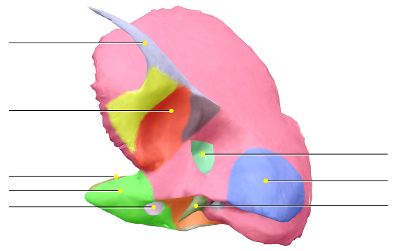
Temporal bone: Mastoid process
Big blue

Temporal bone: Zygomatic process
skinny light purple

Temporal bone: Mandibular fossa
red part

Occipital bone: Foramen Magnum
neon green part
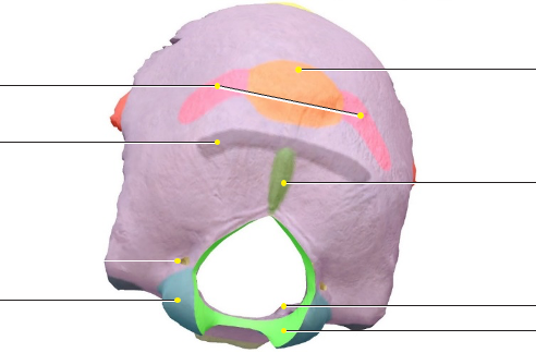
Occipital bone: Occipital condyle
blue part by the foramen magnum (articulates with the atlas (C1))
Foramen magnum — what passes through it?
Spinal cord + vertebral arteries + accessory nerve + anterior & posterior spinal arteries

Sphenoid: Sella Turcica (Turkish Saddle)
Grey part in center. “Turkish saddle”; houses the pituitary gland
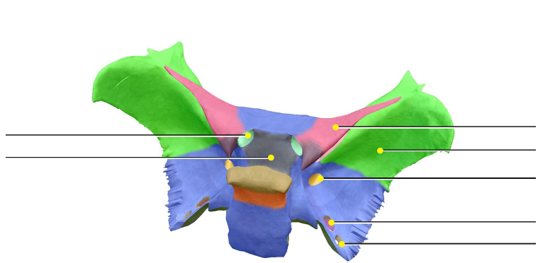
Sphenoid: Optic Foramen
teal/green small part on holes by sella

Sphenoid: Foramen Rotundum-(circular)
bright blue part
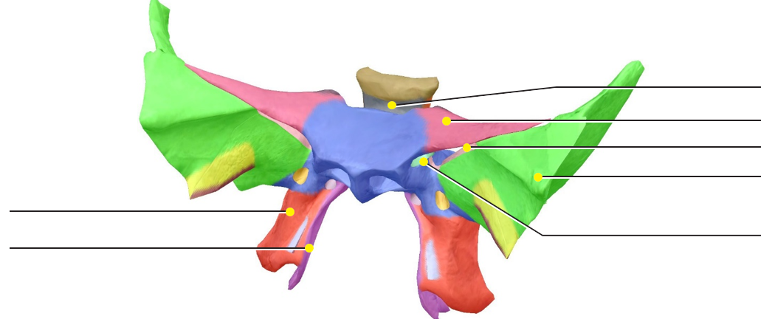
Sphenoid: Pterigoid Plate
bright red/orange
Cranial bone: Ethmoid bone
between the eyes has crista galli + cribriform plate (CN I) + perpendicular plate


Cranial bone: Perpendicular plate
big red

Cranial bone: Crista Gali (Rooster’s Crest)
tiny blue part top

Cranial bone: Cribriform plate (CN I)
light grey
Facial bones
Paired: Lacrimal / Nasal / Inferior nasal conchae/ Maxilla / Palatine / Zygoma
Unpaired: Vomer / Mandible
Which bones are part of the Orbit (eye socket)?
The frontal, sphenoid, zygomatic, maxillae, palatine, ethmoid, and lacrimal bones
Which bone and forms the nasal septum?
The perpendicular plate of the ethmoid bone, vomer, and septal cartilage
Which bone forms the oral cavity?
The maxillae, mandible, and palatine bones
Which bones forms the nasal cavity?
The ethmoid, sphenoid, frontal, maxillae, nasal, and inferior nasal conchae bones

Facial bone Maxilla: Zygomatic Process
dark purple maroon part on the side

Facial bone Maxilla: Hard Palate
not the light pink, but the medium pink on the side

Facial bone Mandible: Body
purple in center front

Facial bone Mandible: mental foramen
Mental nerve is a branch of CN V3, meaning the mandibular division of cranial nerve V (trigeminal nerve)
It provides sensation around the chin/lower lip.
dark grey dots on the body
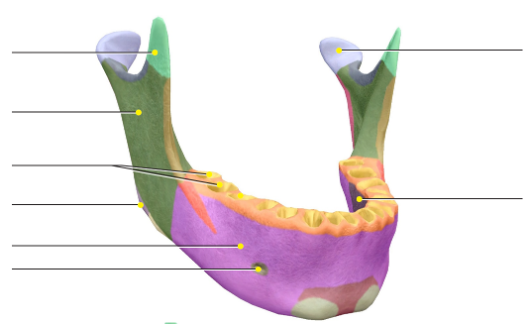
Facial bone Mandible: mental tubercles
two grey stubs by chin

Facial bone Zygomatic: Temporal Process
purple

Facial bone Zygomatic: Maxillary Border
blue

Facial bone Zygomatic: Frontal process
green

Hyoid bone
“floating” bone with lots of muscle/ligament attachments

Precentral gyrus
Primary motor cortex; Houses the primary motor cortex voluntary movement

Central Sulci:
structural boundary between precentral and postcentral

Postcentral gyrus:
primary somatosensory cortex. It receives and processes tactile information from the body, including touch, pressure, temperature, vibration, and pain
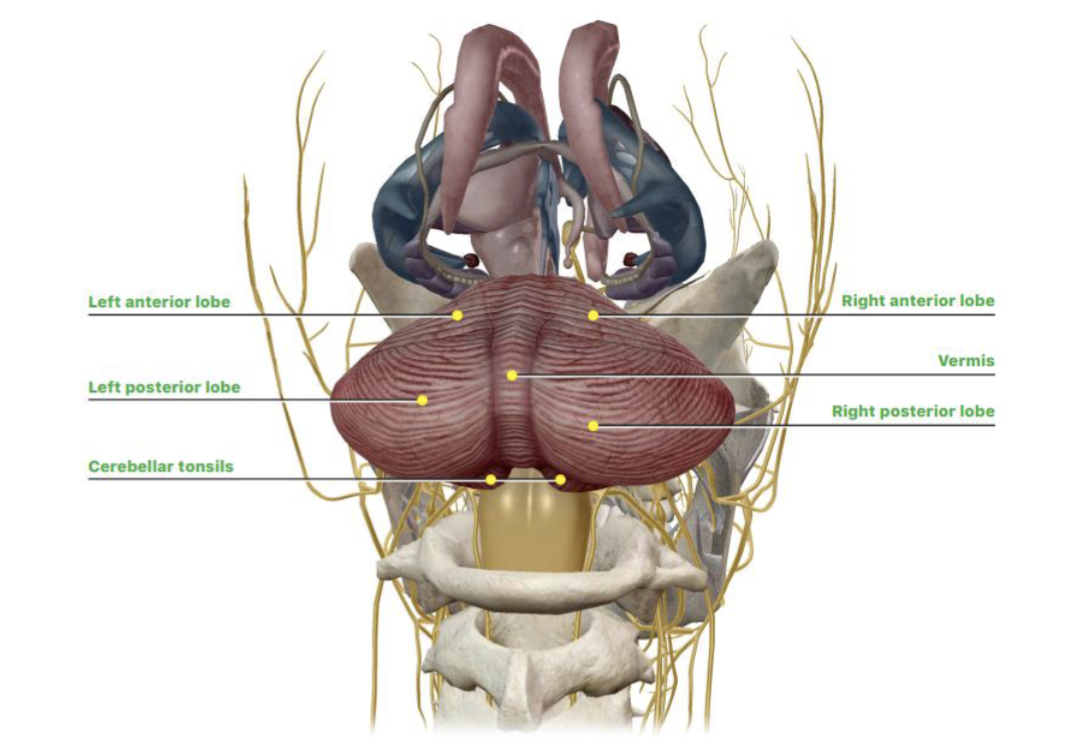
Cerebellum
Balance and fine-tuning/coordination of movement
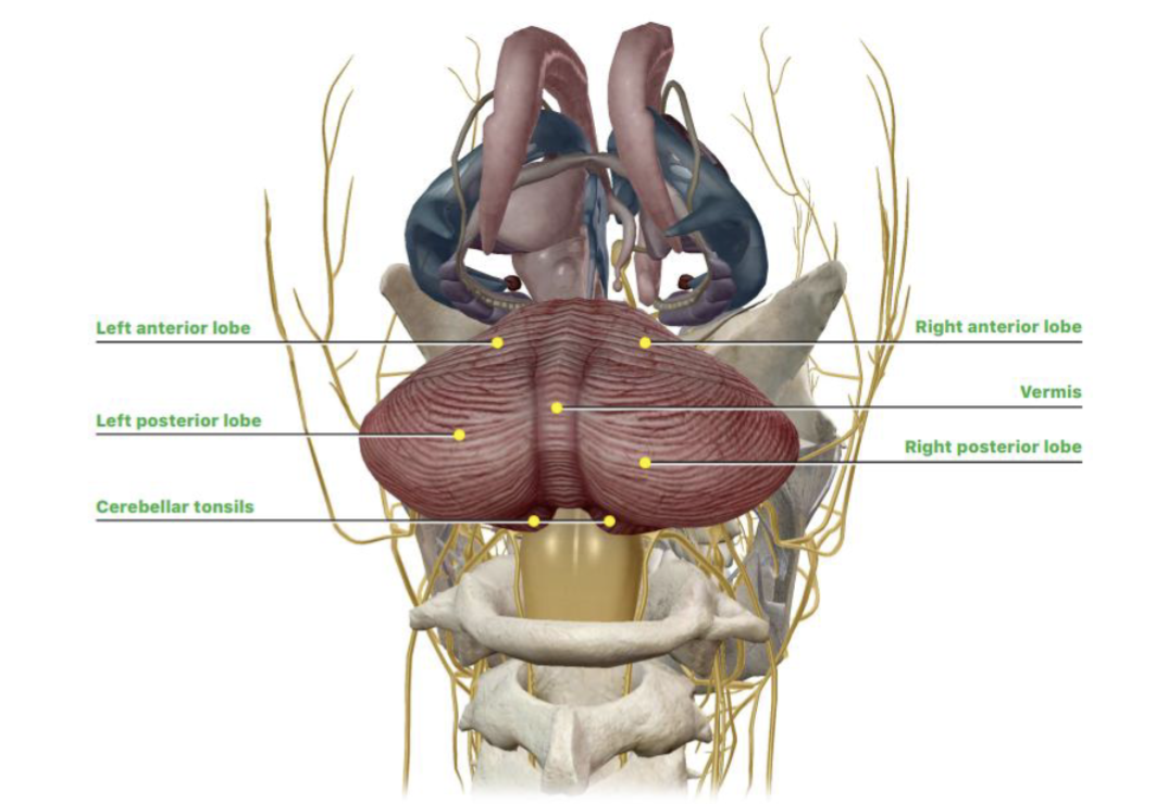
Vermis
Separates/connects the right and left portions of the cerebellum

Arbor vitae
Tree-like white matter of the cerebellum (mostly axons, especially myelinated axons. These carry information from one area to another.)

Dentate nucleus
Gray matter within the cerebellum (mostly neuron cell bodies (plus dendrites/synapses). This is where lots of information is processed.)

4 Ventricles. what is the path of CSF
Lateral ventricles → 3rd ventricle → cerebral aqueduct → 4th ventricle → subarachnoid space or central canal (Think of the ventricles as connected, fluid-filled chambers inside the brain.)

Thalamus function
Mediates sensation; involved in memory and learning
Corpus callosum — function (and is it white or grey matter)
Allows communication between the left and right cerebral hemispheres. white
Brainstem — what structures form it?
Midbrain + pons + medulla oblongata
Forebrain structures
Cerebrum + diencephalon (diencephalon = thalamus + hypothalamus +
epithalamus)
Midbrain
mesencephalon (sensory processing)
Hindbrain
medulla oblongata + pons + cerebellum
what are the four paranasal sinuses and what are there 2 functions
Frontal, maxillary, sphenoid, ethmoid
Lighten the skull + warm/moisten inhaled air

Meninges — superficial → deep
Dura → Arachnoid → Pia (DAP)

Meninges: Dura
Strongest / part of periosteum
• 2 Layers separated by VenousSinus
• dural venous sinus - drain venous blood from brain (valve-less)
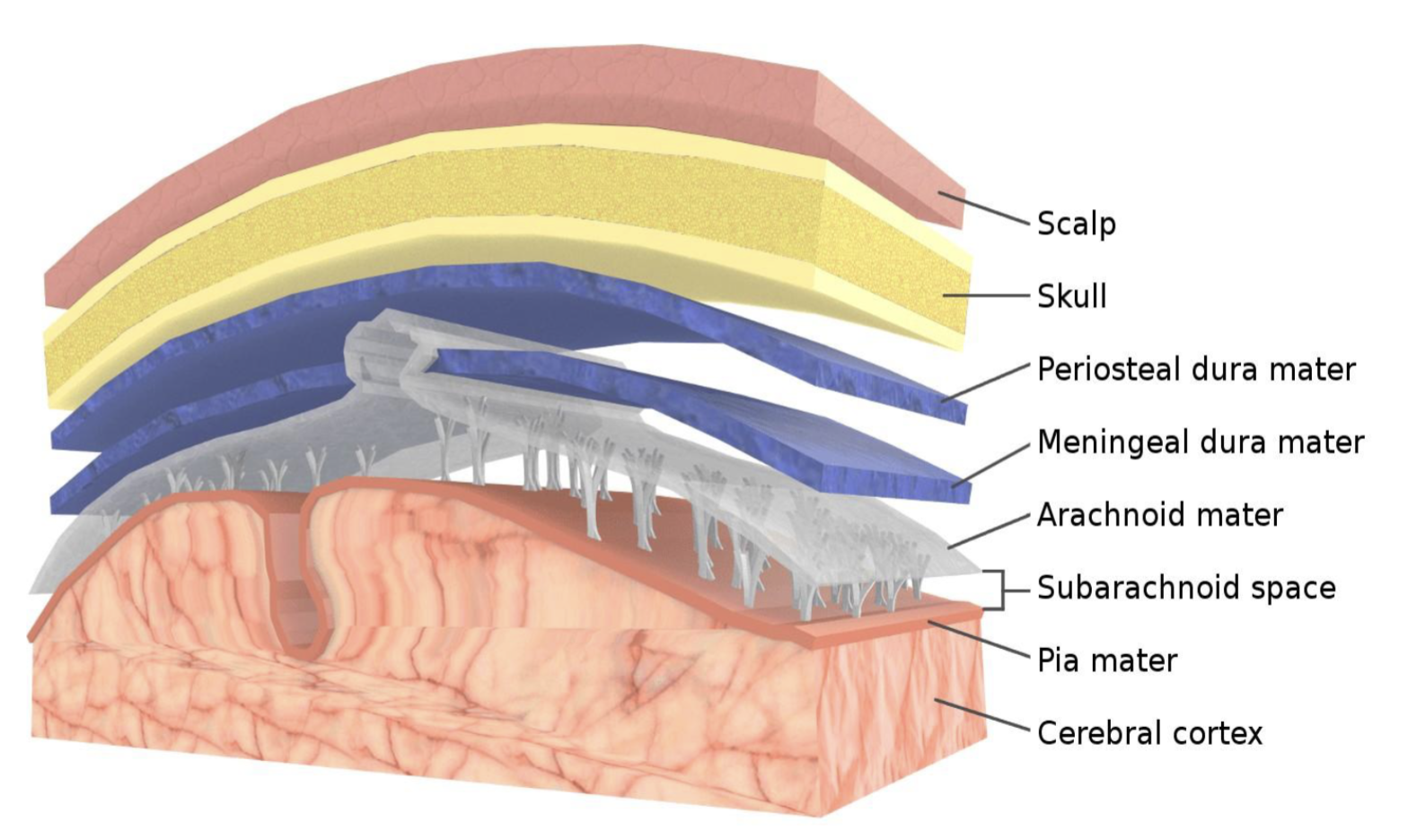
Arachnoid and What helps hold arachnoid to dura?
Web-like connective tissue (CSF)

Pia (tender mother)
Delicate layer that directly covers the brain surface