Microscope Skills
 Note
Note Call Kai
 Learn
Learn Practice Test
 Spaced Repetition
 Match
Match Flashcards
 Knowt Play
1/38

There's no tags or description
Looks like no tags are added yet.
Name | Mastery | Learn | Test | Matching | Spaced | Call with Kai | Chat |
|---|
No analytics yet
Send a link to your students to track their progress
39 Terms
Horizontal stage adjusting knob
Stage Knob
Total Magnification
Objective Lens Power x Ocular Lens (Eyepiece) Power.
Which knob sharply focuses an image?
Fine Focus Knob
Which objective should be in place when putting the microscope up for storage?
The 4x objective.
Stereo Microscope
Also known as a dissecting microscope, it provides magnification up to 300x and is used to view large objects that are too small to see with the naked eye.
Confocal Microscope
Uses laser light and a dichromatic mirror to create 3D images, producing clearer resolution than compound microscopes.
Scanning Electron Microscope (SEM)
Uses electrons to create a 3D image from a vacuum-sealed and coated specimen, favored in research for its high magnification.
Transmission Electron Microscope (TEM)
Similar to SEM, uses electrons to produce a 2D image of specimens, commonly used for observing metals and nanotechnology.
Microscope care principle 1
Carry microscopes with two hands: one around the arm and one under the base.
Microscope care principle 2
Start experiments with the objective lens set to the lowest power and the stage on the lowest setting.
Microscope care principle 3
Look at the microscope from the side when adjusting to avoid damage.
Microscope care principle 4
Always clean microscope objective lenses with special lens paper before use.
Microscope care principle 5
After finishing, return the stage to the lowest position and the objective lens to scanning power before removing slides.

1.
low objective

2.
Revolving Nosepiece

3.
High Power Objective
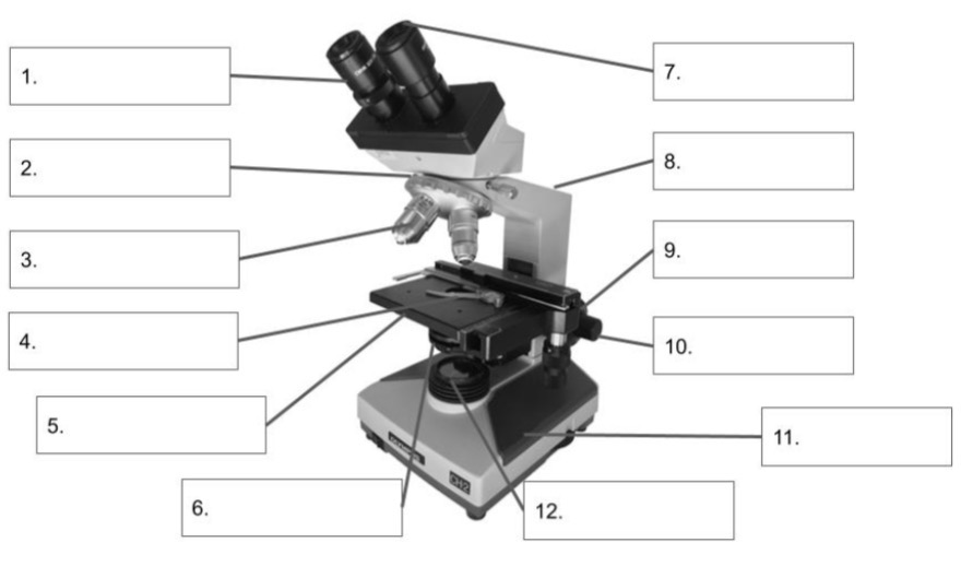
4.
Stage Control Knobs

5.
Mechanical Stage

6.
Iris Diaphragm

7.
Eye Piece

8.
Arm

9.
Coarse Adjustment

10.
Fine Adjustment

11.
Base

12.
Substrate Illuminator
Arm
Connects the head to the base, the place you hold onto when carrying the microscope
Base
The bottom of the microscope, a hand should be under this portion when carrying the microscope
Coarse Adjustment
Large knob on the arm of the microscope, moves the stage up or down in large increments.
Condenser
Located under the stage, directs the beam of light onto the specimen
Eye Piece
The part of the microscope you look into, contains the ocular lens
Fine Adjustment
Smaller knob in the middle of the coarse adjustment, moves the stage up or down in small increments
High Power Objective
Objective lens that is identified as the 40x
Iris Diaphragm
Controls the amount of light that enters the specimen, opens and closes
Low Power Objective
Objective lens that is identified as the 10x
Mechanical Stage
Platform that supports the slide and has clips or a clamp to hold the slide in place
Revolving Nosepiece
Rotating piece below the head that controls the movement of the objective lens
Scanning Objective
Objective lends that is identified as the 4x
Stage Control Knobs
Knobs that move the stage from right to left, and toward and away from the viewer
Substage Illuminator
Light source located in the microscope base