RADT 150 FEMUR, HIP, PELVIS, SI JOINTS
 Call Kai
 Learn
 Practice Test
 Spaced Repetition
 Match
 Flashcards
 Knowt Play
1/27

There's no tags or description
Looks like no tags are added yet.
28 Terms
AP (MId and Distal) Femur
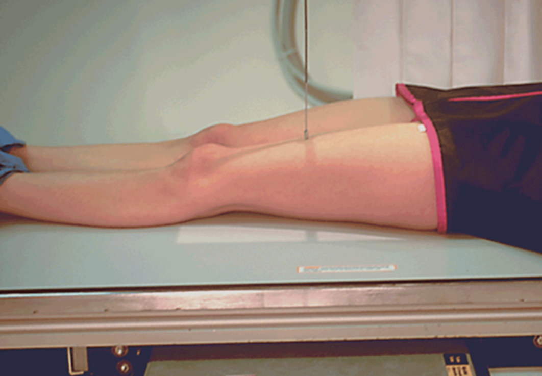
CR to midpoint of IR
40” SID
75 – 85 kVp
Leg extended
Rotate leg 5o internally
Bucky or grid
Shield and maintain modesty
Use full length of IR
*Separate AP Hip (proximal femur) included on smaller field size
Collimate to sides of femur
Include knee joint 2” within edge of IR
AP (MId and Distal) Femur Radiographic Criteria
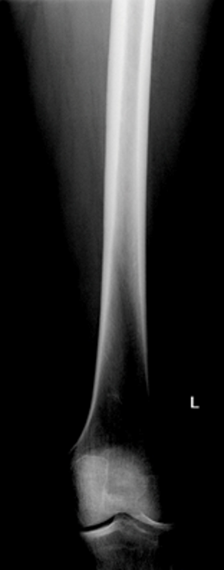
Knee joint included
No Rotation
Lateral (MId and Distal) Femur

CR to midpoint of IR
40” SID
75 – 85 kVp
True lateral
Lay on affected side
Leg bent approximately 45o
Align length of femur to length of table
Bucky or grid
Shield and maintain modesty
Use full length of IR
Separate Lateral Hip (proximal femur) included on smaller field size
Collimate to sides of femur
Include knee joint 2” within edge of IR
Lateral (MId and Distal) Femur Radiographic Criteria

Knee joint included (minimum)
No Rotation
Lateral (MId and Proximal) Femur Radiographic Criteria

Proximal femur not superimposed
True lateral
Suspend respiration
AP Pelvis

CR midway between level of ASIS and symphysis pubis & MSP
40” SID
80 – 90 kVp
Supine
Legs extended and slightly apart
Rotate legs medially 15 – 20o (if non-trauma)
Suspend respiration
Bucky or grid
Shield* and maintain modesty
Collimate 14 x 17 landscape
AP Pelvis Radiographic Criteria
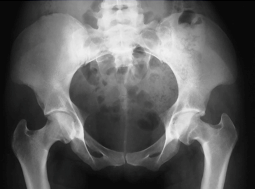
Entire pelvis and proximal femurs included
No rotation of pelvis
Lesser trochanters not visible
Hip and Proximal Femur (AP unilateral Hip)

CR perpendicular to femoral neck
40” SID
80 – 85 kVp
Supine
Legs extended and slightly apart
Rotate affected leg medially 15 – 20o(if non-trauma)
Suspend respiration
Bucky or grid
Shield* and maintain modesty
Collimate 10 x 12 portrait (extend length for history of prosthesis)
Hip and Proximal Femur (AP unilateral Hip) Radiographic Criteria

Proximal 1/3 of femur included
Hip joint space and acetabulum visualized
Lesser trochanter not visible or only slightly visible
Total existing orthopedic appliance demonstrated
Unilateral "Frog-Leg” (Non-Trauma)
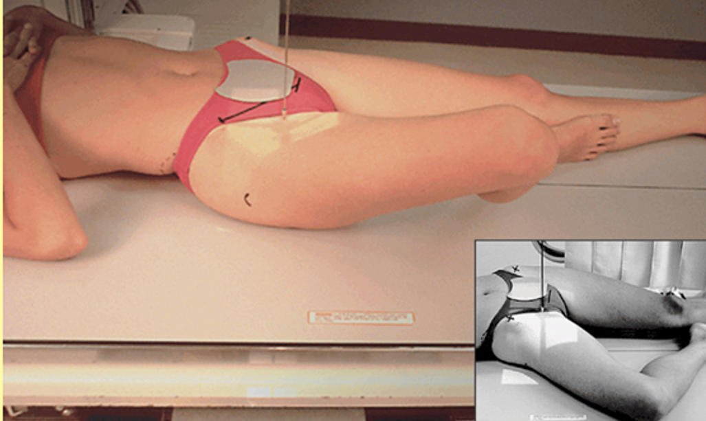
CR perpendicular to mid femoral neck/femoral neck
40” SID
80 – 85 kVp
Supine
Place the heel (plantar surface) against knee
Abduct femur 40-45o (best demonstrated, abduct femur to 90o from vertical)
Abduct affected leg 40-45o if non-trauma
Suspend respiration
Bucky or grid
Shield* and maintain modesty
Collimate 10 x 12 landscape (extend length for history of prosthesis)
Unilateral "Frog-Leg” (Non-Trauma) Radiographic Criteria

Entire femoral head, neck, and trochanters centered to IR
Femoral head and neck in profile
AP Bilateral “Frog-Legs” (Non-Trauma)

CR 3” below level of ASIS & MSP
CR 3 in. below level of ASIS
40” SID
80 – 90 kVp
Supine
Abduct femurs 40-45o
Abduct femurs 40 – 45o if non-trauma
Place plantar surface of both feet together
Suspend respiration
Shield* and maintain modesty
Bucky or grid
Collimate 14 x 17 landscape
AP Bilateral “Frog-Legs” (Non-Trauma) Radiographic Criteria

Pelvic girdle centered horizontally
No rotation of pelvis
Lesser trochanters equal in size
Greater trochanters superimposed over femoral neck
Axiolateral (Inferosuperior) - Danelius-Miller Method)

CR perpendicular to mid femoral neck
Axiolateral Hip (Bedside Mobile Procedure)

CR perpendicular to femoral neck and IR (level of mid thigh)
40” SID
80 – 95 kVp
Place IR with grid landscape, top edge of IR above Iliac crest
Center IR parallel to femoral neck (perpendicular to CR)
Raise unaffected leg (maintain modesty*)
Prop under affected hip
Suspend breath
Axiolateral Hip Radiographic Criteria

Entire femoral head, neck, and acetabulum visualized
No visible grid lines
Modified Axiolateral Projection (Hip and Proximal Femur)- Clements-Nakayama Method

CR perpendicular to femoral neck with 15 – 20 degrees downward angle
40” SID
80 - 90 kVp
Place patient near edge of table
Place grided IR landscape on Bucky tray parallel with femoral neck and perpendicular to IR
Suspend breath
Collimate 10 x 12
Modified Axiolateral Hip Radiographic Criteria

Entire femoral head, neck, and trochanters centered to IR
Femoral head and neck in profile
Posterior Oblique (Acetabulum)- Judet Method

CR 2 in. distal and medial to downside ASIS (RPO)
CR 2 in. distal to upside ASIS (LPO)
40” SID
80-90 kVp
Both obliques of the affected side, 45 degrees
Suspend respiration
Collimation 10 x 12 portrait
Posterior Oblique (Acetabulum)- Judet Method Radiographic Criteria
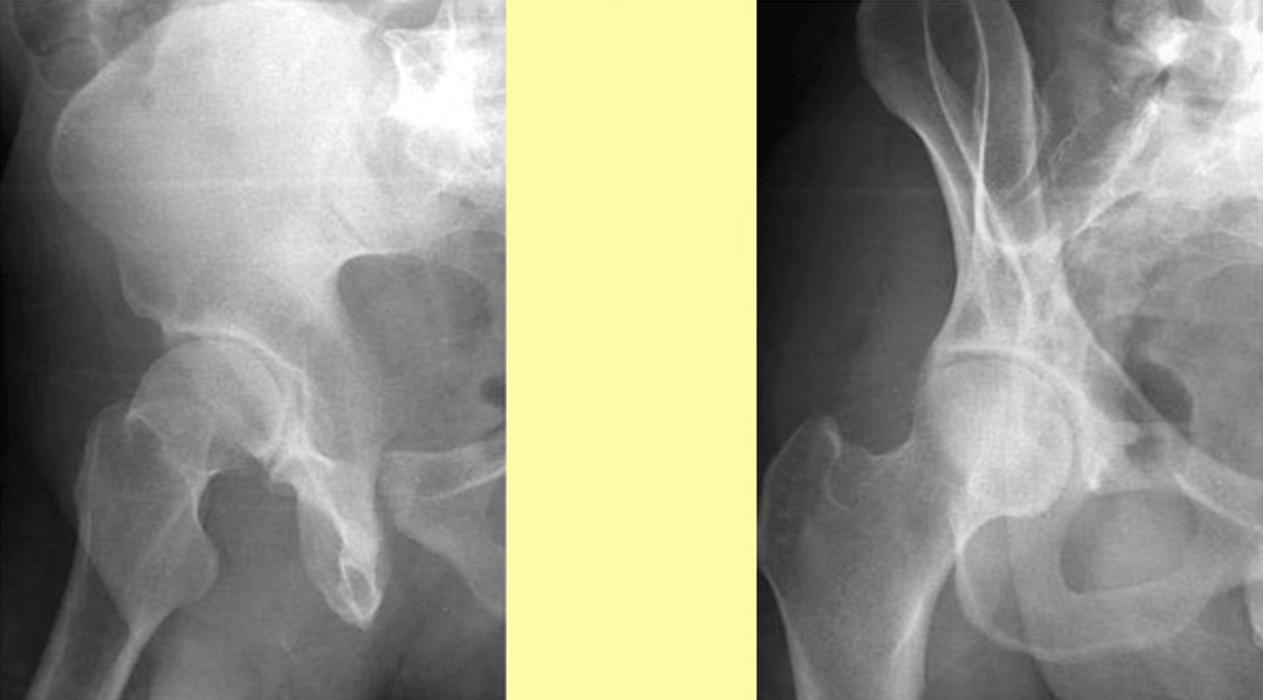
RPO
Anterior rim and posterior ilioschial column demonstrated
LPO
Posterior rim and anterior iliopubic column demonstrated
AP Axial “Outlet” Projection (Taylor Method)

CR 20 – 35 degrees cephalad males, 30 – 45 degrees cephalad females
CR 1 – 2” distal to symphysis pubis & MSP
40” SID
80 – 90 kVp
Suspend respiration
Bilateral assessment of pubis and ischium for fractures and displacement
Collimate landscape to area of interest
AP Axial “Outlet” Projection of Pelvis

Elongated and magnified public and ischial bones
No rotation of pelvis
Pubic and ischial bones centered to collimation field
AP Axial “Inlet” Projection

CR 40 degrees caudal, at level of symphysis pubis & MSP
40” SID
80 – 90 kVp
Suspend respiration
Assessment of pelvic trauma for posterior displacement or inward or outward rotation of anterior pelvis
Collimate landscape to area of interest
AP Axial “Inlet” Projection Radiographic Criteria
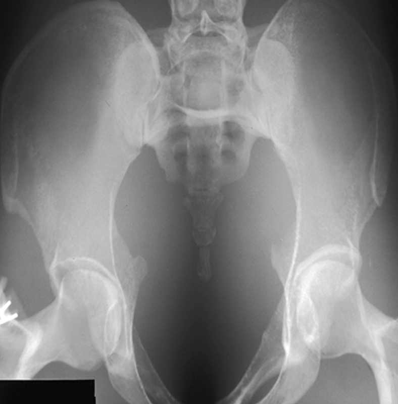
Ischial spines demonstrated and equal
Pelvic inlet centered
Lateral collimation evident
AP Axial SI Joints

CR 30 – 35 degrees cephalad
CR to MSP, midway between level of ASIS and symphysis pubis
40” SID
80 – 95 kVp
Supine, legs extended, support under knees
Suspend respiration
Collimate 10 x 12 landscape to area of interest
AP Axial SI Joints Radiographic Criteria

Sacroiliac joint centered to collimation field
Sacroiliac joint spaces and L5-S1 junction open
Posterior Oblique SI Joints


CR perpendicular, 1” medial to upside ASIS
40” SID
80 – 95 kVp
Elevate affected side 25 – 30 degrees
Suspend respiration
Collimate 10 x 12 portrait to area of interest
RPO to evaluate left SI joint
LPO to evaluate right SI joint
Posterior Oblique SI Joints Radiographic Criteria

Sacroiliac joint of interest is open
Ala of ilium not overlapped over sacrum