CH. 23 Digestive System PT 1- Overview, gross anatomy
 Call Kai
 Learn
 Practice Test
 Spaced Repetition
 Match
 Flashcards
 Knowt Play
1/70

There's no tags or description
Looks like no tags are added yet.
71 Terms
organs of the digestive system fall into two groups
alimentary canal (gastrointestinal or GI tract or gut)
acessory digestive organs

alimentary canal (gastrointestinal or GI tract or gut)
continuous muscular tube that runs from the mouth to anus
digests food: breaks down into smaller fragments
absorbs fragments through lining into blood
organs: mouth, pharynx, esophagus, stomach, small intestine, large intestine, anus

accessory digestive organs
teeth
tongue
gallbladder
digestive glands: produce secretions that help break down food (salivary glands, liver, pancreas)

processing of food involves six essential activities
ingestion
propulsion
mechanical breakdown
digestion
absorption
defecation

ingestion
eating

propulsion
movement of food through the alimentary canal, which includes:
swallowing
peristalsis: major means of propulsion of food

mechanical breakdown
includes chewing, mixing food w/ saliva, churning food in stomach, and segmentation (local constriction of intestine that mixes food with digestive juices)

digestion
series of catabolic steps that involves enzymes that break down complex food molecules into chemical building blocks

absorption
passage of digested fragments from lumen of GI tract into blood or lymph

defecation
elimination of indigestible substances via anus in form of feces

perstalsis
adjacent segments of the alimentary canal organs alternately contract and relax
food is moved distally along the tract
primarily propulsive; some mixing may occur
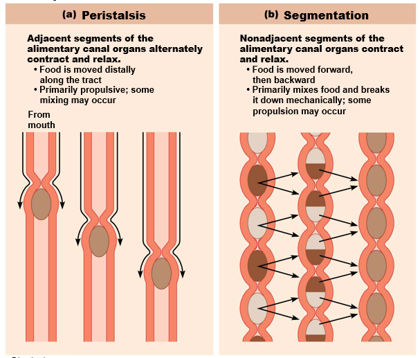
segmentation
nonadjacent segments of the alimentary organs contract and relax
food is moved forward, then backward
primarily mixes food and breaks it down mechanically; some propulsion may occur

peritoneum
serous membranes of abdominal cavity that consists of
visceral vs parietal
peritoneal cavity
fluid-filled space between two peritoneums to lubricate mobile organs
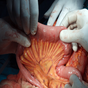
mesentery
double layer of peritoneum; layers are fused back to back
extends from body wall to digestive organs
provides routes for blood vessels, lymphatics, and nerves
holds organs in place and also stores fat
intraperitoneal (peritoneal) organs
organs that are located within the peritoneum

retroperitoneal organs
located outside, or posterior to, the peritoneum
includes most of pancreas, duodenum, and parts of large intestine

mucosa
tunic layer that lines lumen
secretes mucus, digestive enzymes, and hormones
absorbs end products of digestion
protects against infectious disease
made up of three sublayers
epithelium
lamina propria
muscularis mucosae

epithelium in mucosa
contains mucus secreting cells in most of tract that secretes mucus to:
protect digestive organs from enzymes and eases food passage

lamina propria in mucosa
rich supply of capillaries located here
also contains lymphoid follicles that help defend against microorganisms

muscularis mucosae in mucosa
smooth muscle that produces local movements of mucosa

submucosa
contains blood and lymphatic vessels, lymphoid follicles, and submucosal nerve plexus that supply surrounding GI tract tissues
has abundant amount of elastic tissues that help organs to regain shape after storing large meal

muscularis externa
muscle layer responsible for segmentation and peristalsis
contains inner circular muscle layer and outer longitudinal layers
circular layer thickens in some areas to form sphincters

serosa
outermost layer, which is made up of the visceral peritoneum
formed from areolar connective tissue covered with mesothelium (single layer of squamous epithelium) in most organs
replaced by fibrous adventitia in esophagus (dense connective tissue that holds esophagus to surrounding structures)

splanchnic circulation
includes arteries that branch off aorta to serve digestive organs
hepatic, splenic, and left gastric arteries
inferior and superior mesenteric arteries
hepatic portal circulation
drains nutrient-rich blood from digestive organs
delivers blood to liver for processing

GI tract has its own nervous system, referred to as
enteric nervous system (gut brain)
enteric nervous system (gut brain)
made up of enteric neurons that communicate extensively with each other
short reflexes: mediated by enteric nerve plexuses (gut brain); respond to stimuli in GI tract
long reflexes: respond to stimuli arising inside or outside of gut, such as from autonomic nervous system
autonomic nervous system:
parasympathetic system enhances digestive process
sympathetic system inhibits digestion

three key concepts regulate GI activity
digestive activity is provoked by a range of mechanical and chemical stimuli
receptors located in walls of GI tract organs
respond to stretch, changes in osmolarity and pH, and presence of substrate and end products of digestion
effectors of digestive activity are smooth muscle and glands
neurons (intrinsic and extrinsic) and hormones control digestive activity
- Nervous system control:
intrinsic controls: involve short reflexes (enteric nervous system)
extrinsic controls: involve long reflexes (autonomic nervous system)
- Hormonal controls
mouth
where food is chewed and mixed with enzyme-containing saliva that begins process of digestion, and swallowing process is initiated
also called the oral (buccal) cavity

oral (buccal) cavity
bounded by lips anteriorly, cheeks laterally, palate superiorly, and tongue inferiorly
palate: forms the roof of the mouth and has two distinct parts
hard palate
soft palate

hard palate
formed by palatine bones and palatine processes of maxillae

soft palate
fold formed mostly of skeletal muscle
closes off nasopharynx during swallowing

tongue
occupies floor of mouth, composed of interlacing bundles of skeletal muscle
functions include:
gripping, repositioning, and mixing of food during chewing
formation of bolus, mixture of food and saliva
initiation of swallowing, speech, and taste
anatomical structures:
lingual frenulum: attachment to floor of mouth
superior surface bears papillae, peglike projections of underlying mucosa
terminal sulcus: groove located posterior to vallate papillae
-marks division between
body: portion of tongue that resides in oral cavity
root: posterior third residing in oropharynx
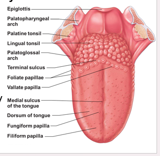
lingual frenulum
attachment to floor of mouth

terminal sulcus
groove located posterior to vallate papillae
marks division between:
body: portion of tongue that resides in oral cavity
root: posterior third residing in oropharynx

saliva function
cleanses mouth
dissolves food chemicals for taste
moistens food; compacts into bolus
begins breakdown of starch w/ enzyme amylase
compostion of saliva
mostly water (97-99.5%)
slightly acidic (pH 6.75 to 7.00)
electrolytes: Na+, K+, Cl-, PO4²-, HCO3-
salivary amylase and lingual lipase
proteins: mucin, lysozyme, and IgA
metabolic wastes: urea and uric acid
lysozyme, IgA, defensins, and nitric oxide from nitrates in food
major salivary glands include
parotid: anterior to ear and external to masseter muscle
submandibular: medial to body of mandible
sublingual: anterior to submandibular gland under tongue

salivary glands are composed of two types of secratory cells
serous cells: produce watery secretion, enzymes, ions, bit of mucin
mucus cells: produce mucus

teeth
lie in sockets in gum-covered margins of mandible and maxilla
mastication
deciduous and permanent teeth
mastication
prcoess of chewing that tears and grinds food into smaller fragments
primary dentition of teeth
20 deciduous teeth (baby teeth)
erupt between 6 and 24 months of age
permanant teeth
32 permanant teeth
enlarge and develop while roots of milk teeth are resorbed from below, causing them to loosen and fall out
occurs around 6-12 years of age
all but 3rd molars (wisdom teeth) are in by end of adolescence
teeth shape classification
according to shape
incisors
canines
premolars
molars

incisors
chisel shaped for cutting

canines
fanglike teeth that tear or pierce

premolars
(bicuspids) broad crowns with rounded cusps used to grind or crush

molars
broad crowns, rounded cusps, best grinders
during chewing, upper and lower molar lock together, creating tremendous crushing force

tooth structure
two major regins
crown
root
______
cement
peridontal ligament
dentin
apical foramen
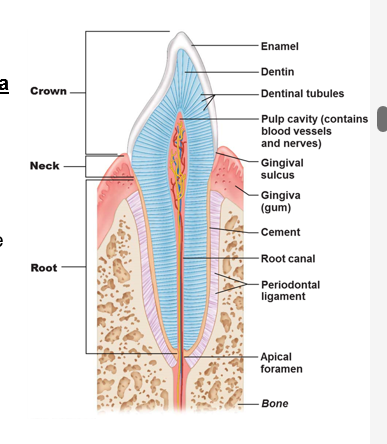
crown of tooth
exposed part above gingia (gums)
covered by enamel
heavily mineralized with calcium salts and hydroxyapatite crystals

root of tooth
portion embedded in jawbone
connected to crown by neck

cement
calcified connective tissue
covers root; attaches it to periodontal ligament
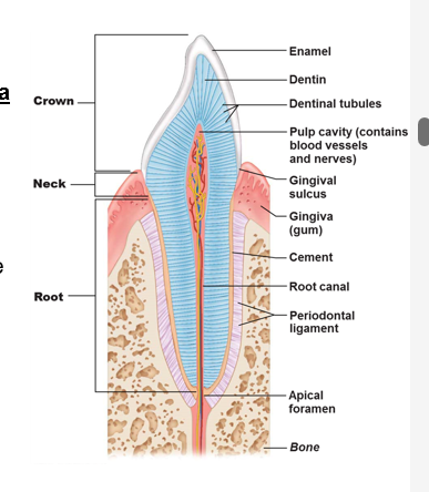
periodontal ligament
forms fibrous joint called gomphosis

dentin
bonelike material under enamel
maintained by odontoblasts of pulp cavity

apical foramen
at proximal end of root
entry for blood vessels, nerves, etc

the pharynx
food passes from mouth into oropharynx and then into laryngopharynx
external muscle layers consists of two skeletal muscle layers
inner layer of muscles run longitudinally
other pharyngeal constrictors encircle wall of pharynx
esophagus
flat muscular tube that runs from laryngopharynx to stomach
joins stomach at cardial orfice
gastroesophageal (cardiac) sphincter (esophageal sphincter)
surrounds cardial orfice > keeps orifice closed when food is not being swallowed and helps protect esophagus from acid reflux
major function of both pharynx and esophagus
propulsion that starts with deglutition (swallowing)


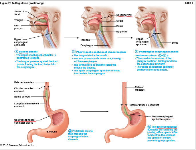
deglutition
swallowing; deglutition involves coordination of 22 muscle groups and two phases:
buccal phase: voluntary contraction of tongue
pharyngeal-esophageal phase: involuntary phase that primarily involves vagus nerve (sm. muscle)
stomach
temporary storage tank that starts chemical breakdown of protein digestion
converts bolus of food to paste-like chyme (digesta and stomach juices)

major regions of the stomach
pyloric part: wider and more superior portion of pyloric region, antrum, narrows into pyloric canal that terminates in pylorus
pylorus is continuous with duodenum through pyloric valve (sphincter controlling stomach emptying)
not as important, know sphincters!

muscularis externa in the stomach
has circular and longitudinal smooth muscle layers, but also has extra third layer
inner oblique layer allows stomach not only to churn, mix, and move chyme, but also to pummel it, which increases physical breakdown

mucosa layer of the stomach
dotted with gastric pits, which lead into gastric glands.
gastric glands produce gastric juice
glands include secretory cells
mucous neck cells
parietal cells
chief cells
enteroendocrine cells

mucous neck cells
secrete thin, acidic mucus of unknown function

parietal cells
hydrochloric acid (NCI)
pH 1.5-3.5; denatures protein, activates pepsin, breaks down plant cell walls, and kills any bacteria
intrinsic factor
glycoprotein required for absorption of vit B12 in small intestine

chief cells
pepsinogen: inactive enzyme that is activated to pepsin by HCI and by pepsin itself (positive feedback mechanism)
lipases
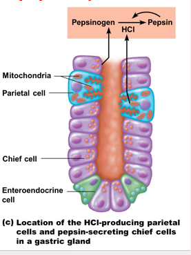
enteroendocrine cells
secrete chemical messengers into lamina propria
act as paracrines (serotonin and histamine)
hormones (somatostatin (also acts as paracrine) and gastrin)

processes carried out by stomach
carries out breakdown of food
serves as holding area for food
delivers chyme to small intestine
denatures proteins by HCI
pepsin carries out enzymatic digestion of proteins (milk protein (casein) is broken down by rennin in infants)
lipid-soluble alcohol and asprin are absorbed into blood
only stomach function essential to life is secretion of intrinsic factor for vitamin B12 absorption (B12 needed for RBCs to mature)


gastric mucosa secretions and regulation
>3L of gastric juice/day and are regulated by:
neural mechanisms
vagus nerve stimulation increases secretion
sympathetic stimulation decreases secretion
hormonal mechanisms
gastrin stimulates enzyme and HCI secretion
gastrin antagonists are secreted by small intestine
