Introduction to intestinal worms and parasitic gut diseases: ruminants
 Call Kai
 Learn
 Practice Test
 Spaced Repetition
 Match
 Flashcards
 Knowt Play
1/29

There's no tags or description
Looks like no tags are added yet.
30 Terms
why intestinal worms?
• Highly prevalent infections of animals and humans
• Impact animal welfare and cause production losses → impact on food security
• Often zoonotic with significant environmental reservoirs
• Chronic morbidity in humans
• Stunting and wasting in children

nematodes

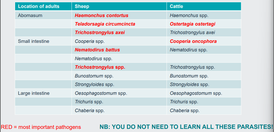
ruminant gut nematodes (gins-gastrointestinal nematodes)
typical liecycle of ruminant gins

gins in cattle
• Worldwide distribution (temperate & subtropical)
• Larvae develop in gastric glands of abomasum & mature on mucosal surface
• Prepatent period (PPP) = 3 weeks
• Up to 2 grazing seasons to acquire immunity
• Immunity not complete, develops when cattle graze infected pasture
prepatent period - the time elapsed between when a host is first infected with a parasite and when the parasite's presence can be detected in the body (such as by finding eggs, larvae, or oocysts in the feces or blood).

type I disease of bovine ostertagiosis
• Calves in first grazing season
• Ingest larvae, develop disease 3-4 weeks later
• Occurs from mid-July
• Profuse watery diarrhoea (green), weight loss; mortality rare if treated early

type II disease of bovine ostertagiosis
• Yearlings, late winter/spring following first grazing season
• Larvae ingest in autumn & arrest (hypobiosis)
• Larvae emerge together in late winter months
• Disease prevalence low, but high mortality rate
• Profuse diarrhoea, anorexia & thirst, hypoalbuminaemia → submandibular oedema
• Poor treatment response

what is hypobiosis?
• In nematodes of ruminants & horses
• Development arrested at L3 or L4 stage
• Metabolic rate of arrested larvae decreases
Initiating factors
• Seasonal influences on larvae on pasture
• Environment/genetically programmed clock (??) trigger to resume development
• Host immune responses
• Overcrowding → feedback inhibition
biological importance of hypobiosis
• Survival during hostile environmental conditions
• Serious outbreaks of disease
• Pasture contamination when environmental conditions favourable
• Larvae depressed metabolically - may be less susceptible to some anthelmintics
ostertagiosis - mechanism of pathogenesis
• Larvae grow ~100 fold in gastric glands
• Erosion of secretory epithelium, swelling of gland & loss of secretory cells
• Osmotically active material in gut lumen, transfer of fluid & hence diarrhoea
• ↑ permeability of abomasal epithelium, leakage of pepsinogen into circulation &
loss of plasma proteins to gut lumen → hypoalbuminaemia

epidemiology of ostertagiosis (dairy)
1) Overwintered L3 infect calves after turn-out → sub-clinical infection, eggs deposited
2) Overwintered L3 die during spring
3) Eggs develop to L3, become infective by mid-June, can → type I disease in July-Oct
4) As temps↓ larvae arrest at EL4. By late autumn calves can harbour 1000s of EL4
5) Maturation of EL4 larvae in winter & early spring → type II disease

epidemiology in beef herds
Spring calving
• Immune adults graze alongside calves & ingest larvae
• Little ostertagiosis, egg production by adults is low
• Overwintered L3 die before suckling calves ingest much grass
• Low numbers of L3 available on pasture later in the year
Autumn/winter calving
• Ostertagiosis in following grazing season if grazed on contaminated pasture & insufficient immunity as young calves

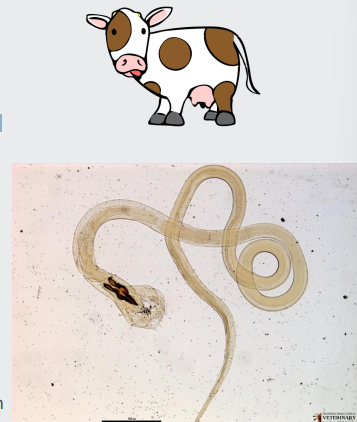
cooperia oncophora
• Inappetence/poor weight gain in calves
• Adults develop on surface of intestinal mucosa
• Partial immunity develops after 1 grazing season
• Epidemiology similar to Ostertagia
gut nematodes in sheep
Four main diseases in UK:
• Nematodirosis (Nematodirus battus)
• Ostertagiosis or teladorsagiosis (Teladorsagia circumcincta)
• Haemonchosis (Haemonchus contortus)
• Trichostrongylosis (Trichostrongylosus spp.)
nematodirosis
• Nematodirus battus
• Important in lambs
• Development to L3 takes place in eggshell
• Hatching requires prolonged chill, then mean temp >10oC (late spring)
• L3 larvae penetrate mucosa of small intestine, 2 moults then enter lumen

nematodirosis pathogenesis
• Due to larvae
• Cause severe damage to villi & erosion of mucosa → villous atrophy & fusion
• Impaired ability of intestine to exchange fluids & nutrients

nematodirosis - clinical features and diagnosis
• Yellowy-green diarrhoea
• Dehydration & inappetence, reduced growth rate
• Mortality high if untreated
• PM: carcass dehydrated, acute enteritis, thin twisted worms look like cotton wool (>10000 worms)

unique epidemiology of nematodirosis
• Eggs survive on pasture for long periods
• Lots of L3 appear on pasture at same time
• Disease outbreak if hatching coincides with grazing of lambs (often April to June)
• Cool, wet summers prolong L3 survival – may get outbreaks in older lambs later
• Lamb-to-lamb transmission, one generation of parasites each year
• Adult sheep play little role in transmission
haemonchosis
• Haemonchus contortus – worldwide distribution
• Increasing problem in the UK – spreading north
• Females - “barber’s pole” appearance
• Larvae exsheath in rumen & moult 2x in gastric glands
• Develop piercing lancet, obtain blood from mucosal vessels
• No acquired immunity

clinical features of haemonchosis
Acute disease (>2000 worms)
• Anaemia, ↓↓ packed red cell volume, loss of iron & protein to GI tract
• Oedema e.g. submandibular & ascites
• Inappetence, lethargy, dark coloured faeces, falling wool
• Mortality
Chronic haemonchosis (100s of worms)
• Continual loss of blood → weight loss, weakness & inappetence
• Particularly in areas where pasture deficient in nutrients

epidemiology of haemonchosis
• Larval development depends on high temp and humidity
• High egg output so rapid pasture contamination
Temperate areas – changing epidemiology
• Ewes deposit eggs in spring, L3s ingested by ewes & lambs in early summer,
• Disease in lambs and adults in summer sometimes extending to late autumn
• Sometimes disease in spring due to hybobiosis
• Associated with warm, wet periods
• Increased geographical spread
ostertagiosis
• Teladorsagia circumcincta
• Clinical signs: weight loss & intermittent diarrhoea, poor protein & fat
deposition, particularly in lambs
• Disease similar to type I bovine ostertagiosis in July-Sept
• Occasionally type II type in young adults in late winter/early spring
• Immunity acquired slowly – requires exposure over two grazing seasons
trichostrongylosis
• Trichostrongylus axei (abomasum) & Trichostrongylus spp. (small intestine)
• T. axei causes similar changes to Teladorsagia circumcincta
• Eggs/L3 larvae survive in environment under adverse conditions
• Larvae on pasture ↑ in late summer/autumn → clinical problems in lambs in
Nov-Dec
• Occasionally disease in spring due to survival of eggs & L3, also hypobiosis
• Rapid weight loss & diarrhoea (dark, foul-smelling)
• Inappetence & poor growth rates (lower levels of infection)
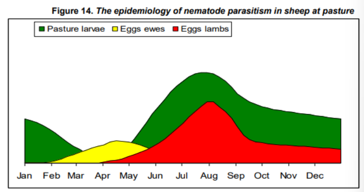
epidemiology of Teladorsagia and Trichostrongylus
Larvae numbers increase on pasture in summer/early autumn due to:
1. Eggs passed by ewes in spring
2. Eggs passed by lambs after ingesting overwintered larvae
3. Eggs passed by lambs after ingesting eggs deposited by ewes
More Teladorsagia larvae in early summer and more Trichstrongylus in late summer/ early autumn
diagnosing gins - considerations
• Host species e.g. cow, sheep, goat
• Host age
• Season
• Grazing history
• History of parasite infection on farm
• Clinical signs
• Faecal egg counts (NB not for early diagnosis of type II ostertagiosis/ nematodirosis)
• Other tests
• Post-mortem examination – worms & pathology
parasite forecast
Runs in spring/ summer
Predicts hatch date for Nematodirus battus based on temperature
identifying strongylids to genus
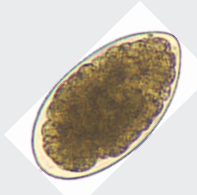
• All “strongyle” eggs look very similar apart from Nematodirus battus
• N. battus eggs – 160 μm long, “strongyle” eggs – ~80 μm long
• Culture eggs so that larvae hatch out - possible to identify larvae to genus or species
• Now also molecular approaches e.g. “nemabiome”

other tests
• Plasma pepsinogen levels in ostertagiosis & haemonchosis
• Anti-Ostertagia ostertagi (T. circumcincta) antibody levels can measured in milk
• Blood haemoglobin, packed cell volume & red blood cell count ↓ in haemonchosis


summary table - sheep gins
good to know the days and weeks (PPP)

other gut worms - rumen fluke
they look similar to fasciola hepatica eggs - but difference is on the right rumen fluke stays blue under staining
• AKA paramphistomes e.g. Calicophoron daubneyi
• Trematodes
• Young flukes in duodenum, adults in forestomachs
• Disease - heavy infestations of immature fluke in intestine
• Diarrhoea, anorexia, anaemia, ill-thrift
• Emerging parasite in Europe, now common in the UK
• Diagnosis – FECs (faecal egg count) using sedimentation, PME
• Treatment – no licenced treatment in UK (oxyclozanide – off label)
metacircarae are like cysts


tapeworms
Moniezia benedeni & M. expansa
• Cestode parasites – tapeworms
• Adults in small intestine
• Not highly pathogenic, solid mass of tapeworms could occlude intestinal lumen in lambs
• Common in calves & lambs, seasonal fluctuation – due to active periods of oribatid (forage) mites
