Non-inflammatory Exocrine Pancreatic and SI Disorders (Maldigestion and Malabsorption)
 Call Kai
 Learn
 Practice Test
 Spaced Repetition
 Match
 Flashcards
 Knowt Play
1/53

There's no tags or description
Looks like no tags are added yet.
54 Terms
Maldigestion(EPI)
Not enough enzymes to digest food as insufficient pancreas AND ↓ intrinsic factor
→ Impaired cobalamin absorption
→ Hypocobalaminemia(Low B12)
Malabsorption
Impaired intestinal absorption of improper / properly digested food – Loss of receptors → cannot absorb IF–Cobalamin complex
Dysbiosis / SIBO: Bacteria consume cobalamin → deficiency
Maldigestion (EPI) vs Malabsorption
Both manifested by Weight Loss and Chronic Diarrhea
Polyphagia
Causes of Exocrine Pancreatic Insufficiency EPI (Maldigestion): Non-Inflammatory disease
Mainly due to pancreatic acinar atrophy (wasting away)
Juvenile atrophy (immune mediated) – young GSD
Acquired (chronic pancreatitis / fibrosis), Iatrogenic or idiopathic
Causes of Exocrine Pancreatic Insufficiency EPI (Maldigestion): Rare Pancreatic neoplasia
exocrine pancreatic adenoma/adenocarcinoma (usually focal not diffuse – thus do not destroy enough exocrine tissue to cause EPI
EPI only develops when?
exocrine capacity < 10 – 15 % normal pancreas
Pathogenesis of maldigestion: Inherited: Pancreatic acinar atrophy
Acquired: GSD
idiopathic; neoplasia; chronic pancreatitis in cats
>85 loss of acinar cells
Pathogenesis of maldigestion: Loss of pancreatic secretions
• ↓Lipase*
• ↓Amylase*
• ↓Protease
• ↓HCO3
Pathogenesis of maldigestion: maldigestion
Fecal loss of nutrients
Nutrients are used by gut pathogens
Clinical Presentation of EPI: Clinical signs
Severe weight loss, could be gradual
Thin hair coat
Voluminous greasy malodorous feces - steatorrhea, Undigested fat in feces
Ravenous appetite, +/-coprophagia
EPI: Species and breed variation
More common in dogs than cats
German shepherd, Cavelier King Charles Spaniels, Chows
Routine Clinical Pathology Abnormalities EPI
CBC: Usually within reference intervals
Serum Chemistry: Usually within reference intervals
Usually normal pancreatic enzymes (amylase & lipase) → Further testing: TLI
Trypsin-like Immunoreactivity (TLI): samples
• Serum preferred but heparin plasma may be used
• Fasting 12 - 18 hours
• TLI can be falsely increased by lipemia
TLI: analysis
ELISA (and RIA) species specific antibody
Measures trypsin and trypsinogen (mostly)
TLI: diagnostic properties
Species-specific: cTLI, fTLI at TAMU & equine TLI
• Excellent sensitivity and specificity for EPI
TLI: Low values (Diagnostic)
x < 2.5μg/L
Decreased exocrine pancreatic mass (EPI)
TLI: high values
Pancreatitis
Renal azotemia
Equivocal results - can be due to:
Recent feeding - Inadequate fasting < 12 hours
Recovering function, Heat exposure in transit, Early EPI
Repeat in 1 month
Best practices for TLI testing: High TLI
Usually indicates pancreatitis – NOT used for this purpose
PLI is the test of choice for the diagnosis of pancreatitis
Trypsin only enters blood via lymphatics or peritoneal fluid when pancreatic acinar cells are damaged:
Has a very short half-life
No greater advantage than serum lipase or amylase
Small Intestinal Disease(SID)(Gastro-intestinal Disease (GID / Gut disease)
Failure of the SI to absorb digested food: Malabsorption
What causes SID?
Damage to intestinal epithelium
Decreased absorption surface
Intestinal lymphangiectasia
SID: Location / Distribution
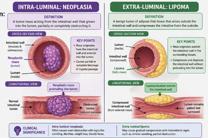
Inter-luminal, Intra-luminal, Extra-luminal
Lymphangiectasia
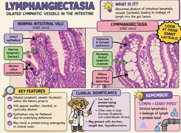
Interluminal vs intraluminal lesions

Intra vs extraluminal
SID differential list: Degenerative
Lymphangiectasia
SID differential list: A
Angiopathic: Includes torsion, thromboembolism, and hemorrhage.
Anomalous: (Congenital) primary lymphangiectasia
SID differential list: M
Metabolic: EPI, Small intestinal bacterial overgrowth (SIBO)
– Tylosin / antibiotic resp,
Endocrine : Addison’s, Hyperthyroidism
SID differential list: N
Neoplasia: Lymphoma, Gastric or adenocarcinoma, Leiomyoma / leiomyosarcoma, Mast Cell tumor (cats), Plasmacytoma
SID differential list: I
Infection: Salmonella, Giardia, histoplasmosis, Fungal, Viral, Worms,
Inflammation: Immune-mediated (eosinophilic, lymphocytic/ plasmacytic), IBD, idiopathic
SID differential list: T
Trauma: foreign body, Intussusception, Surgery
Toxic: Dietary indiscretion, Drugs (NSAIDs), Chemo agents
SID clinical signs
weight loss
vomiting +/-hematemesis
diarrhea +/-melena
Intestinal Malabsorption-Dysbiosis(SIBO) or antibiotic-responsive enteropathy
Abnormal quality and/ or quantity of intestinal microbiome
Bacteria produce excess folate → increase serum folate
Bacteria utilize cobalamin → decreased serum cobalamin
Causes of SIBO
EPI, antibiotics, abnormal GI motility
Canine microbiota dysbiosis index (DI): Quantifies 8 groups of gut bacteria using quantitative PCR
↑Strep, ↑E.coli, ↓clostridium, ↓Fusobacterium, ↓Turicibacter, etc..
<0 = normal gut microbiota
0-2 = ambiguous or mild change in the microbiome
>2 = intestinal dysbiosis
PLE: not a specific disease, but generally refers to
GI neoplasm (lymphoma), parasites, microorganisms; bacteria, rickettsia, Johne’s dz,
PLE test: Fecal α-1 protease inhibitor
Usually found in plasma, but not feces
Presence in feces » leakage from blood to GI
Resists degradation by proteases - Inhibits activity of proteases including trypsin when in feces
Limitation: sample must be maintained under frozen conditions
Increased fecal α-1 protease inhibitor
Protein-losing enteropathy or chronic blood loss in GI; has the same molecular weight as albumin
False positive: sample collected using thermometer
SID hematology
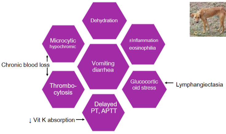
SID: serum chem

Limitations of Routine Laboratory Tests in the Diagnosis of SID
low sensitivity in detecting SID → may be WRI in severe SID
low specificity for detecting SID → Non-SID can cause similar abnormal laboratory results
Diagnosis of SID requires additional specialized testing
Decreased serum cobalamin: Distal SID
Absorbed in ileum (lower SI or distal SI
Decreased serum cobalamin: EPI
Failure to release cobalamin from R-proteins due to lack of HCO3
Measure serum B12 in any dog with poor response to enzyme replacement therapy for EPI
Decreased serum cobalamin: Small intestinal bacterial overgrowth (SIBO)
Intestinal bacteria bind/ utilize cobalamin
Decreased serum cobalamin: Breeds
Giant schnauzers, Beagles, Shar Pei, Austr Shepherds, Border Collies
Selective hereditary deficiency / cobalamin malabsorption
Increased serum cobalamin
Hepatocellular injury / neoplasia
Clinical signs of Cobalamin (B12) deficiency
Chronic wasting / failure to thrive/ weight loss
Inappetance and lethargy typically present at 3-6 months of age
● GIT signs - diarrhea
● Worsening outcome of chronic enteropathies
Cobalamin deficiency: Many cats with clinical signs of chronic GI disease (diarrhea)
have decreased serum cobalamin concentrations
show good response to Vit B12 therapy
Serum Folate
Absorbed in duodenum (upper SI or proximal SI)
Decreased serum folate: Proximal SID
Dietary deficiency
Cobalamin deficiency required for conversion to N5 –methylhydro-folate
Increased serum folate
Small intestinal bacterial overgrowth (SIBO): Due to increased synthesis of folate
EPI: Impaired synthesis and secretion of HCO3 due to Low pH
Hemolysis: RBCs are rich in folate; spun serum sample of choice
Summary vit. B12 and folate patterns

Main Causes for Decreased Serum B12
1. EPI
2. Ileal disease - intestinal villus atrophy
3. intestinal bacterial overgrowth (IBO)
Low B12 is an indication for checking serum TLI
Not all dogs with these conditions will have a low B12 concentration