Lower limb
 Call Kai
 Learn
 Practice Test
 Spaced Repetition
 Match
 Flashcards
 Knowt Play
1/124

There's no tags or description
Looks like no tags are added yet.
125 Terms
The ____ is directly anchored to the axial skeleton by
a sacroiliac joint and by strong ligaments, which link the
pelvic bone to the sacrum.
Lower limb

The lower limb is separated from the abdomen, back, and perineum by a _____
continuous line

Continuous line that joins the pubic tubercle with the anterior superior iliac spine (position of the inguinal ligament) and then continues along the iliac crest to the posterior superior iliac spine to separate the lower limb from the _____
anterior and lateral abdominal walls

Continuous line that passes between the posterior superior iliac spine and along the dorsolateral surface of the sacrum to the coccyx to separate the lower limb from the _____
muscles of the back

Continuous line that joins the medial margin of the sacrotuberous ligament, the ischial tuberosity, the ischiopubic ramus, and the pubic symphysis to separate the lower limb from the ____.
perineum

The lower limb is divided into the ___, ____, ____, and ___ on the basis of major joints, component bones, and superficial landmarks
gluteal region, thigh, leg, and foot

The _____ [region of the lower limb] is posterolateral and between the iliac crest and the fold of skin (____) that defines the lower limit of the buttocks.
gluteal region; gluteal fold

The _____ is between the inguinal ligament and the knee joint [region of the lower limb].
anterior thigh

the ____ is just inferior to the middle third of the inguinal ligament [region of the lower limb].
hip joint

the ____ is between the gluteal fold and the knee [region of the lower limb].
posterior thigh

The ____ is between the knee and ankle joint [region of the lower limb]
leg

The _____ is distal to the ankle joint [region of the lower limb]
Foot

The ____ and ____, as well as the _____, are important areas of transition through which structures pass between regions
femoral triangle; popliteal fossa; tarsal tunnel

The _____ [area of transition] is a pyramid-shaped depression formed by muscles in the proximal regions of the thigh and by the inguinal ligament, which forms the base of the ***. The major blood supply and one of the nerves of the limb (____) enter into the thigh from the abdomen by passing under the inguinal ligament and into the ***
femoral triangle; femoral nerve

The ____ [area of transition] is posterior to the knee joint and is a diamond-shaped region formed by muscles of the thigh and leg. Major vessels and nerves pass between the thigh and leg through the ***.
popliteal fossa

Most nerves, vessels, and flexor tendons that pass between the leg and foot pass through a series of canals (collectively termed the ____) on the posteromedial side of the ankle. The canals are formed by adjacent bones and a flexor retinaculum, which holds the tendons in position.
tarsal tunnel

Functions of the lower limb
Support the body weight and Locomotion
A major function of the lower limb is to support the weight
of the body with minimal expenditure of energy. When
standing erect, the center of gravity is anterior to the edge
of the ___ vertebra in the pelvis
SII

The vertical line through the center of gravity is slightly posterior to the ____ and anterior to the ____ and ____.
hip joints; knee and ankle joints

The vertical line through the center of gravity is directly over the almost circular support base formed by the ____ on the ground and holds the ____ and ____ in extension.
feet; knee and hip joints

The organization of ligaments at the hip and knee joints,
together with the shape of the articular surfaces, particularly at the knee, facilitates “____” of these joints into
position when standing, thereby reducing the muscular
energy required to maintain a standing position.
locking

A second major function of the lower limbs is to move the body through space(____). This involves the integration of movements at all joints in the lower limb to position the foot on the ground and to move the body over the foot.
Locomotion

Movements at the hip joint are _______.
flexion, extension, abduction, adduction, medial and lateral rotation, and circumduction.

The knee and ankle joints are primarily ____ joints.
hinge
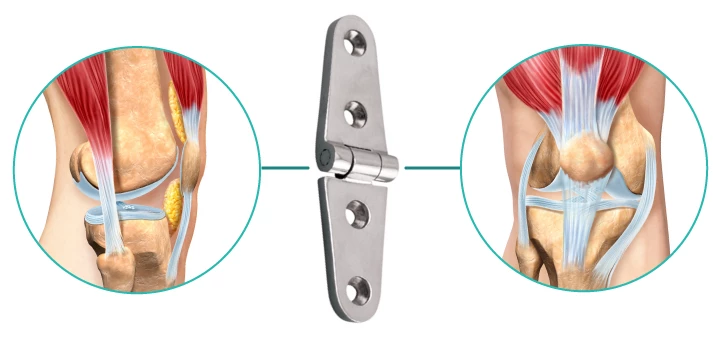
Movements at the knee are mainly _____
flexion and extensIon

Movements at the ankle are _____
dorsiflexion (movement of the dorsal side of the foot toward the leg) and plantarflexion

During walking, many anatomical features of the lower limbs contribute to minimizing fluctuations in the body’s center of gravity and thereby reduce the amount of energy needed to maintain locomotion and produce a smooth, efficient ____.
Gait

Determinants of gait include ____ in the
coronal plane, _____ in the transverse plane,
movement of the ___ toward the midline, ___ of the
knees, and complex interactions between the ___,____, & ____. As a result, during walking, the body’s center of gravity normally fluctuates only ___ cm in both vertical and lateral directions.
pelvic tilt; pelvic rotation; knees; flexion; hip, knee, and ankle; 5 cm

The bones of the gluteal region and the thigh are the _____
pelvic bone and the femur

The large ball and socket joint between these two bones is the ____.
hip joint

The ____ is the bone of the thigh.
femur

At its distal end(femur), its major weight-bearing articulation is with the ___, but it also articulates anteriorly with the ____.
tibia; patella (knee cap).

The ____ is the largest sesamoid bone in the body and is embedded in the quadriceps femoris tendon.
patella

The joint between the ___ and ____ is the principal
articulation of the knee joint, but the joint between
the ___ and ____ shares the same articular cavity.
femur & tibia; patella & femur

Although the main movements at the knee are flexion and
extension, the knee joint also allows the femur to rotate on
the ___. This rotation contributes to “____” of the knee
when fully extended, particularly when standing
tibia; locking
The leg contains 2 bone
Tibia & fibula

The ____ is medial in position, is larger than the laterally
positioned fibula, and is the weight-bearing bone.
tibia

The ___ does not take part in the knee joint and
forms only the most lateral part of the ankle joint—
proximally, it forms a small synovial joint (superior tib-
iofibular joint) with the inferolateral surface of the head
of the tibia
fibula

The tibia and fibula are linked along their lengths by an _____ membrane, and at their distal ends by a _____ joint, and little movement occurs between them.
interosseous; fibrous inferior tibiofibular;

The distal surfaces of the tibia and fibula together form a ____. The ankle joint is formed by this recess and part of one of the tarsal bones of the foot (____), which projects into the recess. The ankle is most stable when ____.
deep recess; talus; dorsiflexed.

The bones of the foot consist of the:
tarsal bones, the metatarsals, and the phalanges

There are [number] tarsal bones, which are organized in two rows with an intermediate bone between the two rows on the medial side.
seven

Inversion and eversion of the foot, or turning the sole of the foot inward and outward, respectively, occur at joints between the ___ bone
tarsal bone

The tarsal bones articulate with the _____ at
_____ joints, which allow only limited sliding
movements.
metatarsals; tarsometatarsal

Independent movements of the metatarsals are
restricted by _____, which
effectively link together the distal heads of the bones at the
_______.
deep transverse metatarsal ligaments;metatarsophalangeal joint

There is a metatarsal for each
of the five digits, and each digit has ____ phalanges except
for the _____, which has only ___
three; great toe (digit I); two

The ______ joints allow flexion, exten-
sion, abduction, and adduction of the digits, but the range
of movement is more restricted than in the hand.
metatarsophalangeal

The ____ joints are hinge joints and allow
flexion and extension
interphalangeal joints

The bones of the foot are not organized in a single plane so that they lie flat on the ground. Rather, the metatarsals and tarsals form _____
longitudinal and transverse arches

The longitudinal arch is highest on the medial side
of the foot. The arches are flexible in nature and are supported by muscles and ligaments. They absorb and transmit forces during walking and standing.
medial

Muscles of the gluteal region consist predominantly of:
extensors, rotators, and abductors of the hip joint
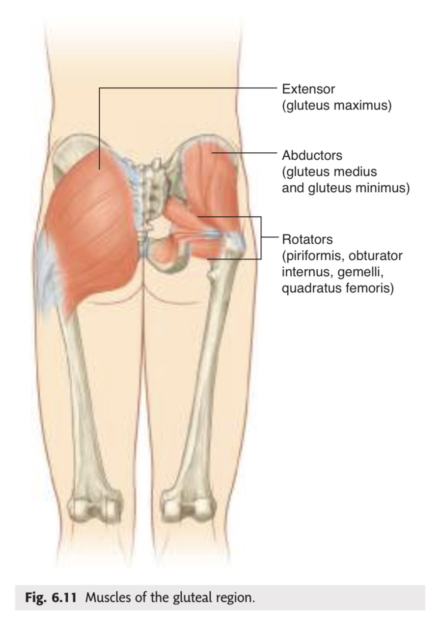
In addition to moving the thigh on a fixed pelvis, these muscles also control the movement of the pelvis relative to the limb bearing the body’s weight (____) while the other limb swings forward (___) during walking.
weight-bearing or stance limb; swing limb

Major flexor muscles of the hip (_____) do not originate in the gluteal region or the thigh. Instead, they are attached to the posterior abdominal wall and descend through the gap between the inguinal ligament and pelvic bone to attach to the proximal end of the femur.
iliopsoas—psoas major and iliacus

Muscles in the thigh and leg are separated into three
compartments by layers of:
fascia, bones, and ligaments

In the thigh, there are _____ compartments.
medial (adductor), anterior (extensor), and posterior (flexor)

Most muscles in the ____ compartment of the thigh act mainly on the hip joint
medial

The large muscles (____) in the _____ compartment of the thigh act on the hip(extension) and knee(flexion) because they attach to both the pelvis and bones of the leg
hamstrings; posterior

Muscles in the ____ compartment (____) of the thigh predominantly extend the knee
anterior; (quadriceps femoris)

Muscles in the leg are divided into ____ compartment
lateral (fibular), anterior, and posterior

Muscles in the ____ compartment of the leg predominantly evert the foot.
lateral

Muscles in the ____ compartment of the leg dorsiflex the foot and extend the digits
anterior

Muscles in the ____ compartment of the leg plantarflex the foot and flex the digits; one of the muscles can also flex the knee because it attaches superiorly to the femur.
posterior

Muscles found entirely in the ____
modify the forces produced by tendons entering the toes
from the leg and provide dynamic support for the longitu-
dinal arches of the foot when walking, particularly when
levering the body forward on the stance limb just before
toe-off.
foot (intrinsic muscles)

Unlike in the upper limb where most structures pass between the neck and limb through a single axillary inlet, in the lower limb, there are ____ major entry and exit points between the lower limb and the abdomen, pelvis, and perineum
four

Apertures of communication between the lower limb and other regions:
Gap between inguinal ligament & pelvic bone, greater sciatic foramen, obturator canal(at top of obturator foramen), & lesser sciatic foramen
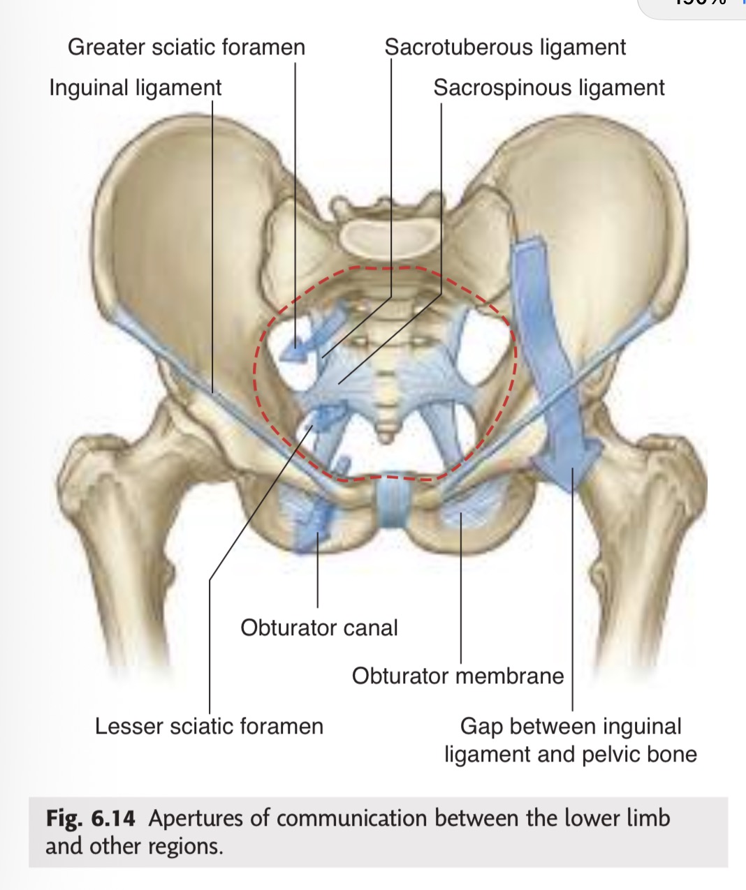
The lower limb communicates directly with the abdomen
through a gap between the ____ and the ____
pelvic bone; inguinal ligament

Structures passing through the gap between the pelvic bone and inguinal ligament include:
muscles—psoas major, iliacus, and pectineus;
nerves—femoral and femoral branch of the genitofemoral nerves, and the lateral cutaneous nerve of the thigh;
vessels—femoral artery and vein; and
lymphatics.

This gap between the pelvic bone and the inguinal ligament is a weak area in the abdominal wall and often associated with abnormal protrusion of the abdominal cavity and contents into the thigh (____). This type of *** usually occurs where the lymphatic vessels pass through the gap (the ____).
femoral hernia; femoral cana

Structures within the pelvis communicate with the lower
limb through two major apertures
Greater and lesser sciatic foramen

Posteriorly, structures communicate with the gluteal
region through the greater sciatic foramen and include:
a muscle—piriformis;
nerves—sciatic, superior and inferior gluteal, and pudendal nerves; and
vessels—superior and inferior gluteal arteries and veins, and the internal pudendal artery.

The ____ is the largest peripheral nerve of the
body and is the major nerve of the lower lim
sciatic nerve

Anteriorly, the obturator nerve and vessels pass between the pelvis and thigh through the ***. This canal is formed between bone at the top of the obturator foramen and the obturator membrane, which closes most of the foramen during life
obturator canal

Structures pass between the perineum and gluteal region through the _____
lesser sciatic foramen

The most important structure passing through the lesser sciatic foramen with respect to the lower limb is the ____
tendon of the obturator internus muscle.
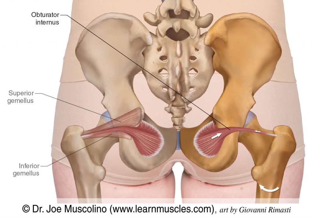
The nerve and artery of the perineum (_____) pass out of the pelvis through the greater sciatic foramen into the gluteal region and then immediately pass around the ischial spine and sacrospinous ligament and through the lesser sciatic foramen to enter the perineum.
the internal pudendal artery and pudendal nerve
Somatic motor and general sensory innervation of the
lower limb is by peripheral nerves emanating from the
____ plexuses on the posterior abdominal and
pelvic walls.
lumbar and sacral
These plexuses that provide somatic motor and general sensory innervation of the lower limb and are formed by the ____
anterior rami of L1 to L3 and most of L4 (lumbar plexus) and L4 to S5 (sacral plexus).

Nerves originating from the lumbar and sacral plexuses
and entering the lower limb carry fibers from spinal cord
levels ___
L1 to S3

Nerves from lower sacral segments innervate the ____.
perineum
Terminal nerves exit the abdomen and pelvis through a number of apertures and foramina and enter the limb. As a consequence of this innervation, lumbar and upper sacral nerves are tested clinically by examining the ___.
lower limb
Dermatome region over the inguinal ligament
L1

Dermatome region on the lateral side of the thigh:
L2

Dermatome region of the lower medial side of the thigh:
L3

Dermatome region on the medial side of the great toe (digit I):
L4

Dermatome region of the medial side of digit II:
L5

Dermatome region of the little toe (digit V):
S1

Dermatome region of the back of the thigh:
S2

Dermatome region of the skin over the gluteal fold:
S3

The dermatomes of ____ are tested in the perineum.
S4 and S5

Selected joint movements are used to test ___
myotomes

Flexion of the hip is controlled primarily by ___
L1 and L2.

Extension of the knee is controlled mainly by ___
L3 and L4.

Knee flexion is controlled mainly by ___
L5 to S2.

Plantarflexion of the foot is controlled predominantly by
___
S1 and S2.

Adduction of the digits is controlled by ____
S2 and S3

In an unconscious patient, both somatic sensory and somatic motor functions of spinal cord levels can be tested using ____
A tap on the patellar ligament at the knee tests predominantly ____
L3 and L4.

A tendon tap on the calcaneal tendon posterior to the ankle (tendon of gastrocnemius and soleus) tests ____
S1 and S2.

Large muscles in the gluteal region are innervated by
the ______
superior and inferior gluteal nerves.

Most muscles in the anterior compartment of the thigh
are innervated by the ____ (except the tensor
fasciae latae, which are innervated by the ____)
femoral nerve; superior gluteal nerve
