where inflammation from an infection accidentally activates immune cells that attack pancreatic β-cells.
 Call Kai
 Learn
 Practice Test
 Spaced Repetition
 Match
 Flashcards
 Knowt Play
1/51

There's no tags or description
Looks like no tags are added yet.
52 Terms
Factors affecting HR (3)
• autonomic innervation
• hormones
• age
Factors affecting SV (4)
• heart size
• contractility (force, duration)
• preload (end diastolic volume)
• afterload (vascular resistance)
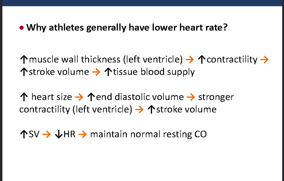
Why athletes generally have lower heart rate
what is Preload
(stretch, filling)
volume of blood in ventricles at end of diastole (EDP)
what is Afterload
(vascular resistance)
Left ventricle must overcome resistance to circulate blood
when does preload increase
- hypervolemia
- regurgitation of cardiac valves
- heart failure
when does after load increase?
↑afterload = ↑cardiac workload
afterload increases in
- hypertension
- vasoconstriction
what are factors that affect Contractility

what factors affect Preload

what is Ejection fraction?
The fraction of blood ejected from ventricle in each heart beat (contraction)
EF= SV/EDV = (~70 ml) / (~140 ml) = (~ 50%)
what does stroke volume depend on
preload
after load
Contractility
If preload increases and afterload decreases this will increase stroke volume. If contractility increases will increase stroke volume
higher stroke volume means higher cardiac output
what happens to the heart in congestive heart failure (CHF)
• fails to pump the blood efficiently
• not able to maintain normal blood flow to organs
• unable to provide adequate perfusion to meet
metabolic needs of peripheral tissues
what are two major types of heart failure?
systolic failure
Diastolic failure
what is systolic failure?
The heart loses its ability to contract or pump blood
into the circulation
It cannot eject enough blood during systole.
More blood remains in the ventricle after each contraction.
what is Diastolic failure
The heart loses its ability to relax because it becomes
stiff and cannot fill properly between each beat.
Less blood can enter the ventricle at normal filling pressures.
Ejection fraction may remain normal despite impaired filling.
CHF is divided into 2 categories based on changes in what?
Ejection fraction
- heart failure with preserved EF (≥50%)
- heart failure with reduced EF (≤40%
Common causes of Congenital heart failure (7)
• heart attack (myocardial infarction)
• coronary heart disease
• hypertension
• congenital heart defects
• heart valve defects (mitral, aortic)
• cardiac myopathy (heart muscle disease)
• lung disease
Four major compensatory mechanisms in CHF to help maintian bp
Elevated heart rate (activation of SNS
Elevated preload
Ventricular hypertrophy (enlarged heart
Vasoconstriction (endothelin, RAAS, SNS
what are pros and cons of Elevated heart rate (activation of SNS
- Pros: helps maintain cardiac output
- Cons:↓filling time, ↑O 2 demand, ↑risk of arrhythmia
pros and cons of Elevated preload
- Pros: ↑stroke volume; ↑cardiac output
- Cons: pulmonary/peripheral edema, ↑O2 demand
whats the method used to elevate preload?
↑renin/angiotensin/aldosterone system → Na+ and water
retention
pros and cons of Ventricular hypertrophy
- Pros: helps to maintain cardiac output
- Cons: ↑risk of ischemia, arrhythmias, fibrosis
pros and cons of Vasoconstriction (endothelin, RAAS, SNS)
- Pros: maintains BP, recruit blood to heart and brain
- Cons: ↑afterload (resistance) → ↓stroke volume
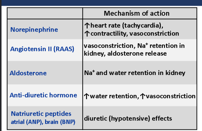
functions of NE, RAAS, ALdosterone, Anti diuretic hormone, Natriuretic peptides
Signs and symptoms of CHF
Symptoms depends on the type of heart failure (left-sided
or right-sided) as a side note
Fluid retention and edema (peripheral/pulmonary
Shortness of breath (dyspnea, orthopnea, cough)
Fatigue (especially with activity)
Cyanosis (insufficient oxygen in blood)
Confusion (if blood flow to brain is reduced
what does fluid retention and edema cause (4)
– weight gain (mainly due to edema)
– pitting edema (squishy feet/ankles)
– elevated JVP (jugular venous pressure)
– crackling sounds in lungs (on auscultation)
what are two types of heart failure and why they happen
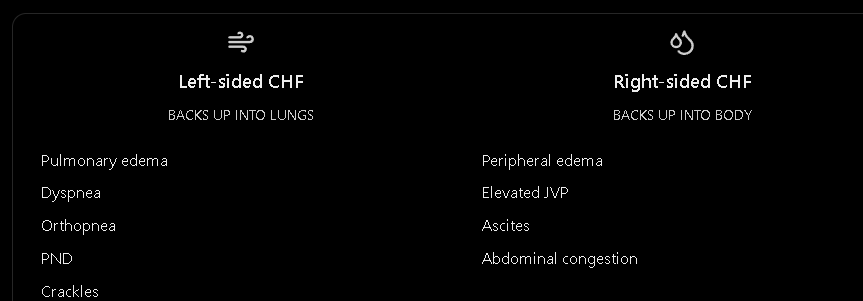
left sided heart failure - left side of heart (mainly left ventricle) fails; blood back up in lungs
Right sided heart failure - - also called “Cor Pulmonale”
- right side of heart fails; blood back up in abdominal organs and peripheral tissues
Symptoms of left-sided CHF
Symptom | Simple explanation |
|---|---|
Exertional dyspnea | Shortness of breath during exercise |
Orthopnea | Difficulty breathing when lying flat |
Paroxysmal nocturnal dyspnea (PND) | Suddenly waking up at night struggling to breathe |
Crackles | Abnormal lung sounds caused by fluid in the lungs |
Tachypnea | Increased breathing rate |
Tachycardia | Increased heart rate |
Fatigue | Reduced blood delivery to tissues |
Cyanosis | Bluish discoloration due to inadequate blood oxygenation |
how do diagnose CHF by looking at Physical assessment?
Assessment of JVP
Presence of ascites
Peripheral edema (pitting edema
what is Jugular venous pressure (JVP)
JVP is an estimate of pressure in the right atrium, assessed using the jugular veins in the neck.
Normal: Generally no more than about 3–4 cm above the sternal angle, depending on measurement technique.
Elevated JVP: Suggests increased right atrial pressure and possible systemic congestion.
In simple terms, if pressure is backing up into the veins, the jugular veins may become more distended.
what are Ascites
Ascites = Abnormal fluid accumulation in the peritoneal cavity (abdomen).
In right-sided heart failure, systemic venous pressure increases, promoting fluid accumulation in the abdomen.
what is peripheral edema
Peripheral edema = Fluid accumulation in peripheral tissues, especially the legs, ankles, and feet.
Your slide mentions pitting edema.
This means that when pressure is applied to the swollen skin, a temporary indentation remains.
LS CHF symp vs RS CHF symp
what imaging techniques can you use to diagnose CHF
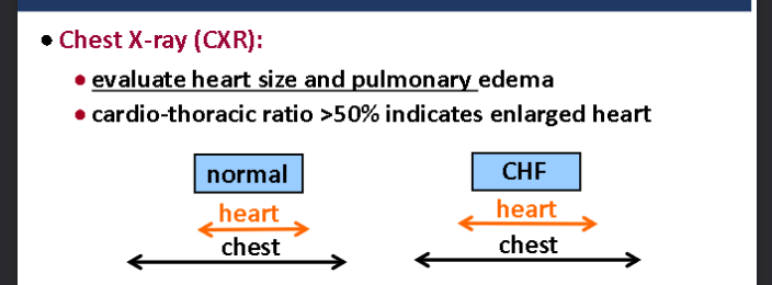
Chest X-ray (CXR)
Echocardiogram (ECHO)
what do you evaluate in Chest X-ray (CXR)
Heart size: Determine whether the heart is enlarged (cardiomegaly).
Pulmonary edema: Detect fluid accumulation in the lungs due to heart failure.
cardio-thoracic ratio >50% indicates enlarged hea r
what is Echocardiogram (ECHO):
• ultrasound waves used to image heart
• detect heart structural & functional abnormalities
• assess left ventricular EF (normal = 55-70% ; HF = <40%)
Strategies used to improve CHF
control diet - less sodium and restrict fluids to 2L/day
Modify lifestyle - stop smoking, lose weight if obese, exercise only after symptoms are controlled
what is Thrombosis
Formation of blood clot comprised of
platelets, fibrin, & entrapped RBCs in
the vascular system which is attached
to vascular endothelium
what is Embolism
A small portion of the clot breaks off, travels, and becomes trapped in a small vessel, causing occlusion leading to ischemia and infarction
what is the Virchow’s Triad?
Three factors that promote venous thrombosis
Venous Stasis - abnormality of blood flow
Hypercoagulability - abnormality in clotting
componentEndothelial Injury
what is Thrombosis in heart called
Thrombus on the wall of a heart chamber (ventricle or atria) is called mural thrombus
Thrombosis of Arterial system. Common locations include?
• cerebral
• coronary
• mesenteric
• renal arteries
thrombosis Venous system. Common locations include
• large deep veins of pelvis & lower extremities
• is referred to as deep venous thrombosis (DVT)
• may result in pulmonary embolism
how does Arterial thrombosis happen
It usually results from endothelial injury & platelet activation
(main cause: atherosclerosis)
Complications of arterial thrombosis
- vessel occlusion
- ischemic injury
- organ infarction (myocardial infarction,
cerebrovascular accident
how does Pulmonary embolism occur
Blood clot (or fat, air, tumor) forms in a vein and breaks free from vessel wall
Embolus travels through the bloodstream to the hear
Embolus travels through the heart & blocks blood vessels in the lung
Clinical features of Deep vein thrombosis
• pain
• edema
• diaphoresis
• erythema
clinical symptoms of Pulmonary embolism
• shortness of breath
• chest pain
• cough
• syncope
• hemoptysis
what is used to Diagnosis of deep vein thrombosis?
Ultrasonography
Duplex ultrasonography
what is used to Diagnos of pulmonary embolism
Ventilation-Perfusion Scan
A) Ventilation scan: inhalation of radioactive compound
B) Perfusion scan: injection of radioactive compound
Treatment of pulmonary embolism (5)
• anticoagulants
• vena cava filters
• thrombolysis
• embolectomy
• prevention strategies for high-risk patients
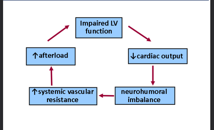
Consequences of impaired LV function