human anatomy exam #2
 Call Kai
 Learn
Learn Practice Test
 Spaced Repetition
 Match
Match Flashcards
 Knowt Play
1/125

There's no tags or description
Looks like no tags are added yet.
Name | Mastery | Learn | Test | Matching | Spaced | Call with Kai | Chat |
|---|
No analytics yet
Send a link to your students to track their progress
126 Terms
bone functions
structural support for entire body
framework for attachment of soft tissues/organs
storage of minerals and lipids (stored in the form of adipose tissue within yellow bone marrow)
blood cell production; red bone marrow (within medullary cavity of many bones) is responsible for production of RBC, WBC, and other blood elements
protection: ribs, skull, vertebral column, pelvis
leverage and movement: joints and bones function as levers; change magnitude and direction of forces generated by skeletal muscles
what type of tissue is bone
connective tissue
bone is made of…
cells + collagen & elastic fibers + calcified ground substance
ground substance of bone
mineral crystals, calcium phosphate, calcium carbonate
creates hard, calcified matrix
cells of bone
osteoblasts, osteoclasts, osteocytes
protein fibers
collagen fibers → strong & flexible
extracellular matrix
aka bone matrix aka lamellae
made of protein fibers + ground substance
hard/dense due to calcium salts deposited around collagen protein fibers
this combination gives bone its strength: very strong but still somewhat flexible, highly resistant to shattering under high pressures but still susceptible to certain forces (twisting, bending, sudden impacts)
osteogenic cells
“mesenchymal stem cells”; divide and differentiate into osteoblasts
location: inner cellular layer of periosteum; endosteum lining medullary cavities and BV passageways
osteoblast
immature bone cell
responsible for osteogenesis (process for producing new bone matrix)
osteocytes
mature bone cells; osteoblasts surrounded by bone matrix; makes up most of bone cell population
location: lacunae (pocket btw layers of matrix)
functions: maintain protein/mineral content of surrounding matrix and repair damaged bone
osteoclasts
bone-removing cells
perform osteolysis
origin: derived from same stem cells that produce monocytes/macrophages
lamellae
“bone matrix”
protein fibers + ground substance
osteogenesis
ossification— bone production
2 steps: (1) osteoid production (2) trigger Ca deposition → bone
hematopoiesis
process of producing new blood cells from stem cells
osteolysis
bone destruction/breakdown
metaphysis
region in long bone between epiphysis and diaphysis
location: of epiphyseal cartilage of developing long bone
compact bone
dense bone containing parallel osteons
forms protective outer layers of all bones
spony bone
contains open network of struts/plates resembling 3D lattice frame
does not contain large central cavity or bone marrow between lattice framework
osteon
main structural unit of compact bone
osteocytes organized within matrix around central canal
separated by concentric lamellae
central canal
“haversian canal”
longitudinal along bone
bv and nerves
volkmann’s canal
“perforating canal”
perpendicular to bone surface
bv/nerves
connections between adjacent osteons
canaliculi
narrow passageways throughout matrix
extend between lacunae and osteocytes
passageway for BV to travel throughout bone— allows diffusion of nutrients/waste to and from osteocytes
lamellae
layers of bone matrix
form lacunae (pockets within matrix in which osteocytes are located)
types: concentric, circumferential, interstitial
concentric lamellae
forms rings around individual osteons
circumferential lamellae
form larger rings around entire bone structure just deep to periosteum
interstitial lamellae
within spaces between concentric and circumferential lamellae
periosteum
outer covering of all bone surfaces (except joints)
outer fibrous region + inner cellular region
function: isolates boens from surrounding tissues; provides route for BV/nerves to enter/leave bone
collagen fibers of periosteum interwoven with fibers of tendons/ligaments attached to bone (extremely strong attachment)
endosteum
inner covering of all bone surfaces → lines medullary cavity and trabeculae
incomplete cellular layer: active during bone growth/repair and remodeling; simple flattened layer of osteoprogenitor cells; covers most of bone matrix (osteoclasts and osteoblasts work at these exposed areas)
compact bone functional characteristics
supports most of heavy weight-bearing of the body
resists twisting/bending/linear stresses on bone created from movement
protection
compact bone structural characteristics
cells and lamellae arranged in repeating cylindrical units
dense and solid
located on outer later of all bones, and the diaphysis of long bones
spony bone functional characteristics
reduces overall weight of skeleton
redistributes weight and shock-absorption
houses most of red bone marrow
spongy bone structural characteristics
lamellae form parallel sheets
osteocytes still reside in lacunae and communicate via canaliculi
no osteons or central/perforating canals
porous and light-weight
located in epiphysis of long bones and the center of flat, irregular, and spongy bones
trabeculae
meshwork of supporting bundle of fibers that form matrix of spongy bone
branch and cross-brace to create connected, open network
red marrow
children: present in all bones
adults: axial skeleton → ribs, skull, sternum, vertebrae, pelvis, and part of humerus/femur
function: RBC production
yellow marrow
adults: appendicular skeleton → replaces red bone marrow throughout skeletal maturation
function: fat (adipocyte) storage
able to convert back into red bone marrow if necessary
skeletal development
starts during gestation and continue until skeletal maturity (age 25)
ossification
formation of bone
endochondral ossification
bone tissue replaces existing hyaline cartilage
most bones of the body
intramembranous ossification
bone tissue replaces existing fibrous connective tissue
bones of skull, face, clavicle, and pelvis
interstitial growth
bone lengthening
occurs via endochondral ossification at the epiphyseal plate
occurs until skeletal maturity
appositional growth
bone widening
osteoblast activity on outside of bone
osteoclast activity on inside of bone
occurs throughout lifetime
interstitial growth PROCESS
1) chondroctyes within hyaline cartilage of growth plate multiply on epiphyseal side
2) new cartilage moves towards diaphysis
3) mineral is deposited and cartilage ossifies
4) cartilage of epiphyseal plate eventually replaced with spongy bone
appositional growth PROCESS
1) inner cellular layer of periosteum differentiates into osteoblasts → deposits superficial layers of matrix
2) eventually surrounded by matrix → differentiate to osteocytes
3) additional layers form circumferential lamellae → deeper layers of circumferential lamellae eventually recycled/replaced by compact bone
4) osteoclasts removing matrix at inner surface of bone → medullary cavity gradually enlarges
bone remodeling
dynamic process involving bone resorption (osteoclasts) and bone formation (osteoblasts) occurring at the same site
functions: maintain bone integrity, manage mineral homeostasis, adjust bone architecture to meet mechanical needs, repair micro-damage
carried out via osteocytes (continuously removing/replacing surrounding calcium matrix), osteoclasts (constantly removing old matrix), osteoblasts (always adding new matrix and developing into osteocytes)
crest
raised edge

tuberosity
rough, raised bump; usually for muscle attachment

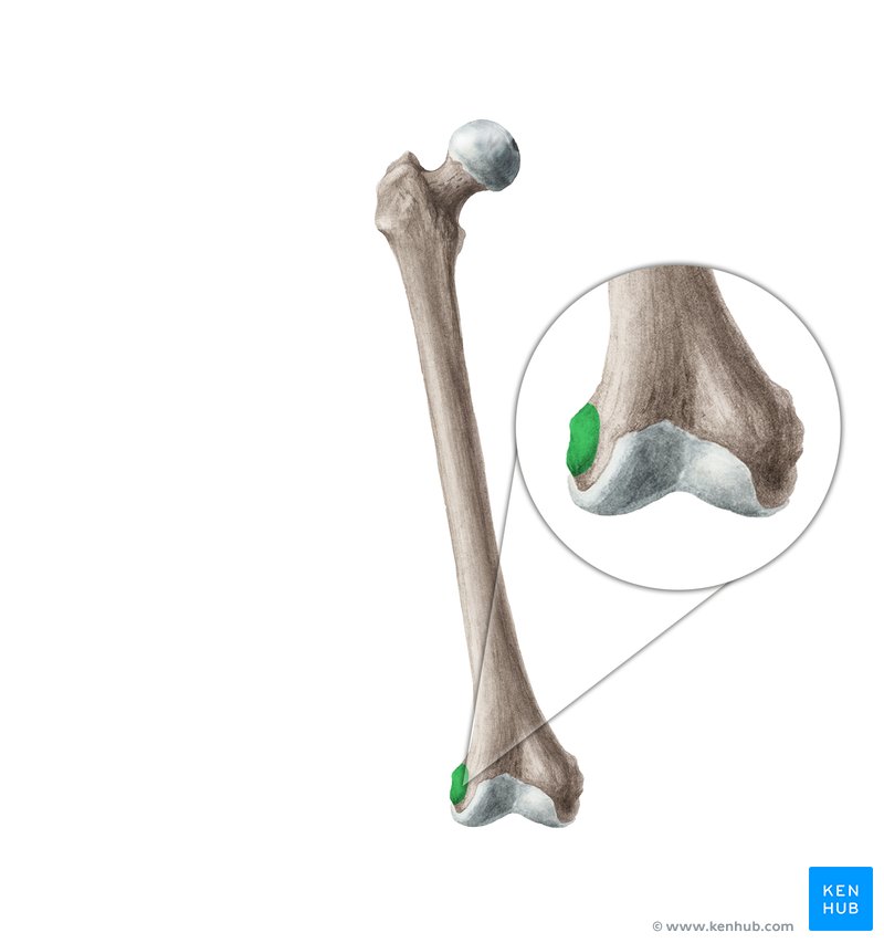
epicondyle
bump superior to a condyle
process
projection/raised area

trochanter
large process; only found on femur

spine
sharp, pointed process
tubercle
small, rounded process

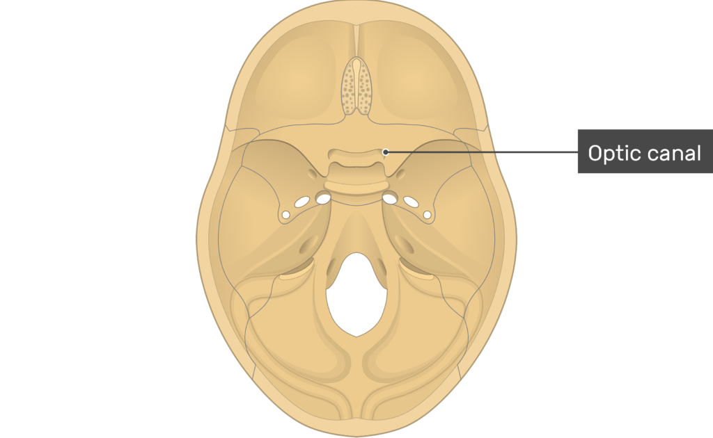
canal
tunnel through bone
fissure
long slit for blood vessels/nerves

foramen
round opening; usually passageway for nerves/blood vessels

meatus
tube-like opening

sinus
cavity within a bone

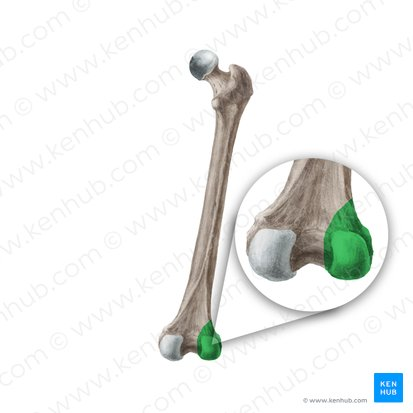
condyle
rounded knob; usually fits into fossa on another bone
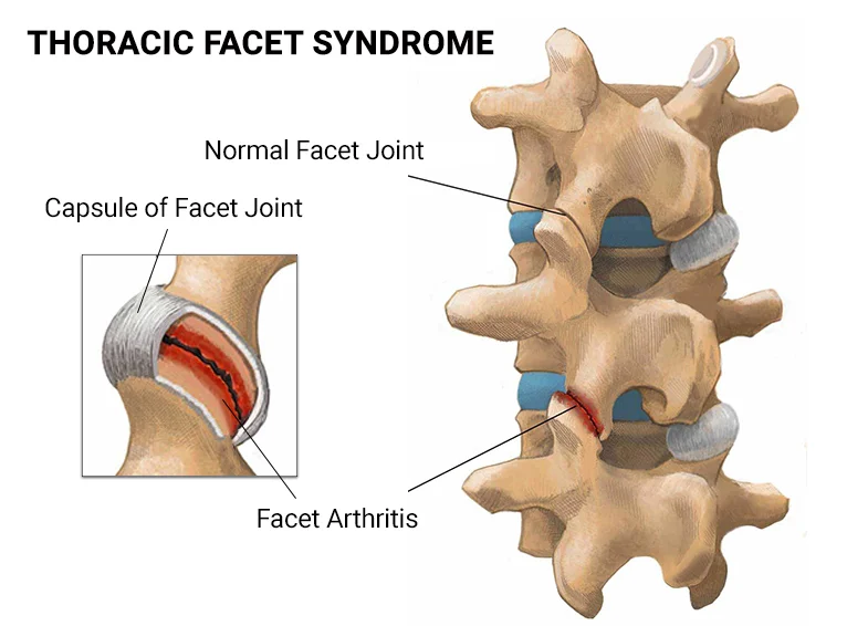
facet
flat surface
head
prominent, expanded end of a bone

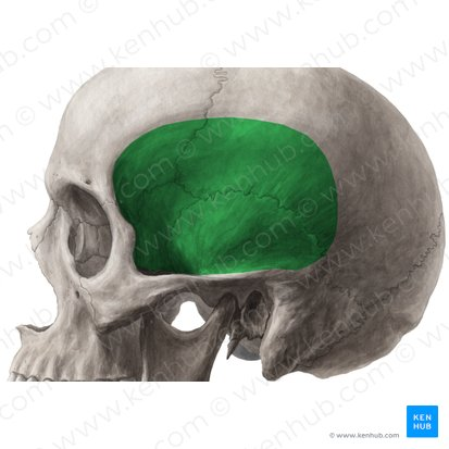
fossa
wide, shallow, basin-like groove
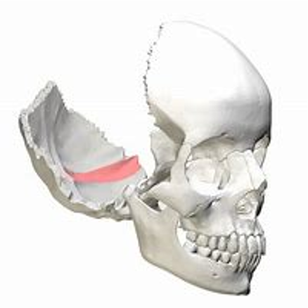
sulcus
narrow, deeper, elongated groove
fovea
small pit, indent

axial skeleton
skull, vertebral column, sacrum, ribs, sternum (80 bones)
support and protection of brain, SC, thoracic/abdominal organs
provide attachment areas for ligaments of muscles: head/neck/trunk position, breathing movement, stabilize/position limbs
joints of axial skeleton: limited movement, very strong and heavily reinforced
appendicular skeleton
limbs, pelvic bones, scapula, clavicle (126 bones)
movement and locomotion
attachment areas for ligaments of limb muscles
protection: organs of pelvic girdle
weight distribution and support
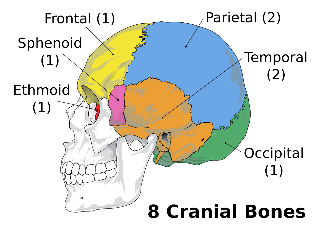
cranial bones
8
frontal, parietal (2), temporal (2), occipital, sphenoid, ethmoid
facial bones
14
nasal (2), maxillary (2), zygomatic (2), lacrimal (2), palatine (2), inferior nasal concha (2), mandible, vomer

primary vertebral curves
thoracic & sacral
develop before birth
provide space
secondary curves
cervical & lumbar
develop after birth
support weight
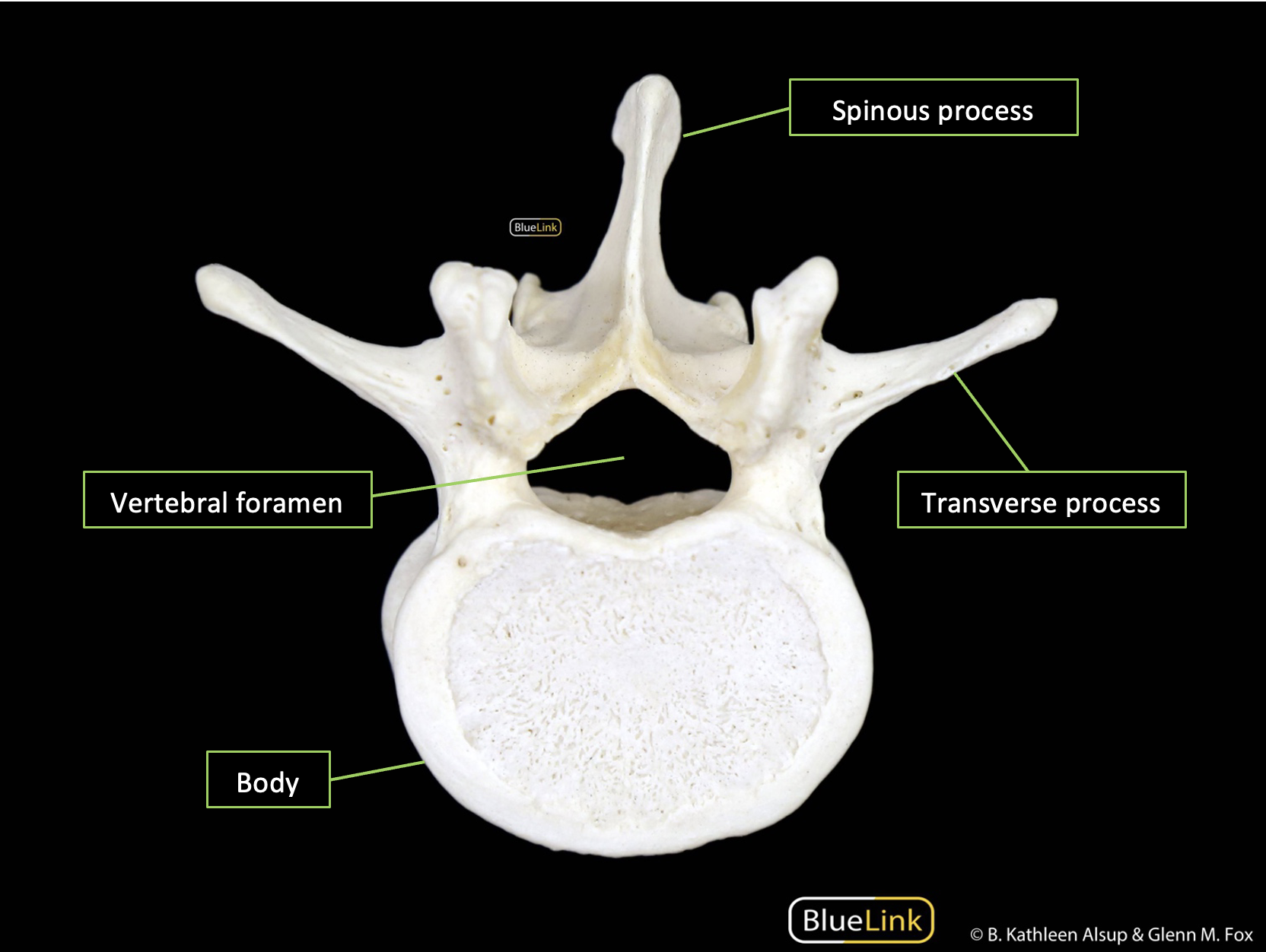
vertebrae parts
spinal canal: spinal cord travels in this space
vertebral arch: forms posterior margin of spinal canal; pedicles & laminae; anchor for vertebral processes
vertebral body: weight-bearing area of the vertebrae; forms anterior margin of spinal canal
transverse processes: attachment sites; articulates with ribs in thoracic region
articular processes: articulation area for adjacent vertebrae; inferior and superior on each
intervertebral discs: fibrocartilage pads separating bodies of vertebrae
intervertebral foramina: gaps between pedicles of successive vertebrae; allow passage of spinal nerves
vertebral canal: formed by continuous line of all vertebral foramina
vertebral foramen
spinal canal
vertebral artery
travels through transverse foramen
regional vertebrae characteristics
as move down vertebral column: spinal cord diameter decreases, vertebral arch diameter decreases, vertebral bodies enlarge to compensate for increasing load
cervical vertebrae
small body
large vertebral foramen
transverse foramen
notches spinous process
thoracic vertebrae
heart-shaped body
smaller vertebral foramen
transverse costal facets (transverse process)
costal facet (vertebral body)
articulates with ribs
lumbar vertebrae
large body
support weight of body
smaller vertebral foramen
atlas
C1
articulates with occipital condyles: joint allows neck flexion/extension
transverse ligament: binds dens of axis within vertebral foramen
structurally unique: no body, no spinous process, large round vertebral foramen
axis
C2
dens (ondontoid process):
body of atlas fuses to body of axis
large spinous process = attachment point for many neck muscles
transverse ligament: binds dens within atlas to allow rotation of atlantoaxial joint (shake head no)
sacrum
consists of 5 fused sacral vertebrae
function: protects organs within abdominopelvic cavity; attaches axial and appendicular skeleton at pelvic girdle
coccyx
3-5 fused coccygeal vertebrae
fuse after age 25-26
may fuse with sacrum later in life
rib structure
head (capitulum): articulates with vertebral column; superior and inferior articular facets
tubercle: small dorsal projection next to neck; inferior portion contains facets that articulate with thoracic vertebrae
body: curves toward the sternum; costal groove is a prominent indentation on inferior internal surface that is a pathway for BV and nerves
sternum
manubrium: widest, most superior portion
jugular notch: shallow indentation on superior surface between clavicular articulations
body
xyphoid process: attachment sites for diaphragm and rectus abdominis; palpable landmark for CPR
femur
longest/heaviest bone in body
head/epiphysis: articulates with pelvic bone in acetabulum
fovea capitis: attachment area for ligament between femur and acetabulum
medial and lateral epicondyles: small raised bump just above condyles
patellar surface: smooth area on inferior/anterior surface between medial/lateral condyles; articular surface for patella
patella
large sesamoid bone
forms within tendon of quadriceps femoris
anterior surface: rough and convex; attachment area for quadriceps tendon and patellar ligament
posterior surface: smooth and concave; area of articulation with medial and lateral femoral condyles
tibia
shinbone
medial and lateral condyles: articualte with condyles of the femur; intercondylar eminence (ridge separating condyles)
body → tibial tuberosity: rough, raised bump on anterior surface of tibia just distal to condyles; attachment site of patellar ligament
medial malleolus: large process at distal, medial tibial border; forms large medial ankle bump; supports ankle joint on medial aspect
fibula
does not bear weight
more important for muscle attachment site and lateral support of ankle
lateral malleolus: widened, distal end of fibula; forms large lateral ankle bump; supports ankle joint on lateral aspect
talus
tarsal bone
transfers weight of body from tibia towards toes
trochlea of talus: spool-shaped articular process
calcaneus
largest tarsal bone
transfers weight of body from talus to ground while standing
posterior portion: rough, knob shaped projection
coracoid process
smaller, anterior process extending above anterior surface of shoulder joint
attachment site for ligaments/tendons of shoulder joint
acromion process
larger, posterior process extending superior to shoulder joint
continuous with spine of scapula
articulates with acromial end of clavicle
attachment site for ligaments/tendons of shoulder joint
glenoid cavity
cup-shaped depression at lateral angle that forms glenohumeral joint
scapula-humerus articulation
anterior (costal) surface
smooth and concave
attachment site for many tendons/ligaments
subscapular fossa: depression in anterior surface
posterior surface
spine of scapula: ridge crossing posterior surface from acromion and to medial border
clavicle
s-shaped bone originating at superior, later border of manubrium (sternum) lateral to jugular notch
sternal end: forms sternoclavicular joint
acromial end: broad distal end that articuales with acromion process of scapular
humerus
head: articular with scapula in glenoid cavity
greater Tubercle: Rounded projection on lateral head of humerus
lesser Tubercle: Smaller rounded projection on anterior, medial surface of head of humerus
Both: attachment point for rotator cuff muscles
Body:
Medial and Lateral Epicondyles
Medial Epicondyle - Much larger
Ulnar Nerve crosses posterior surface, this is what you hit when you bump your “Funny Bone”
Condyle:
Articular surfaces:
trochlea: spool shaped medial portion of condyle that articulates with ulna; coronoid Fossa to Olecranon Fossa
capitulum: rounded surface on lateral trochlea that articulates with radius
Coronoid Fossa:
Shallow depression on anterior, proximal condyle
Fits Coronoid Process of Ulna during elbow flexion
Olecranon Fossa:
Shallow depression on posterior, proximal condyle
Fits Olecranon Process of Ulna during elbow extension
Radial Fossa:
Shallow depression on anterior condyle superior to capitulum
Fits radial head during elbow flexion
ulna
Medial bone of forearm
Connected to Radius via Interosseous membrane (Fibrous connective tissue)
Olecranon:
Point of the elbow
Trochlear Notch: Groove on anterior surface that articulates with Trochlea of Humerus
Olecranon Process: Superior point of trochlear notch
Fits into Olecranon fossa on humerus during elbow extension
Coronoid Process: Inferior point of trochlear notch
Fits into Coronoid fossa on humerus during elbow flexion
Radial Notch
Area of articulation with Head of Radius
Proximal Radio-Ulnar Joint
Ulnar Head
Ulnar Styloid Process: Articulates with distal radius at Distal Radio-Ulnar Joint
Does not directly articulate with carpal bones
Separated from carpal bones by Articular disc
radius
Lateral bone of forearm
Connected to Ulna via Interosseous membrane (Fibrous connective tissue)
Head
Articulations:
Flat superior surface: articulates with capitulum of humerus
Medial surface: articulates with Radial Notch of Ulna
Radial Tuberosity: Medial projection just below radial neck where biceps Brachii tendon attaches
Radial Styloid Process:
Ulnar Notch: Medial surface on distal radius marking articulation site with ulnar head
Radial Styloid Process
Distal surface: articulates with carpal bones
Lateral process: stabilizes wrist joint
carpals
forms 2 rows in wrist (4 proximal, 4 distal)
metacarpals: I-V starting medially
phalanges:
polex → proximal and distal
all others → proximal, middle, and distal
joint
area where adjacent bones or other structures of the skeletal system interact
“articulations”
functions: allow movement, provide stability, proprioception, accommodates growth
structural classification of joints
fibrous: bound together by fibrous CT; immovable/slightly moveable
cartilaginous: joined by cartilage; immovable/slightly moveable
bony: fibrous/cartilaginous joint ossifies; immovable
synovial: joint cavity separates adjacent bones; highly movable
functional classifications of joints
synarthrosis: immovable; fibrous or cartilaginous; axial skeleton
amphiarthrosis: slightly movable; fibrous or cartilaginous; axial skeleton
diarthrosis: highly movable; appendicular skeleton; further classifications (hinge, gliding, condyloid, saddle, pivot, ball-and-socket)