PNB 2264 EXAM 3
 Call Kai
 Learn
 Practice Test
 Spaced Repetition
 Match
 Flashcards
 Knowt Play
1/173

There's no tags or description
Looks like no tags are added yet.
174 Terms
Paraplegia
Paralysis of the lower extremities
Central nervous system (CNS)
The brain and Spial cord make up what system?
Peripheral nervous system
All the neurons that are completely or partially outside of the central nervous system make up what system?
True
True or False: The PNS is composed of sensory neurons and motor neurons that control muscle (somatic) and organs (autonomic).
Brain
Receives and processes sensory information, initiates responses, stores memories, generates thoughts and emotions
Spinal Cord
Conducts signals to and from the brain, controls reflex activities
Sensory Neurons
Sensory organs to CNS
Motor Neurons
CNS to muscles and glands
PNS
Is the Sympathetic Division (Fight or Flight system) PNS or CNS?
Autonomic Nervous System
What division are the Sympathetic Division and Parasympathetic Division part of?
Somatic Nervous System
Controls voluntary movements
Motor Neurons
What are the Autonomic Nervous system and Somatic Nervous System part of?
Peripheral Nervous System
What are Sensory Neurons and Motor Neurons part of?
afferent
Sensory information is sent to the CNS through _______ pathways.
efferent
True or False: Sensory information carried to the PNS through ________ pathways
False
The CNS is comprised of the sensory and motor neurons, while the PNS is comprised of the brain and spinal cord
Hierarchy of organization
What is it called where the lower levels (the spinal cord) relay messages to and from the upper levels (the cerebral cortex)?
The Brain
What is the ultimate destination of most sensory information?
Structure-Function Relationships
What is it called when neurons with similar functions are found in the same physical place?
Ex: The brainstem controls reflexes and involuntary processes. The Occipital lobe processes visual information. The Cerebellum coordinates movement, balance, and posture.
Topographical organization
The organization of neurons reflects body structures.
Ex. Neurons that control the fingers are next to other neurons that control fingers.
Neuroplasticity
What is it called when the CNS changes (on the millisecond timescale) in response to internal and external influences?
b
Which of the following best illustrates the hierarchical nature of information flow through the nervous system?
a. All brain functions are initiated in the spinal cord.
b. Sensory information flows from the spinal cord to the brain.
c. The brain collects sensory information and relays it to the spinal cord.
d. There is no communication between the PNS and CNS.
d
Which of the following statements best highlights differences between the CNS and PNS?
a. The CNS is part of the nervous system, while the PNS is not.
b. The CNS and PNS have identical functions, but are different anatomically.
c. The CNS can communicate with the PNS, but the PNS cannot communicate with the CNS.
d . The CNS is composed of the brain and spinal cord, while the PNS is composed of sensory and motor neurons.
False
True or False: The CNS is integrated with the rest of the body.
Cranial cavity
Where is the brain located?
Meningus
Three connective tissue layers that cover and protect the surface of the brain and spinal cord.
Dura mater
What is the first layer of the meninges?
Arachnoid mater
What is the second layer of the meninges?
Pia mater
What is the deepest layer of the meninges?
a
Starting with the skin, which of the following describes the correct order of structures that a neurosurgeon performing brain surgery would encounter?
a. bone, dura mater, arachnoid mater, pia mater, nervous tissue
b. dura mater, arachnoid mater, bone, pia mater, nervous tissue
c. bone, pia mater, dura mater, arachnoid mater, nervous tissue
d. bone, arachnoid mater, dura mater, pia mater, nervous tissue
Dura mater
Thick (0.3,,) fibrous connective tissue that is adjacent to the inner skull and vertebrae
Arachnoid mater
A thin (50µm) transparent membrane composed of fibrous tissue. Contains large blood vessels that form capillaries within the deeper mater.
Pia mater
Delicate layer (<30µm) of connective tissue attached to the surface of the brain and psinal cord.
Epidural space (Where Adipose tissue and larger blood vessels reside)
What sits between the dura mater and surrounding bone?
Subdural space
What sits between the dura mater and arachnoid mater?
Subarachnoid space
What sits between the arachnoid mater and pia mater?
Subarachnoid hemorrhage
Bleeding that occurs between the arachnoid mater and pia mater either spontaneously or as a result of trauma.
Subdural hematoma
Area of blood that collects between the dura and arachnoid mater when the small veins that connect them are torn as a result of trauma
Epidural hematoma
When blood collects between the dura mater and the surrounding skull bone
Cerebrospinal fluid
A salt solution produced by the cells of the choroid plexus that is constantly secreted into the ventricles of the brain. It helps to cushion the brain and protect it from chemical and immunological damage.
Homeostatic ion regulation, support and cushioning
What are three properties of Cerebrospinal fluid?
Homeostatic
Maintenance of the environment within narrow limits around set-points that support life.
1400g
How much does the average brain weigh without cerebrospinal fluid?
60g
How much does the average brain weigh with cerebrospinal fluid?
b
Which substances are present at higher concentrations in CSF than in plasma?
a. proteins
b. sodium ions
c. potassium ions
d. glucose
b
Why would levels of protein be lower in CSF than in plasma?
a. Proteins are not necessary for neurons to function.
b. Having fewer protein buffers makes CSF more sensitive to changes in pH.
c. Membranes like the blood-brain barrier are permeable to proteins.
d. Unlike ions, proteins have no effect on osmolarity.
b
What conclusion can be drawn from the observation that CSF is colorless?
a. CSF must be a thin layer covering the surface of the CNS.
b. Hemoglobin is not typically found in CSF.
c. Oxygen is stored inside neurons.
d. CSF and blood have identical compositions.
Choroid plexus
Formed from invaginations of the pia mater, this specialized tissue produces the cerebrospinal fluid
Ependymal cells
Neuroglia that maintains the blood-brain barrier and secretion of cerebrospinal fluid. Makes up the Choroid plexus.
ventricles
Hollow chmbers
Hypertonic
What does the choroid plexus pumping Na+ make the CSF?
In
Is water drawn into the CSF or out when Na+ is pumped into it?
500mL/day
How much CSF is produced per day?
150mL
How much CSF is within the brain and spinal cord at any given point?
Foramina of Luschka
Paired apertures located in the lateral recesses of the fourth ventricle. Drains the CSF into the subarachnoid space and travels into venous blood or lymphatic fluid.
hydrocephalus
Blockage of the foramina of Luschka
Lateral Ventricles
Large, C-shaped cavities separated from one another by the thin septapellucida which lies between them
Intraventricular foramina
A small opening that joins the left and right ventricles with the third ventricle
cerebral aqueduct
A narrow canal, allows CSF to travel from the third ventricle into the fourth ventricle.
True
True or False: The central canal extends almost the full length of the spinal cord
80
What percent of our CSF is produced in the ventricles by the choroid plexus
Skin
What layer of the Skull is this?

Aponeurosis
What layer of the Skull is this?

Periosteum
What layer of the Skull is this?
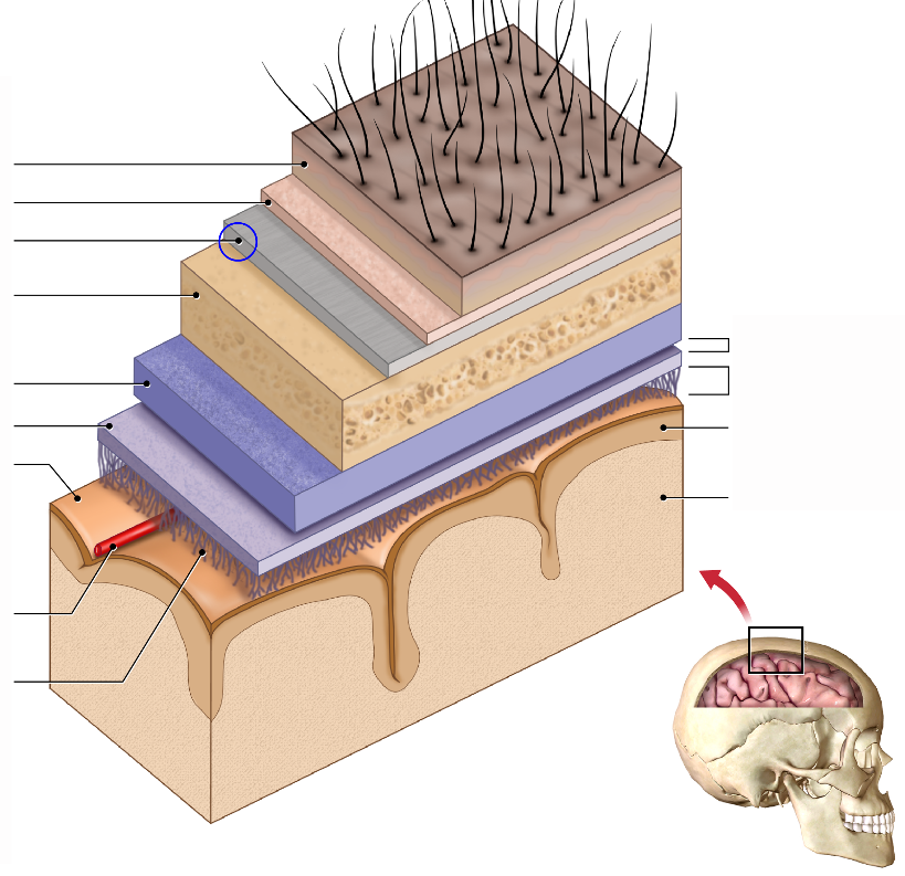
Bone
What layer of the Skull is this?

Dura mater
What layer of the Skull is this?

Arachnoid mater
What layer of the Skull is this?

Pia mater
What layer of the Skull is this?
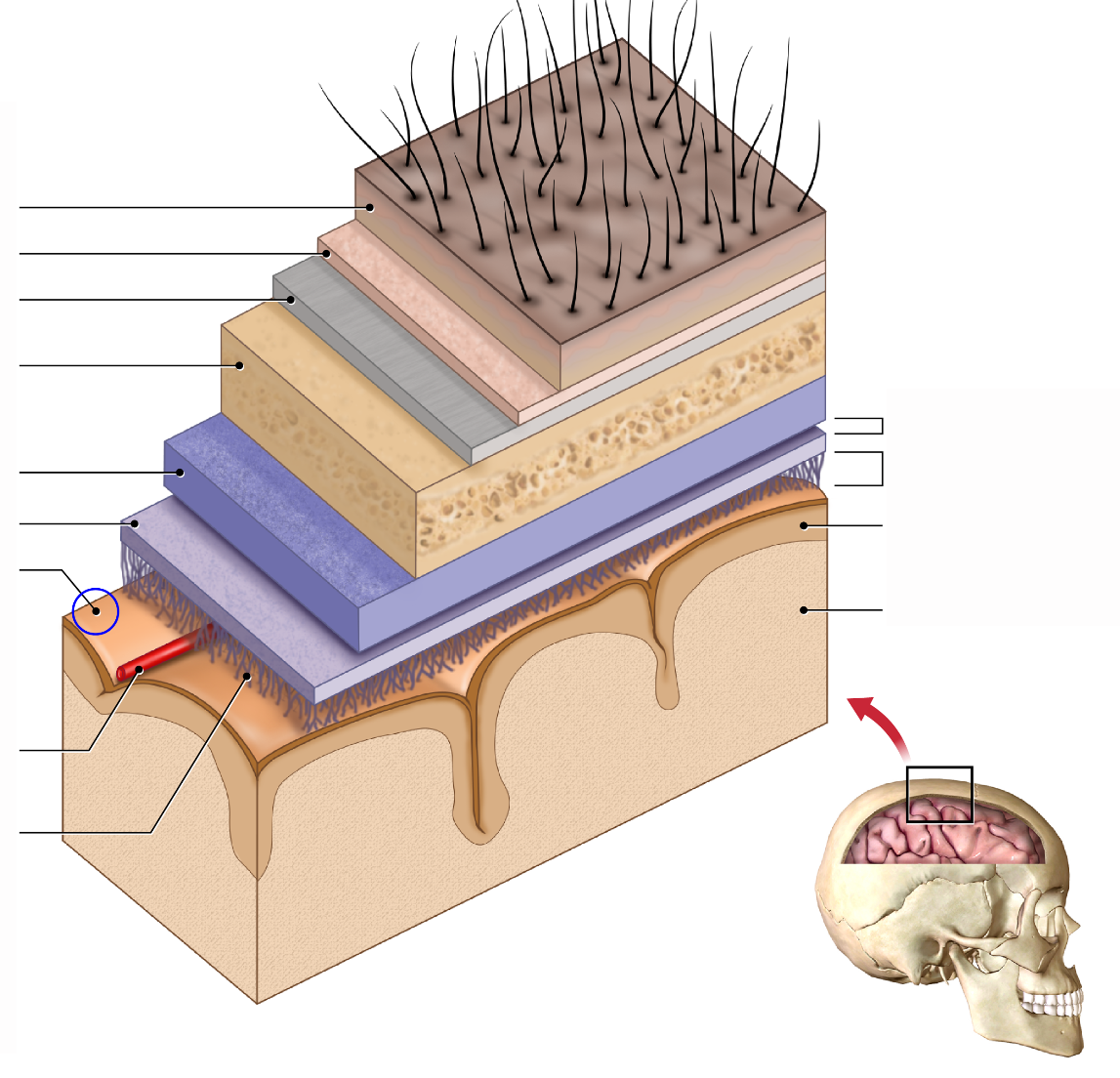
Blood vessel
What layer of the Skull is this?

Arachnoid trabeculae
What layer of the Skull is this?

Subdural space
What layer of the Skull is this?

Subarachnoid space
What layer of the Skull is this?

Cerebral cortex
What layer of the Skull is this?

White matter
What layer of the Skull is this?

Superior sagittal sinus
What segment of the CSF flovw is this?

Arachnoid granulation
What segment of the CSF flow is this?

Subarachnoid space
What segment of the CSF flow is this?
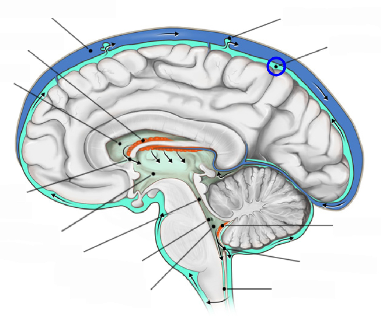
Choroid plexus
What segment of the CSF flow is this?
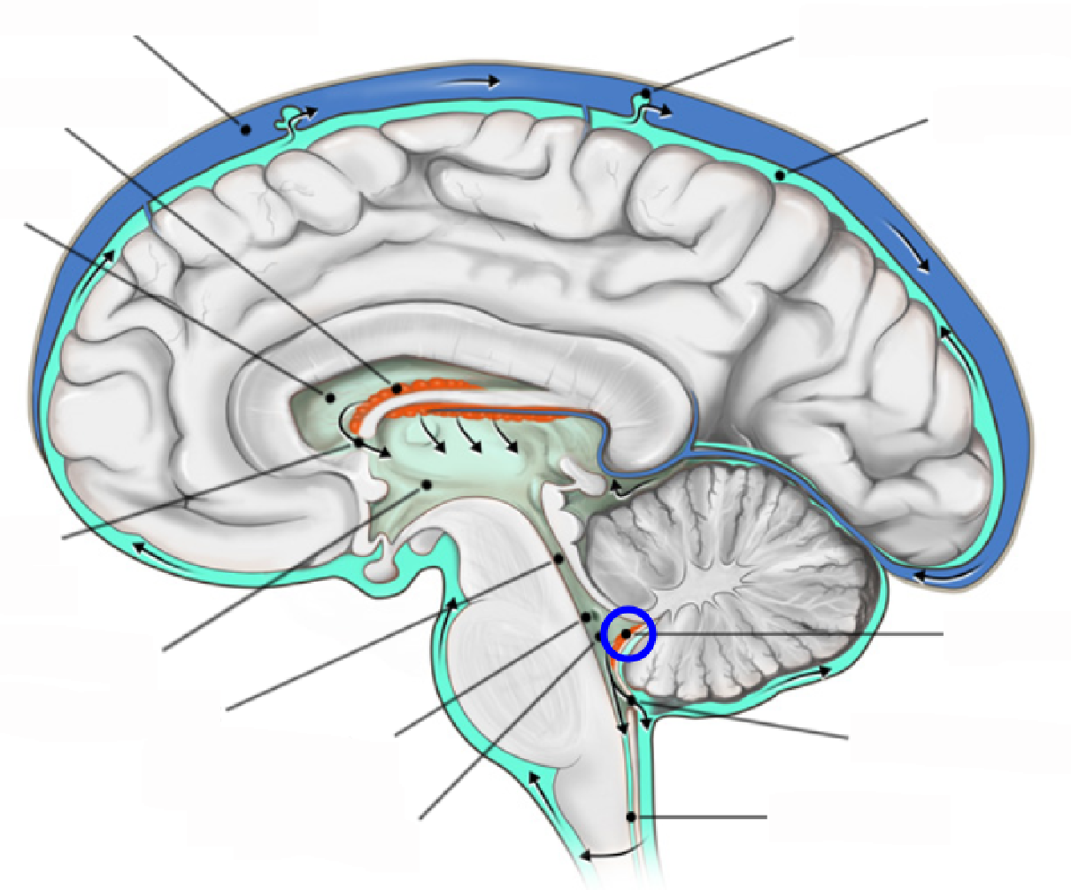
Median aperture
What segment of the CSF flow is this?

Central canal
What segment of the CSF flow is this?

Fourth ventricle
What segment of the CSF flow is this?

Lateral aperture (Formina of Luscka)
What segment of the CSF flow is this?

Cerebral aqueduct
What segment of the CSF flow is this?
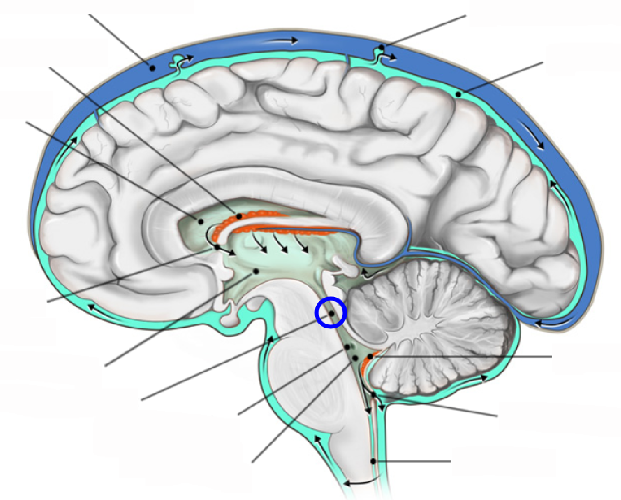
Third ventricle
What segment of the CSF flow is this?
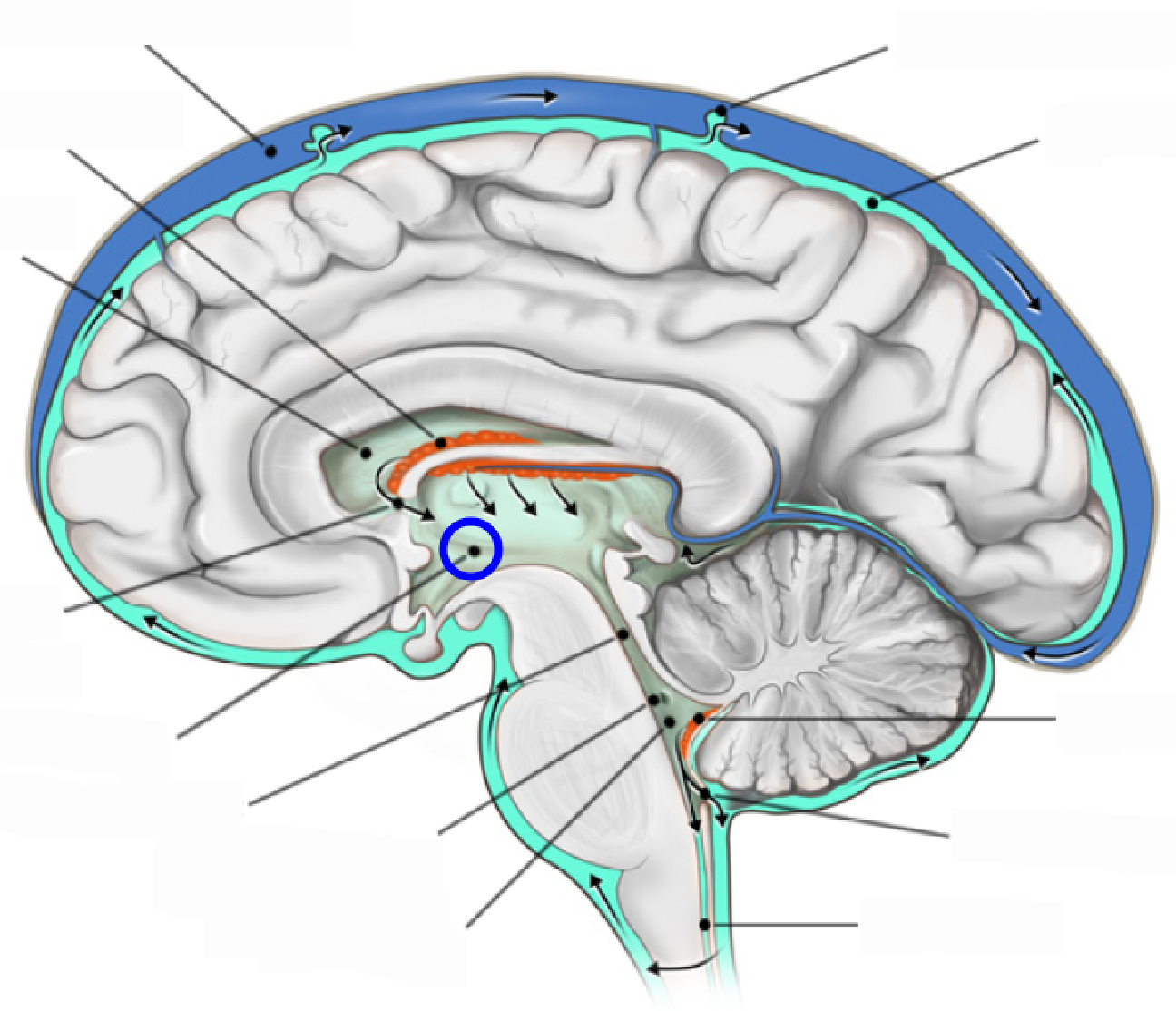
Interventricular foramen
What segment of the CSF flow is this?

Lateral ventricle
What segment of the CSF flow is this?

Choroid plexus
What segment of the CSF flow is this?

Brain
What part of the Brain ventricle is this?
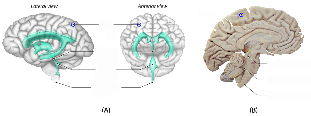
Cerebral aqueduct
What part of the Brain ventricle is this?

Fourth ventricle
What part of the Brain ventricle is this?

Spinal cord
What part of the Brain ventricle is this?

Brain interstitial system (ISS)
The other 20% of CSF produced between neurons and capillaries.
Brain interstitial fluid (ISF)
Similar in composition to CSF, and is optimized for CNS neuronal functioning,
ISS
What system operates between the CSF and capillaries/neurons? ISS or CNS?
Blood-brain barrier
Selectively semi-permeable barrier between blood in cerebral vessels and cerebrospinal fluid
False
True or False: Ethanol, THC, and nicotine affect the CNS because they are large and fat-insoluble and get stuck before the BBB.
Neurovascular unit
Endothelial cells are tightly cemented together by tight junctions, which prevents solutes from passing paracellularly between cells.
Pericytes
Vascular mural cells that surround the capillary endothelium and regulate the flow of nutrients and waste products.
A smooth muscle cell
Astrocytes
Star-shaped cells with various functions, including maintenance of extracellular ion balance, nutrient storage, and release, and repair of damaged nerves.
A type of neuroglia.
Red blood cell (erythrocyte)
What part of the neurovascular unit is this?
