Practical 3
 Call Kai
 Learn
 Practice Test
 Spaced Repetition
 Match
 Flashcards
 Knowt Play
1/67

There's no tags or description
Looks like no tags are added yet.
68 Terms

Muscle Cell Model
1.Sarcolemma
2.Endomysium
3.Axon (Myelin Sheath)
4.Dark A Band
5.Light I Band
6.Nucleus
7.Axon Terminal
8.Myofibrils
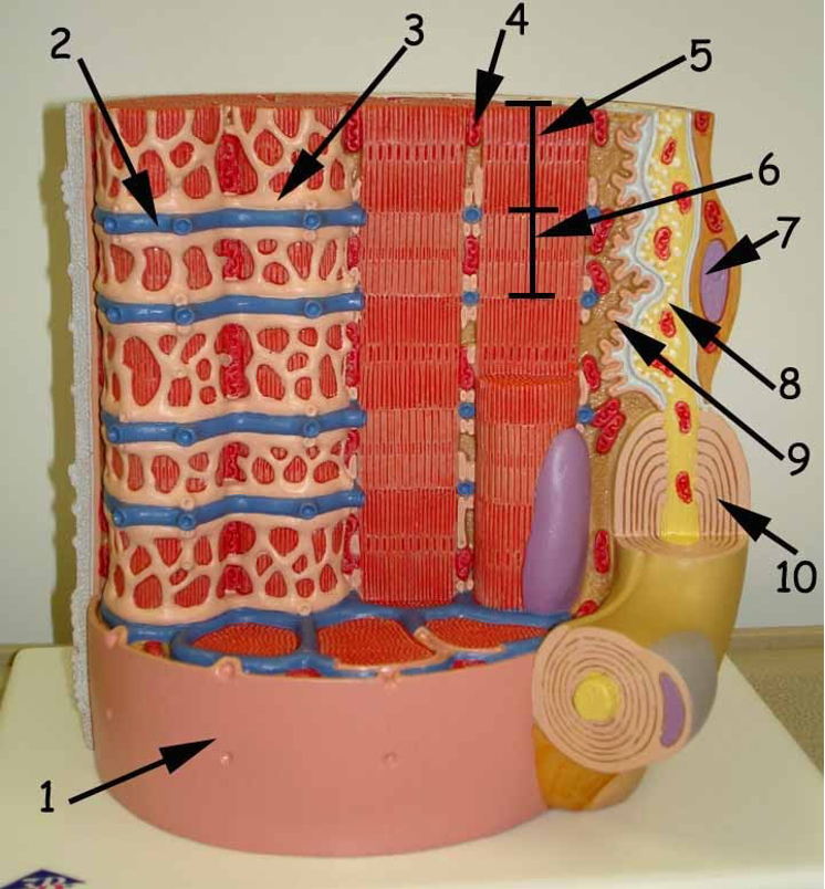
Muscle Cell Model
1.Sarcolemma
2.T Tubules
3.Sarcoplasmic Reticulum
4.Mitochondrion
5.Dark A Band
6.Light I Band
7.Nucleus
8.Axon terminal
9.Synaptic cleft
10.Myelin sheath

Head Anterior
1.Epicranius (Frontal Belly)
2.Orbicularis oris
3.Sternocleidomastoid
4.Sternohyoid
5.Orbicularis oculi

Head Lateral
1.Temporalis
2.Masseter
3.Sternocleidomastoid
4.Epicranius (Occipital Belly)

Head Inferior
1.Mylohyoid
2.Digastric
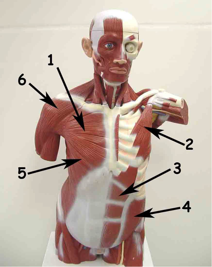
Torso Anterior
1.Pectoralis major
2.Pectoralis minor
3.Rectus abdominis
4.Internal oblique
5..External oblique
6.Deltoid

Torso Lateral
1.Pectoralis minor
2.Serratus anterior
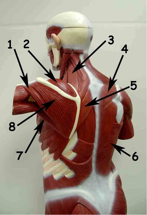
Torso Posterolateral
1.Teres minor
2.Supraspinatus
3.Levator scapulae
4.Trapezius
5.Rhomboid (Major)
6.Latissimus dorsi
7.Teres major
8.Infraspinatus
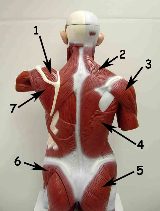
Torso Posterior
1.Supraspinatus
2.Trapezius
3.Deltoid
4. Latissimus dorsi
5.Gluteus maximus
6.Gluteus medius
7.Infraspinatus

Upper Limb Anterior
1.Brachioradialis
2.Pronator teres
3.Biceps brachii
4.Subscapularis
5.Triceps brachii
6.Flexor carpi radialis
7.Palmaris longus
8.Flexor carpi ulnaris

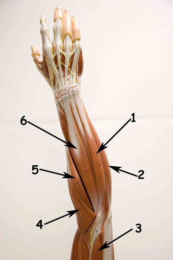
Upper Limb Anterior
1.Brachioradialis
2.Biceps brachii
3.Pronator teres
4.Flexor carpi radialis
5.Flexor carpi ulnaris
6.Palmaris longus
Upper Limb Posterior
1.Extensor carpi ulnaris
2.Flexor carpi ulnaris
3.Triceps brachii
4.Extensor carpi radialis longus
5.Extensor carpi radialis brevis
6.Extensor digitorum

Upper Limb Lateral
1.Extensor digitorum
2.Extensor carpi radialis brevis
3.Triceps brachii
4.Deltoid 5.Brachialis
6.Biceps brachii
7.Extensor carpi radialis longus

Upper Limb Posterior / Lateral
1.Flexor carpi ulnaris
2.Extensor carpi ulnaris
3.Triceps brachii
4.Deltoid
5.Brachialis
6.Biceps brachii
7.Extensor carpi radialis longus
8.Extensor carpi radialis brevis
9.Extensor digitorum

Upper Limb Posterior / Lateral
1.Triceps brachii
2.Teres major
3.Infraspinatus
4.Deltoid
5.Brachialis
6.Biceps brachii
7.Extensor carpi radialis brevis
8.Extensor digitorum

Lower Limb Anterior
1.Psoas major
2.Tensor fasciae latae
3.Vastus lateralis
4.Fibularis longus
5.Extensor digitorum longus
6.Tibialis anterior
7.Vastus medialis
8.Rectus femoris 9.Sartorius
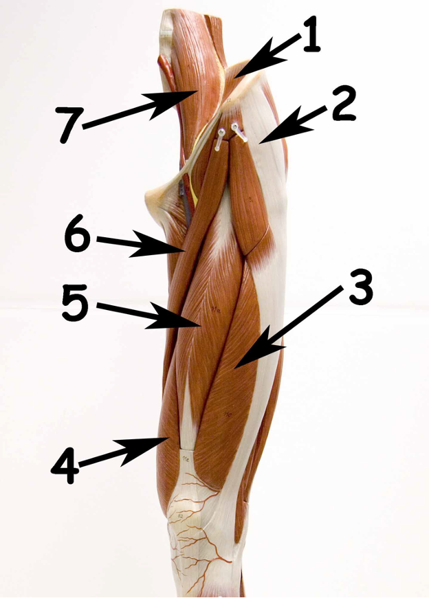
Thigh Anterior
1.Iliacus
2.Tensor fasciae latae
3.Vastus lateralis
4.Vastus medialis
5.Rectus femoris
6.Sartorius
7.Psoas major

Leg Anterior
1.Tibialis anterior
2.Fibularis longus
3.Extensor digitorum longus
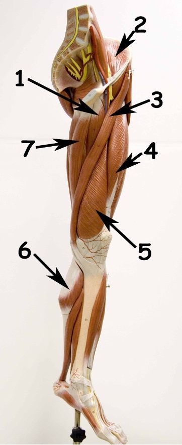
Lower Limb Medial
1.Adductor longus
2.Iliacus
3.Sartorius
4.Rectus femoris
5.Vastus medialis
6.Gastrocnemius
7.Gracilis

Thigh Medial
1.Iliacus
2.Sartorius
3.Rectus femoris
4.Vastus medialis
5.Gracilis
6.Adductor longus

Thigh Medial
1.Psoas major
2.Iliacus
3.Sartorius
4.Retus femoris
5.Vastus lateralis
6.Vastus medialis
7.Gracilis
8.Adductor longus
9.Pectineus
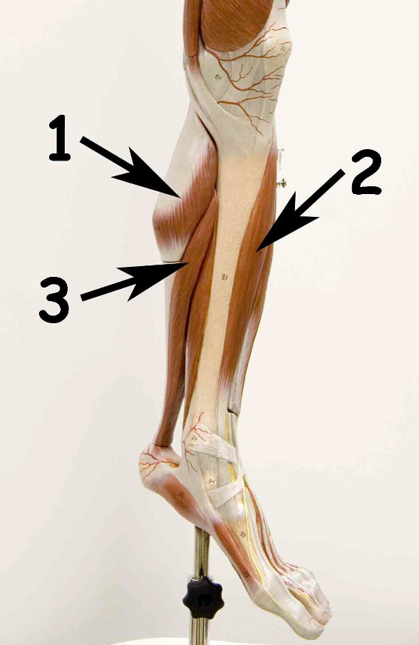
Leg Medial
1.Gastrocnemius
2.Tibialis anterior
3.Soleus

Lower Limb Posterior
1.Gluteus maximus
2.Adductor magnus
3.Gracilis
4.Gastrocnemius
5.Semimembranosus
6.Semitendinosus
7.Biceps femoris

Thigh Posterior
1.Gluteus maximus
2.Adductor magnus
3.Gracilis
4.Semimembranosus
5.Semitendinosus
6.Biceps femoris

Lower Limb Lateral
1.Gluteus maximus
2.Biceps femoris
3.Gastrocnemius
4.Soleus
5.Extensor digitorum longus
6.Fibularis longus
7.Vastus lateralis
8.Tensor fasciae latae
9.Gluteus medius

Thigh Lateral
1.Gluteus maximus
2.Semitendinosus
3.Biceps femoris
4.Vastus lateralis
5.Tensor fasciae latae
6.Gluteus medius

Leg Lateral
1.Gastrocnemius
2.Soleus
3.Fibularis longus
6 synovial joints
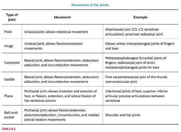
pivot joint (descriptions and examples)
o a rounded portion of a bone is enclosed within a ring formed partially by the articulation with another bone and partially by a ligament; roataion is around a single axis (uniaxial diarthrosis)
ex: atlantoaxial joint, found between the C1 (atlas) and C2 (axis) vertebrae; proximal radioulnar joint

hinge joint (descriptions and examples)
o the convex end of one bone articulates with the concave end of the adjoining bone; only for bending and straightening motions along a single axis (uniaxial)
Ex: elbow joint, knee, ankle, and interphalangeal joints between the phalanx bones of the fingers and toes

condyloid joint (descriptions and examples)
aka ellipsoid joint
o the shallow depression at the end of one bone articulates with a rounded structure from an adjacent bone or bones; biaxial joints
Ex: knuckle (metacarpophalangeal) joints of the hand between the distal end of a metacarpal bone and the proximal phalanx bone; radiocarpal joint of the wrist

saddle joint (descriptions and examples)
o both of the articulating surfaces for the bones have a saddle shape, which is concave in one direction and convex in the other; biaxial joint
Ex: first carpometacarpal joint, between the trapezium (a carpal bone) and the first metacarpal bone at the base of the thumb

plane joint (descriptions and examples)
(aka gliding joint)
o the articulating surfaces of the bones are flat or slightly curved and of approximately the same size, which allows the bones to slide against each other; multiaxial joint
Ex: between the carpal bones (intercarpal joints) of the wrist or tarsal bones (intertarsal joints) of the foot, between the clavicle and acromion of the scapula (acromioclavicular joint), and between the superior and inferior articular processes of adjacent vertebrae (zygapophysial joints)

ball-and-socket joint (descriptions and examples)
o the rounded head of one bone (the ball) fits into the concave articulation (the socket) of the adjacent bone; multiaxial joints
Ex: hip joint and the glenohumeral (shoulder) joint

Flexion and extension
o Flexion and extension are typically movements that take place within the sagittal plane and involve anterior or posterior movements of the neck, trunk, or limbs. For the vertebral column, flexion (anterior flexion) is an anterior (forward) bending of the neck or trunk, while extension involves a posterior-directed motion, such as straightening from a flexed position or bending backward.
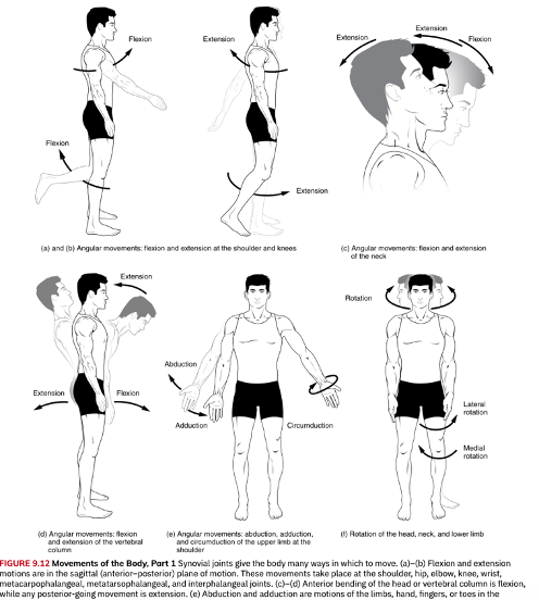
Abduction and adduction
o Abduction and adduction motions occur within the coronal plane and involve medial-lateral motions of the limbs, fingers, toes, or thumb. Abduction moves the limb laterally away from the midline of the body, while adduction is the opposing movement that brings the limb toward the body or across the midline.

Circumduction
o Circumduction is the movement of a body region in a circular manner, in which one end of the body region being moved stays relatively stationary while the other end describes a circle.

Rotation
o Rotation can occur within the vertebral column, at a pivot joint, or at a ball-and-socket joint. Rotation of the neck or body is the twisting movement produced by the summation of the small rotational movements available between adjacent vertebrae. At a pivot joint, one bone rotates in relation to another bone.

Supination and Pronation
o Supination and pronation are the movements of the forearm that go between these two positions. Pronation is the motion that moves the forearm from the supinated (anatomical) position to the pronated (palm backward) position. Supination is the opposite motion, in which rotation of the radius returns the bones to their parallel positions and moves the palm to the anterior facing (supinated) position.

Dorsiflexion and plantar flexion
o Dorsiflexion and plantar flexion are movements at the ankle joint, which is a hinge joint. Lifting the front of the foot, so that the top of the foot moves toward the anterior leg is dorsiflexion, while lifting the heel of the foot from the ground or pointing the toes downward is plantar flexion.

Inversion
o Inversion is the turning of the foot to angle the bottom of the foot toward the midline, while eversion turns the bottom of the foot away from the midline]
![<p><span style="font-family: "Courier New";">o</span><span style="font-family: "Times New Roman"; line-height: normal; font-size: 7pt;"> </span>Inversion is the turning of the foot to angle the bottom of the foot toward the midline, while eversion turns the bottom of the foot away from the midline]</p>](https://assets.knowt.com/user-attachments/1ba75433-cb65-4f26-80b2-fe1aefdf11e1.png)
Protraction and retraction
Protraction and retraction are anterior-posterior movements of the scapula or mandible. Protraction of the scapula occurs when the shoulder is moved forward, as when pushing against something or throwing a ball. Retraction is the opposite motion, with the scapula being pulled posteriorly and medially, toward the vertebral column.

Depression and elevation
o Depression and elevation are downward and upward movements of the scapula or mandible. The upward movement of the scapula and shoulder is elevation, while a downward movement is depression.

Excursion
o Excursion is the side to side movement of the mandible. Lateral excursion moves the mandible away from the midline, toward either the right or left side. Medial excursion returns the mandible to its resting position at the midline.
Superior and Inferior rotation
superior rotation, the glenoid cavity moves upward as the medial end of the scapular spine moves downward
Inferior rotation occurs during limb adduction and involves the downward motion of the glenoid cavity with upward movement of the medial end of the scapular spine
Opposition
o Opposition is the thumb movement that brings the tip of the thumb in contact with the tip of a finger.

functions of skeletal muscle
o to contract and cause movement
o protect internal organs by acting as an external barrier or shield to external trauma and by supporting the weight of the organs
o contribute to the maintenance of homeostasis in the body by generating heat
movement of a pivot joint
Uniaxial joint; allows rotational movement
example of pivot joints
Atlantoaxial joint (C1–C2 vertebrae articulation); proximal radioulnar joint
movement of a hinge joint
Uniaxial joint; allows flexion/extension movements
example of hinge joints
Elbow; ankle; interphalangeal joints of fingers and toes
movement of a condyloid joint
Biaxial joint; allows flexion/extension, abduction/ adduction, and circumduction movements
example of condyloid joint
Metacarpophalangeal (knuckle) joints of fingers; radiocarpal joint of wrist; metatarsophalangeal joints for toes
movement of a saddle joint
Biaxial joint; allows flexion/extension, abduction/ adduction, and circumduction movements
example of saddle joint
First carpometacarpal joint of the thumb; sternoclavicular joint
movement of a plane joint
Multiaxial joint; allows inversion and eversion of foot, or flexion, extension, and lateral flexion of the vertebral column
example of plane joint
Intertarsal joints of foot; superior-inferior articular process articulations between vertebrae
movement of a ball-and-socket joint
Multiaxial joint; allows flexion/extension, abduction/adduction, circumduction, and medial/ lateral rotation movements
example of ball-and-socket joint
Shoulder and hip joints
The 3 layers of connective tissue that provide structure to the muscle:
epimysium: sheath of dense, irregular connective tissue that wraps each muscle, allows a muscle to contract, separate muscle from other tissue/ organs in the area to move independently
perimysium: middle layer of connective tissue; surround the individual bundles of muscle fibers (aka fasicle)
endomysium: thin connective tissue layer of collagen and reticular fibers that encase each muscle fiber in each fasicle
fascicle
individual bundle of muscle fibers; common in muscle of the limbs
sarcolemma
The plasma membrane of muscle fibers
sarcoplasm
cytoplasm of muscle fibers
sarcoplasmic reticulum (SR)
specialized smooth endoplasmic reticulum, which stores, releases, and retrieves calcium ions (Ca++)
sarcomere
the functional unit of a skeletal muscle fiber, a highly organized arrangement of the contractile myofilaments actin (thin filament) and myosin (thick filament), along with other support proteins.
regulatory proteins, troponin and tropomyosin
actin vs myosin
contractile myofilaments
actin (thin filament, bc they form strands that are thinner than the myosin)
myosin (thick filament, bc it has more mass and is thicker)
neuromuscular junction (NMJ).
site where a motor neuron’s terminal meets the muscle fiber
Z-discs (also called Z-lines)
part of the sarcomere, where sctin myofilamnets are anchored