Mycology Week Three Worksheets

0.0(0)
Studied by 0 people
call kaiCall Kai
learnLearn
examPractice Test
spaced repetitionSpaced Repetition
heart puzzleMatch
flashcardsFlashcards
GameKnowt Play
Card Sorting

1/8

encourage image

There's no tags or description

Looks like no tags are added yet.

Last updated 3:21 AM on 10/8/26
Name
Mastery
Learn
Test
Matching
Spaced
Call with Kai
Chat

No analytics yet

Send a link to your students to track their progress

9 Terms

1
New cards

Alternaria sp.

  • Macroscopic:

    • front: cottony, black to grey to olive brown

    • reverse: black-brown

  • Microscopic:

    • conidiophores support mainly chained poroconidia: multi-celled, club-shaped poroconidia contain horizontal or vertical septa


<ul><li><p>Macroscopic:</p><ul><li><p>front: cottony, black to grey to olive brown</p></li><li><p>reverse: black-brown</p></li></ul></li><li><p>Microscopic:</p><ul><li><p>conidiophores support mainly chained poroconidia: multi-celled, club-shaped poroconidia contain horizontal or vertical septa</p></li></ul></li></ul><p></p>
2
New cards

Bipolaris sp.

  • Macroscopic:

    • front: matted center and raise grayish periphery, first grayish brown, becoming black

    • reverse: black

  • Microscopic:

    • bent-knee shaped conidiospore supports multi-celled, canoe-shaped poroconidia (usually more than 6 cells)


<ul><li><p>Macroscopic:</p><ul><li><p>front: matted center and raise grayish periphery, first grayish brown, becoming black</p></li><li><p>reverse: black</p></li></ul></li><li><p>Microscopic:</p><ul><li><p>bent-knee shaped conidiospore supports multi-celled, canoe-shaped poroconidia (usually more than 6 cells)</p></li></ul></li></ul><p></p>
3
New cards

Curvularia sp.

  • Macroscopic:

    • front: wooly, dark olive-green to brown or black

    • reverse: dark

  • Microscopic:

    • bent-knee shaped conidiophore give rise to large, four-fived celled poraconidia (rounded at the ends)

    • poroconidia have over-enlarged central cells


<ul><li><p>Macroscopic:</p><ul><li><p>front: wooly, dark olive-green to brown or black</p></li><li><p>reverse: dark</p></li></ul></li><li><p>Microscopic:</p><ul><li><p>bent-knee shaped conidiophore give rise to large, four-fived celled poraconidia (rounded at the ends)</p></li><li><p>poroconidia have over-enlarged central cells</p></li></ul></li></ul><p></p>
4
New cards

Epidermophyton sp.

  • Macroscopic:

    • front: brownish to yellow to olive-gray or khaki, first lumpy and sparse and then folded in center and groved radically

    • reverse: orange to brownish, sometimes with a thin yellow border

  • Microscopic:

    • long, paddle-like macroconidia (with thin, smooth walls) contain 2 to 6 cells and often produced in clusters growing on conidiophore

    • in older cultures see numerous chlamydoconidia; no microconidia
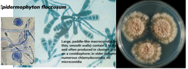

<ul><li><p>Macroscopic:</p><ul><li><p>front: brownish to yellow to olive-gray or khaki, first lumpy and sparse and then folded in center and groved radically</p></li><li><p>reverse: orange to brownish, sometimes with a thin yellow border</p></li></ul></li><li><p>Microscopic: </p><ul><li><p>long, paddle-like macroconidia (with thin, smooth walls) contain 2 to 6 cells and often produced in clusters growing on conidiophore</p></li><li><p>in older cultures see numerous chlamydoconidia; no microconidia </p></li></ul></li></ul><p></p>
5
New cards

Microsporum canis

  • Macroscopic:

    • front: spreading, wooly to cottony, whitish

    • reverse: deep yellow

  • Microscopic:

    • rough, thick-celled wall, spindle-shaped macroconidia contain 6 to 15 cells that are on conidiophores; rare club-shaped microconidia are borne singly along the hyphae

  • Fluorescene results: positive


<ul><li><p>Macroscopic:</p><ul><li><p>front: spreading, wooly to cottony, whitish</p></li><li><p>reverse: deep yellow</p></li></ul></li><li><p>Microscopic:</p><ul><li><p>rough, thick-celled wall, spindle-shaped macroconidia contain 6 to 15 cells that are on conidiophores; rare club-shaped microconidia are borne singly along the hyphae</p></li></ul></li><li><p><mark data-color="yellow" style="background-color: yellow; color: inherit;">Fluorescene results: positive</mark></p></li></ul><p></p>
6
New cards

Nannizzia (formerly Microsporum) gypsea

  • Infects animals very frequently

  • Macroscopic:

    • front: flat and spreading and powdery to granular, tan to cinnamon brown

    • reverse: yellow, orange-tan, brownish red, purpish red in spots

  • Microscopic:

    • rough, thin-walled, ellptical-shaped macroconidia contain 4 to 6 cells; terminal ends are slighty rounded and are conidiophores; club-shaped microconidia are borne singy along the hyphae

  • Flourescence result: positive


<ul><li><p><mark data-color="yellow" style="background-color: yellow; color: inherit;">Infects animals very frequently</mark></p></li><li><p>Macroscopic:</p><ul><li><p>front: flat and spreading and powdery to granular, tan to cinnamon brown</p></li><li><p>reverse: yellow, orange-tan, brownish red, purpish red in spots</p></li></ul></li><li><p>Microscopic:</p><ul><li><p>rough, thin-walled, ellptical-shaped macroconidia contain 4 to 6 cells; terminal ends are slighty rounded and are conidiophores; club-shaped microconidia are borne singy along the hyphae</p></li></ul></li><li><p><mark data-color="yellow" style="background-color: yellow; color: inherit;">Flourescence result: positive</mark></p></li></ul><p></p>
7
New cards

Trichophyton mentagrophytes

  • Major pathogen of the foot

  • Microscopic:

    • front: varies greatly; buff and powdery, white, may become pinkish or yellowish

    • reverse: usually brownish, but may be colorless, yellow, or red

  • Macroscopic:

    • thin, smooth-welled, cigar-shaped macroconidia on a conidiophore; small spherical microconidia are produced in grape-like clusters on the hyphae

    • varying numbers of spherical chlamydoconida and spiral shape may be present

  • Lab results:

    • vitro hair perforation: +, agar 1: 4+, agar 4: 4+, urease: +, fluorescene: negative


<ul><li><p><mark data-color="yellow" style="background-color: yellow; color: inherit;">Major pathogen of the foot</mark></p></li><li><p>Microscopic:</p><ul><li><p>front: varies greatly; buff and powdery, white, may become pinkish or yellowish</p></li><li><p>reverse: usually brownish, but may be colorless, yellow, or red</p></li></ul></li><li><p>Macroscopic:</p><ul><li><p>thin, smooth-welled, cigar-shaped macroconidia on a conidiophore; small spherical microconidia are produced in grape-like clusters on the hyphae</p></li><li><p>varying numbers of spherical chlamydoconida and spiral shape may be present</p></li></ul></li><li><p><mark data-color="yellow" style="background-color: yellow; color: inherit;">Lab results:</mark></p><ul><li><p>vitro hair perforation: +, agar 1: 4+, agar 4: 4+, urease: +, fluorescene: negative</p></li></ul></li></ul><p></p>
8
New cards

Trichophyton rubrum

  • Most frequently isolated dermatophytes infecting humans worldwide affecting the skin and nails

  • Macroscopic:

    • front: granular of fluffy, white to buff

    • reverse: deep red or purplish

  • Microscopic:

    • “birds on telephone wire”

    • thin, smooth-walled cylindrical with blunt ends macroconidia (sparse or absent) on conidiophores teardropped shaped microconidia are borne in singly along the hyphae

    • older clusters may have chlamyoconidia

  • Lab result:

    • vitro hair perforation: 0, agar 1: 4+, agar 4: 4+, urease: 0 or W, fluorescene: negative


<ul><li><p><mark data-color="yellow" style="background-color: yellow; color: inherit;">Most frequently isolated dermatophytes infecting humans worldwide affecting the skin and nails</mark></p></li><li><p>Macroscopic:</p><ul><li><p>front: granular of fluffy, white to buff</p></li><li><p>reverse: deep red or purplish</p></li></ul></li><li><p>Microscopic:</p><ul><li><p>“birds on telephone wire”</p></li><li><p>thin, smooth-walled cylindrical with blunt ends macroconidia (sparse or absent) on conidiophores teardropped shaped microconidia are borne in singly along the hyphae</p></li><li><p>older clusters may have chlamyoconidia</p></li></ul></li><li><p><mark data-color="yellow" style="background-color: yellow; color: inherit;">Lab result:</mark></p><ul><li><p>vitro hair perforation: 0, agar 1: 4+, agar 4: 4+, urease: 0 or W, fluorescene: negative</p></li></ul></li></ul><p></p>
9
New cards

Trichophyton tonsurans

  • Major pathogen of the scalp

  • Macroscopic:

    • front: suedelike with many radical or concentric folds, white, grayish, yellow, or brownish

    • reverse: reddish brown

  • Microscopic:

    • thick, smooth-walled, irregular macroconidia are sparse or absent to conidiophores varying shaped microconidia are borne in grape-like clusters or singly along the hyphae (often unstained by LPCB)

  • Lab results:

    • vitro hair peforation: Ov, agar 1: +_ or +, agar 4: 4+, urease: + , fluoresence: negative


<ul><li><p><mark data-color="yellow" style="background-color: yellow; color: inherit;">Major pathogen of the scalp</mark></p></li><li><p>Macroscopic:</p><ul><li><p>front: suedelike with many radical or concentric folds, white, grayish, yellow, or brownish</p></li><li><p>reverse: reddish brown</p></li></ul></li><li><p>Microscopic:</p><ul><li><p>thick, smooth-walled, irregular macroconidia are sparse or absent to conidiophores varying shaped microconidia are borne in grape-like clusters or singly along the hyphae (often unstained by LPCB)</p></li></ul></li><li><p><mark data-color="yellow" style="background-color: yellow; color: inherit;">Lab results:</mark></p><ul><li><p>vitro hair peforation: Ov, agar 1: +_ or +, agar 4: 4+, urease: + , fluoresence: negative</p></li></ul></li></ul><p></p>



