Visualizing Microbes
 Call Kai
 Learn
 Practice Test
 Spaced Repetition
 Match
 Flashcards
 Knowt Play
1/33

There's no tags or description
Looks like no tags are added yet.
Name | Mastery | Learn | Test | Matching | Spaced | Call with Kai | Chat |
|---|
No analytics yet
Send a link to your students to track their progress
34 Terms
Acid fast bacteria
bacteria that is resistant to decolorization by acids during staining procedures, examples include species of Mycobacterium

Aseptic technique
procedures used in handing cultures, media, and equipment so that only the desired organisms (if any) are present, with no contaminants.

Bacillus
Rod shaped bacteria

Bacillus cereus
gram-positive, rod-shaped, spore-forming bacterium that can be a food born pathogen

Bibulous paper
a special absorbent paper used to dry slides during a gram stain reaction

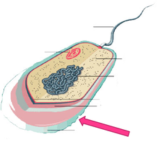
Capsule
sticky layer that surrounds the cell walls of some bacteria, protecting the cell surface and sometimes helping to glue the cell to surfaces.
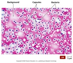
Capsule stain
negative stain method used to identify bacteria with capsules, the capsule remains unstained and forms a white ring around the bacterium
Cell membrane
thin, flexible barrier around a cell; regulates what enters and leaves the cell

Cell wall
rigid structure that surrounds the cell membrane and provides support to the cell

Coccus
spherical bacterium.

Counterstain
provides a contrasting stain to the primary stain. for example - safranin, stains the Gram-negative bacteria pink to red

Crystal violet
Gram stain primary dye, positive charged - penetrates into spaces of peptidoglycan in both gram +/-

Decolorizer
compound which serves to remove the primary stain during staining procedure - 95% ethanol

Escherichia coli
Gram negative bacterium, rod-shaped

Endospore
A thick-walled protective spore that forms inside a bacterial cell and resists harsh conditions.
Gram positive
bacteria that retain the color of the crystal violet stain in the Gram stain. This is characteristic of bacteria that have a cell wall composed of a thick layer of a peptidologlycan

Gram negative
bacteria that lose the crystal violet stain (and take the color of the red counterstain-safranin) in the Gram stain. This is characteristic of bacteria that have a cell wall composed of a thin layer of called peptidoglycan

Gram stain
procedure distinguishes between Gram positive and Gram negative groups by coloring these cells red or violet. Gram positive bacteria stain violet due to the presence of a thick layer of peptidoglycan in their cell walls, which retains the crystal violet these cells are stained with

Gram's iodine
= mordant that helps to trap the crystal violet in the peptidoglycan layer of Gram positive cells, which resist loss of the primary due during decolorization with alcohol; gram negative give up color.

Heat fixation
the process of fixing the bacteria to the slide. This method is when the dry smear on the slide is passed through a bunsen burner flame several times. The cells shrink slightly, but the bacteria ahere to the slide so they do not rinse off in the staining process.
Mordant
Gram's Iodine, helps to trap the crystal violet in the peptidoglycan layer of Gram positive cells, which resist loss of the primary due during decolorization with alcohol; gram negative give up color.

Mycolic acid
waxy lipid, makes cell impermeable to most stains. Common in cell wall of Mycobacteria

Negative stain
simple stain that uses nigrosin or India ink (an acid yielding a negatively charge dye) stains the background dark black but leaves the bacteria or other cells unstained, appearing white

Primary stain
crystal violet, the first stain applied that is retained by peptidoglycan layers

Peptidoglycan
polymer consisting of sugars and amino acids that forms a mesh-like layer outside the plasma membrane of most bacteria, forming the cell wall, presents as a thicker layer in Gram positive and a thinner layer in Gram negative bacteria

Periplasmic space
space between the inner and outer cell membranes in Gram-negative bactera. The peptidoglycan cell wall is found in the periplasmic space, and this space sometimes contains enzymes to degrade antibiotics.

Safranin
positively charged red dye that is used as a counterstain - stains the gram negative bacteria as they lose color

Spore
form taken by certain microbes that enables them to exist in a dormant stage. It is an asexual reproductive cell

Spirochete
spiral-shaped bacteria that have flexible walls and are capable of movement

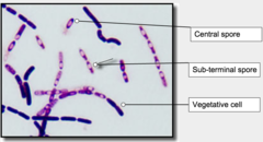
Spore stain
differential stain used to detect spores produced by Bacillus and Clostridium

Staphylococcus aureus
Gram positive bacterium, coccus-shaped

Flagella stain
differential stain that coats the thin flagella, making them visible with the light microscope

Immunofluorescence
method of tagging antibodies with a fluorescent dye to detect or localize antigen-antibody combinations.

Charge of cells
Resting cells are negatively charged on the inside, while the outside environment is more positively charged. This is due to a slight imbalance between positive and negative ions inside and outside the cell. Therefore positive dyes like crystal violet and safranin will diffuse into the cell