Vet Tech Skills Lab
 Call Kai
 Learn
 Practice Test
 Spaced Repetition
 Match
 Flashcards
 Knowt Play
1/155

There's no tags or description
Looks like no tags are added yet.
156 Terms
Hot iron dehorning is performed to
permanently disrupt/stunt horn growth in calves to prevent injury to people and other animals
Dehorning, in this SOP, should be done in calves between what age? Though it can be performed in calves up to what age depending on what?
2-4 weeks age, though up to 8 weeks of age depending on the size of the horn buds
SOP stands for
Standard Operation Procedure
Why would you not do it before 2-4 weeks of age? (Dehorning)
not done before 2-4 weeks of age because they’re considered neonates and are susceptible
What are you worried about when performing in calves after 8 weeks of age? (Dehorning)
Procedure/Method use would be more invasive
Please describe the procedure step by step for dehorning a calf
Prepare hot iron dehorner — attach butane can and turn it on (allowing it to heat up)
Catch and safety restrain the calf (using the gate, place their head through the window). Locate horn buttons on the sides of the poll. If no horn buds are felt, sire of calf is likely polled. If present, clip the hair that is overlying and surrounding each hron bud for visibility and reduce the risk of hair catching fire.
Perform a regional lidocaine block of the corneal nerve on both sides of the calf. Draw up 10 mL of 2% of lidocaine using a 12 mL syringe. Locate the “divot”/depression adjacent to the lateral canthus (outside corner) of the eye and place the needle (18 G 5/8-7’”) under the skin at a perpendicular angle (90°). Ensure the bevel of the needle is facing up. Should feel like a release of pressure (a pop) when the needle has gone through. Aspirate by pulling back plunger of the syringe, making sure you’re not in a blood vessel. If blood enters, gently pull needle back out partially and redirect it at a different angle, re-aspirate before injecting in a new location or if calf moves/struggles. Deposit 5 mL of lidocaine under the skin in this region. If difficult to inject, re-direct and aspirate before deposition of more lidocaine.
Repeat this procedure for the other side so that the both horn buds are desensitized. Use a new needle when drawing up lidocaine from the bottle and for calf (DO NOT reuse needles and change them between each calf). Syringes can be re-used if no blood has entered the syringe during aspiration.
Give meloxicam, pain medication (single dose, 1 mg/kg) by mouth before leaving the hutch and preparing the next calf. Dose is 3 tablets of meloxicam (15 mg/tablet) per 100 pounds of body weight.
Repeat this procedures above (checking for horn buds, clipping, blocking with lidocaine, and administering meloxicam) down a line of calves of appropriate age range that have yet been dehorned.
After 10-15 minutes, the calves can be dehorned using the hot iron. The dehornes must be very hot prior to use and care must be taken to avoid thermal injury.
Properly restrain the calf for the safety of both the animal and the handlers. Place the hot dehorned over the horn bud (with the base of the horn bud completely contacted by the dehorner) and provide direct and firm pressure while rotating the dehorner in a circular motion for 3 seconds at a time. Dehorning should be continued in segments of 3 seconds at a time until there is complete separation of the skin surrounding the base of the entire horn bud. A copper brown ring around the base of the horn bud is seen, dehorning is near completion but must be continued until there is a continuous separation of the skin 360 degree around the horn bud. A white ring should be seen.
If the horn cap comes off and bleeding occurs, place the dehorner directly on the horn bud to cauterize the area and stop the bleeding.
Repeat the process for the second horn bud
Place/spray the dehorned sites with aluminum spray, completely covering the horn bud and the ring of separation that was created. Care must be taken to not spray into the eyes of the calf
Apply a light coating of catron IV fly spray over the poll of the calf and NOT directly over the dehorned sites.
Release the calf and observe for a few minutes to monitor for recovery. Observe calves for bleeding, lethargy, fever, inappetence, or abnormal behavior over the next 12 to 24 hours.
What is the corneal nerve?
nerve that supplies sensation to the horn buds
What is the nerve called that supplies sensation to the horn buds?
Corneal Nerve
What is lidocaine used as?
local anesthetic/numbing agent
What does lidocaine do? (Mode of Action)
Blocks sodium channels in nerve endings, stopping pain signals from reaching the brain
Why would you want the bevel of the needle facing up when injecting?
Creates an opening that allows for smooth, less painful penetration of skin or tissue for injections or blood draws
Why do you not want lidocaine in the blood system?
can lead to local anesthetic systemic toxicity though cows have a high blood tolerance for lidocaine
What is meloxicam, what type of medication?
pain medication and an NASID (cox 2 selective)
Why do you want to avoid the dehorner on the horn bud for more than 3 second?
to prevent thermal injury to the skull and/or brain of the calf
What happens if you do not completely dehorn/separate the horn bud?
spurs
Where are IM injection sites for horses?
neck muscles (within the border of nuchal ligament (dorsal), scapula (caudal), and cervical spine (ventral), pectoral muscle (left and right), gluteal muscles (between the ischium, sacrole, and tuber coxae), and semi muscle/hamstring muscle (between the semimembranosus (medial) and semi tendinosus (lateral)
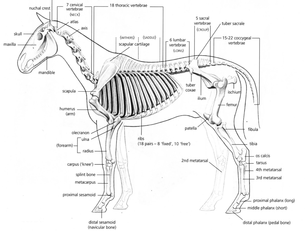
Review skeletal and joint anatomy
skull, mandible, cervical vertebrae, thoracic, lumbar, sacral, coccygeal vertebrae, scapula, scapulohumeral joint, humerus, ulna and radius, carpus/radiocarpal joint, metacarpal and split bones, proximal interphalangeal joint, proximal sesamoids, pastern, distal interphalangeal joint, distal sesamoids, ribcage, pelvis (illium, pubic, ischium) and tuber sacrale, tuber coxae, coccy-fermoral joint, femur, femoral-tibula joint, fibula and tibula, tarsus, meta-tarsus, proximal interphalangeal joint, proximal phalanx, distal interphalangeal joint, distal phalanx
Normal Horses PCV and TP levels
PCV (packed cell volume) — 30-40%
TP (total protein) — 6-8 gams per deciliter
The components of a centrifuged blood sample collected in a purple top tube include the _____ which is the _____ layer, the _____ ____ which is the _____, _____ layer, that is composed of _____ and ____, and the __ ____ ___, which are also known as _____ (medical term)
plasma, liquid layer, buffy coat, middle, thinnest layer, thrombocytes, leukocytes, packed red cells, erythrocytes
Episiotomy
a surgical incision made in the perineum (tissue between the vaginal opening and anus) during childbirth to enlarge the vaginal opening, usually performed with scissors or a scalpel
Caslick
procedure for mares that involves suturing the upper portion of the vulva to prevent fecal contamination, air aspiration, and uterine infections, often due to poor conformation, injury, or to improve fertility
Colic
abdominal pain
Owner’s description of clinical signs is often the first hurdle in a _____ workup
colic
Often described clinical signs from owners for colic
lethargic, inappetence to inability to rise
What in a physical examination can provide useful information for deciding what by going on with your patient suggesting a surgical or severe medical colic conditions?
heart rate greater than 60 beats per min, discoloured mucous membranes (brick red, purplish, or toxic line.
What is a Toxic line? And what is it caused by?
a bright red line on a horse's gums caused by changes in blood flow
Always take a ____ prior to the administration of NSAIDS or _____ _____
temperature, rectal palpation
Typically the procedure that follows physical examination
Nasogastric intubation and reflux
Large volumes (greater than 4 L) of _____ often accompany small intestinal ____, including stangulating ____, non strangulating small intestinal ____, or ____
reflux, lesions, lesion, obstructions, or enteritis
Enteritis
inflammation of the small intestine
Small intestinal strangulation will typically remain ____ even after stomach _____, while enteritis cases may become more _____ but show ____ or ____
painful, decompression, comfortable, depression or lethargy
If you obtain reflux and referral to the hospital is an option for the patient, leave the ____ in place and cover the end with a ____ ___ ____ (taping a ____ with a hole cut in the end). This won’t always prevent a stomach from _____, but provide a route for ____ ____ to exit the stomach while en route
tube, 1-way value, glove, rupturing, excess fluids
Colic workups in the fields include…
physical examination, nasogastric intubation and reflux, rectal palpation, and abdominocentesis
Rectal exam findings that suggest referral or euthanasia include…
distended small intestine or severely distended large colon,.
What would a distended small intestine feel like in rectal palpation?
loops of ballon animals or sausage
colon torsion
acute condition where the large intestine twists on its mesentery, causing obstruction, severe pain, and vascular compromise
It isn’t uncommon for colon torsions to present with ___ ____ severe enough to prevent the ability to perform a _____ ____
colon gas distention, rectal palpation
For colic workups, other diagnostics that can help with decisions-making can include
abdominocentesis and fast localized abdominal sonography (FLASH) ultrasound
Abdominocentesis
minimally invasive veterinary procedure where a needle is inserted into the abdominal cavity to sample or remove excess fluid
For most horses, (abdominocentesis), all that is needed is a ____ inch ____ g needle, ____ gloves, and a ____ top blood tube
1.5 in, 18 g needle, sterile gloves, and a red top blood tube
Abdominocentesis is typically performed at the ___ point of the abdomen when looking at the horse from the side, directly on the ____. An area should be ____ and ____ prepared.
lowest, midline, clipped and aseptically prepared
A ____ amount of local anesthetic can be injected into the proposed site for abdominocentesis.
small
For an average-sized horse, holding the needle ____ for the initial stick will embed enough of the needle that it won’t ___ easily if the horse ___ but will not typically be deep enough to enter the ____
halfway, dislodge, moves, abdomen
Once the needle is seeded in the abdominal wall, it should be ____ advanced until it is within the ____ ______. With ____ advancements you can typically see _______ of the needle as the ____ shifts around it. The needle may need to be advanced slightly ____ but it is very close or within the _____ ____. If no fluid is obtained, the needle should be ____ to ___ the bevel in relation to the _____ and ____. If still no fluid is present, a sterile, empty ____ mL syringe can be used to push a small amount of ___ into the abdomen through the needle. Once ____ is noted, it can be caught used the _____ tube with the ____ removed.
slowly, peritoneal captivity, slow, movement, intestine, further, peritoneal cavity, spun, move, omentum and viscera. 5mL, air. Fluid, blood, stopper
Why would you want to push air into the abdomen through the needle?
may help push omentum off the needle or start a siphon to encourage fluid to exit the needle
(Abdominocentesis) The ____ and ____ of the fluid can provide keen insight into the ___ present in the abdomen. Normal abdominal fluid should be ___ colored and _____. Turbidity of the fluid suggested increased _____. Strangulating lesions result in leakage of ____ from the ____, creating -___ abdominal fluid. Frank blood may suggest a _____ or ___ sampling.
Grossly ___ or ____ fluid with or without feed material may indicated ____ _____ or _________.
color and turbidity, lesion, straw colored and clear, cellularity, cells from the vasculature, creating serosanguinous, hemoabdomen or splenic. brown or green, viscera rupture or enterocentesis.
Normal range (L) of reflux for a horse
4-6 L
Brucellosis vaccination prevents _____ _____ which causes ______/ _____ issues and reduces _____ production
bovine brucellosis, abortions/pregnancy, milk
The brucellosis vaccination is given to eligible _____ between the ages of ___ and ____ months
heifers, 4-12 months of age
Is brucellosis a zoonotic disease?
yes
What are the borders in where the brucellosis vaccination would be given? And where in the borders, is the vaccine given?
the dorsal border is the nuchal ligament, caudal border is the scapular, and in the ventral border is the cervical spine.
In the more caudal-dorsal aspect
How do you administer the Brucellosis vaccination? (nasally, IM, IV, or SubQ) And at what angle?
SubQ, 45 degree angle
When giving an ear tattoo, during brucellosis vaccination, what is the tattoo including? What ear is it on?
R-Strain (on bottle) + shield + year. The right ear
What are the vital signs of cattle?
Temperature: 101.5-103
HR: 100-140
RR: 30-60
CRT: <2 seconds
MM: pink, moist
On (female) calves, where do you check for MM?
vulva tissue
What does BAR stand for?
bright, alert, responsive
Where is skin turgor on cattle performed?
eyelid
The rumen is on what side of the cow and where gut sounds can be heard?
left side
The brucellosis vaccination is a set amount, not body weight based. True or False
True
What is being used to administer the brucellosis vaccination?
pistol-grip syringe
The brucellosis is a modified live vaccination. True or False?
True
Why do we want to change our needle between cows?
We want to prevent the transfer of blood borne diseases
What do you want to keep in mind when tattooing during the brucellosis vaccination?
avoiding the ridges/ribs as theres more blood vessels there
you want a flat and more lengthy area
supernumerary teats
any teat that is in excess of the normal number of teats (4 in cattle)
____ disease is one of the leading causes of illness in young dairy calves
respiratory
Lung damage caused by respiratory disease is claimed to impact..
growth, development, reproduction, and lactation
A calf health scoring system is from ___ to ___ (and dictate which is normal and which dictates several health observation)
0 (normal) to 4 (several health observation)
An objective form of diagnosing lung diseases
utilizing thoracic ultrasonography or lung scanning
Steps to follow when lung scanning claves:
select a group of calves to lung scan.
prepare all necessary equipment: ultrasound machine, 70% isopropyl alcohol, clippers, recording device and method of restraint (halter or headlocks)
properly restrain animal so they’re secured and have access to both sides of calf\
clip hair from both sides of the calf between the 1st and 8th rib spaces on the bottom half of the calf
spray the clipped area with isopropyl alcohol
begin scanning with the linear probe directly behind the shoulder between the space of the first and second ribs
place the linear probe on top of the clipped area and follow the rib space down until you reach the bottom of the clipped area
repeat step 7 for reach rib space up and including the 8th rib space
Step step 5-8 for the opposite site
____ is a good opportunity to scan for lungs
weaning, before moving to group housing because farmers can also monitor heifer growth during the pre-weaning period
Lung Consolidation Score (LSC)
1=no consolidation
2=comet tails on the pleural surface
3=one location of consolidation > or equal to 1 cm but less than 6 cm
4= consolidation > or equal to 6 cm or more than one location of consolidation
Lung consolidation is the term used to describe
damage to the lung tissue
Once the lung has been damaged, consolidation typically cannot be ______
reversed
Consolidation can be thought of as ___ _____ within the lung
scar tissue
How does consolidation affect the lungs?
reduces the lungs capacity and ability to penetrate oxygenate blood
The white horizontal line across the scan shows
the lung tissue lining (pleural surface)
The smooth undisputed line (of the pleural surface) displays a normal, healthy animal or calf with lung damage?
a normal, healthy animal (assuming that includes no comet tails)
LCS of 2 involves
a slight disruption in the pleural surface of the lung tissue, white lines lying vertically to the pleural surface (comet tails or B-lines)
Comet tails are the beginning of
damage to the tissue
Comet tails are commonly found when scanning calves. True or False
True
Lung consolidation scores of 3 and 4 show varying degrees of ____ and should raise ___
disruption, concern
Why is it important to document the lung score on both sides?
Because animals with lung damage on both sides are at high risk of reduced growth than calves with lung damage on only one side.
Calf lung consolidation during the pre-weaning phase lowers
calf performance
Lung consolidation also affects ____ of heifers later in life
performance
Describe the layers when probing.
Skin, muscle and pleural surface
superficial (close to the probe), intercostal muscles, pleural surface, dirty shadow, deep (further from the probe)
Why does the lungs appear like a dirty shadow?
because they’re filled with gas!
What is a sign of the pleural surface?
It is bright white and glide across the screen
When lung scanning, you want to go from ____ to ____ and _____ to ___
cranial to caudal and dorsal to ventral
How can you tell you are looking at ribs and not the pleural surface?
ribs are more superficial and do not glide, it appears like a clean shadow sinces its bone
Lungs cannot or can regenerate
can to some degree, but typically cannot
What are active infection signs you’re watching out for?
increase in respiratory rate, coughing, nasal discharge (purulent), fever, lethargic, oculary discharge, ear positions are droopy
What treatment would you give for active infection (for lungs)?
antibiotics like nuflor/florfenicol which lasts 4 days, SQ, NSAID, melxocian, and vitamin E (injection SQ) which is antioxidant
What does NSAID stand for
non-steroidal anti inflammatory drug
Why are inhalers not used in food animals?
Inhalers and the active ingredients are illegal to use in food animals
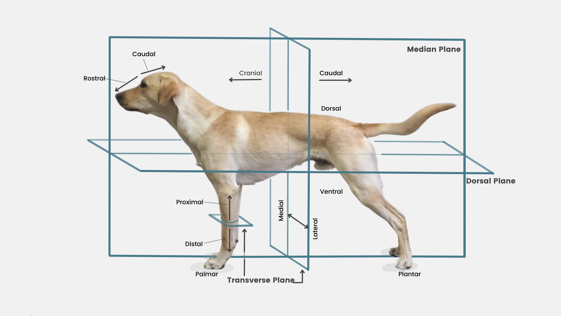
Describe the anatomical terms (caudal, cranial, dorsal, ventral, medial, lateral, proximal and distal)
(caudal, cranial, dorsal, ventral, medial, lateral, proximal and distal)

What surgical instrument is this?
Backhaus towel clamps
How many towel clamps in a basic surgical packs.
4
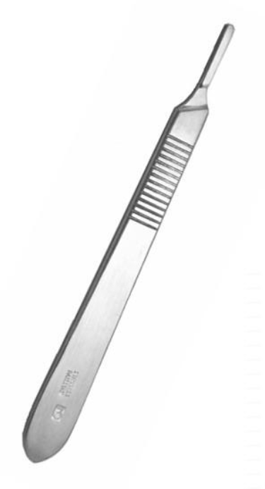
What surgical instrument is this?
scalpel handle

What surgical instrument is this?
Brown adson thumb forceps

What surgical instrument is this?
Rat tooth thumb forceps