HOSA Phlebotomy State
 Call Kai
 Learn
Learn Practice Test
 Spaced Repetition
 Match
Match Flashcards
 Knowt Play
1/593

Earn XP
Description and Tags
HOSA Phlebotomy
Name | Mastery | Learn | Test | Matching | Spaced | Call with Kai | Chat |
|---|
No analytics yet
Send a link to your students to track their progress
594 Terms
Components of Blood
plasma,red blood cells,white blood cells, platelets
Red blood cells
contain hemoglobin, and it's the hemoglobin protein within the red blood cells that binds to and carries oxygen from the lungs to the body's tissues
Main job: carry oxygen to cells and remove CO₂
No nucleus or mitochondria (more room for oxygen)
Biconcave shape → large surface area for gas exchange
List the order of draw
Blood cultures — Sterile bottles.
Light blue — Sodium citrate.
Red / Gold (SST) — Serum or clot activator.
Green — Heparin.
Lavender / Pink — EDTA.
Gray — Fluoride/oxalate.
Order of vein choice
First choice: Median cubital
Well‑anchored (doesn’t roll), low pain, fewer complications.
Second choice: Cephalic
Especially helpful in patients with more fat
Last choice: Basilic
Too close to nerves and the brachial artery → risk of nerve injury or accidental arterial puncture.
Plasma function and importance
the liquid part of blood that transports dissolved proteins (like antibodies, albumin, fibrinogen), nutrients (glucose, amino acids, fats, vitamins), electrolytes (sodium, potassium, calcium), clotting factors, hormones, waste products (urea, CO2), and even blood cells,
helping maintain blood pressure, pH balance, immunity, and fluid balance throughout the body. It acts as a crucial "highway" for delivering essential substances to tissues and removing waste.
~90% water, ~10% proteins, nutrients, and ions
Proteins include:
Albumin: controls water movement (prevents dehydration/swelling)
Fibrinogen: helps clot blood
Globulins: transport fats/lipids
serum importance and contents
is essentially blood plasma minus the clotting factors (like fibrinogen) that are used up during the clotting process, leaving a clear, yellowish liquid rich in antibodies, electrolytes, hormones, and other proteins, used in many diagnostic tests.
white blood cells functions and actions
core of your immune system, constantly patrolling your bloodstream to identify and destroy pathogens (bacteria, viruses, fungi, parasites) and abnormal cells (like cancer cells)
through actions like phagocytosis (engulfing invaders), producing antibodies (B cells), and directly killing infected cells (T cells, natural killer cells), providing both innate and adaptive defenses against illness
Have nuclei, appear white/clear, move like amoebas
Types:
Neutrophils: most common; destroy pathogens by phagocytosis (engulfing and digesting them)
Lymphocytes: control immune responses and make antibodies
Pus = dead WBCs + bacteria from infections
platelets use and function
essential for blood clot formation (hemostasis) as they are the first responders that stick to injured vessels, form a plug, release clotting factors, and create a scaffold for stable fibrin meshwork, stopping bleeding and maintaining vascular integrity
Not actual cells
No nucleus
Main job-help form blood clots
fibrin meshwork
a three-dimensional protein network formed from fibrin, a sticky protein, that acts as the structural scaffold of a blood clot, trapping platelets, red blood cells, and other components to stop bleeding and initiate wound healing
thrombin
a crucial enzyme in blood clotting (hemostasis), formed from inactive prothrombin; it converts soluble fibrinogen into insoluble fibrin strands, creating a mesh that stabilizes blood clots with platelets.
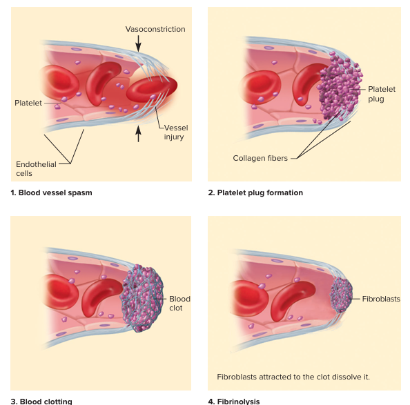
Fibrinolysis
A separate system (fibrinolysis) later breaks down the fibrin mesh, allowing the clot to dissolve once the vessel has healed and restoring blood flow.
Soluble fibrinogen
a crucial, liver-produced protein circulating in the blood that acts as the precursor to insoluble fibrin, forming the structural meshwork of a blood clot to stop bleeding.
spontaneously polymerize into a stable clot
Insoluble fibrin monomers
the fundamental, building-block proteins that spontaneously link together (polymerize) to form insoluble fibrin fibers, creating a stable blood clot,
coagulation
the process where a liquid, like blood or milk, changes into a solid or semi-solid state,
It involves complex steps like platelet plug formation and the coagulation cascade, leading to a stable fibrin mesh.
coagulation cascade
a complex, step-by-step process involving blood proteins (clotting factors) that leads to the formation of a stable blood clot (fibrin mesh) to stop bleeding, following initial platelet plug formation in primary hemostasis, with intrinsic and extrinsic pathways converging to activate factor X, which starts the common pathway, ultimately converting fibrinogen to fibrin to reinforce the clot.
Primary Hemostasis:
forming a temporary platelet plug to stop bleeding through a sequence of platelet adhesion, activation, and aggregation at the site of damage, a process critical for preventing blood loss before the more stable fibrin mesh of secondary hemostasis forms.
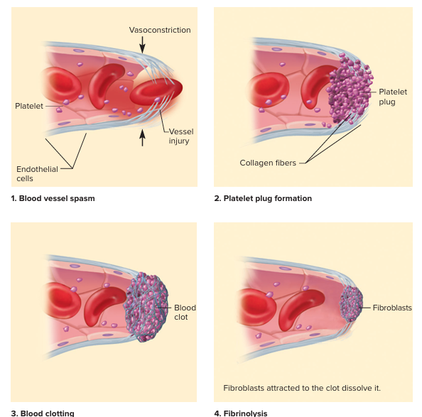
Secondary Hemostasis
the process where the initial platelet plug from primary hemostasis is stabilized by a strong, insoluble fibrin mesh, forming a durable blood clot to stop bleeding, involving sequential activation of clotting factors that convert fibrinogen to fibrin, trapping red blood cells for a robust plug. This intricate enzymatic process ensures a stable clot, especially for larger injuries where the temporary platelet plug isn't enough
platelet adhesion
the crucial first step in stopping bleeding (hemostasis), where platelets stick to the damaged blood vessel wall's exposed collagen and other matrix proteins, initiated by key receptors like the GPIb-IX-V complex
platelet Aggregation
where platelets stick to each other and form clumps (aggregates) at a site of blood vessel injury, creating a temporary plug to stop bleeding (hemostasis
Vasoconstriction
the narrowing of blood vessels from smooth muscle contraction, reducing the inner diameter (lumen) to slow or block blood flow
a vital process for regulating blood pressure, conserving heat in cold, controlling bleeding (hemostasis), and diverting blood to essential organs during stress or exercise, though it can also be triggered by medications, smoking, or underlying conditions like atherosclerosis
venipuncture and typical blood amount
the preferred method for routine laboratory testing, particularly when larger volumes of blood are required (greater than 2 mL) for comprehensive testing, such as hematology or clinical chemistry panels. It is preferred over capillary (fingerstick) sampling due to the higher quality of the specimen, which improves test accuracy and provides better stability usually has 1-5 ml of blood taken
antecubital fossa (also known as the cubital fossa, "elbow pit," or anterior elbow region)
a triangular, shallow depression located on the anterior (front) surface of the elbow joint. It is the primary site for venipuncture (blood draws), intravenous (IV) catheter insertion, and blood pressure measurements because the veins in this area are superficial, large, and easily accessible.
lumen
the open, central space within a hollow tubular structure or organ, such as blood vessels, the intestines, or ducts. It acts as a passageway for fluids, nutrients, or gases, with its diameter and shape varying based on function, such as regulating blood flow or aiding nutrient absorption.
tourniquet
a stretchable band, typically made of rubber, silicone, or fabric, wrapped around a patient's arm to restrict venous blood flow, causing veins to distend for easier needle access. Applied 3-4 inches above the site, it is essential for identifying veins but must be released within one minute to prevent hemoconcentration.
Anticoagulants
reduce the body’s ability to form clots by inhibiting clotting factors or activating natural anti-clotting mechanisms
Examples of anticoagulants
EDTA, Citrate, Heparin
anti-thrombin
crucial blood protein that regulates clotting by inactivating key enzymes like thrombin (Factor II) and Factor Xa, preventing excessive clot formation and maintaining blood flow,
Hemolysis
destruction of red blood cells. It is a continuous, normal physiological process in the body, which becomes a medical condition when red blood cells are destroyed prematurely or at an excessive rate
analyte
the specific component, substance, or chemical species in a sample that is identified and measured during an analytical procedure, such as in laboratory testing, pharmaceutical research, or environmental monitoring
hemolysis, improper anticoagulant use, or delayed processing can…
alter analyte concentrations and compromise diagnostic accuracy
Arterioles
small-diameter blood vessels, typically 10 to 100 μm, that branch from arteries and lead into capillary beds, acting as the primary resistance vessels in the circulation. They possess muscular walls that regulate blood pressure and flow to tissues through constriction or dilation.
Venipuncture targets veins because
they are closer to the skin's surface, possess thinner walls, and contain fewer nerves compared to arteries, making them easier, safer, and less painful also have lower pressure, reducing the risk of excessive bleeding or damage.
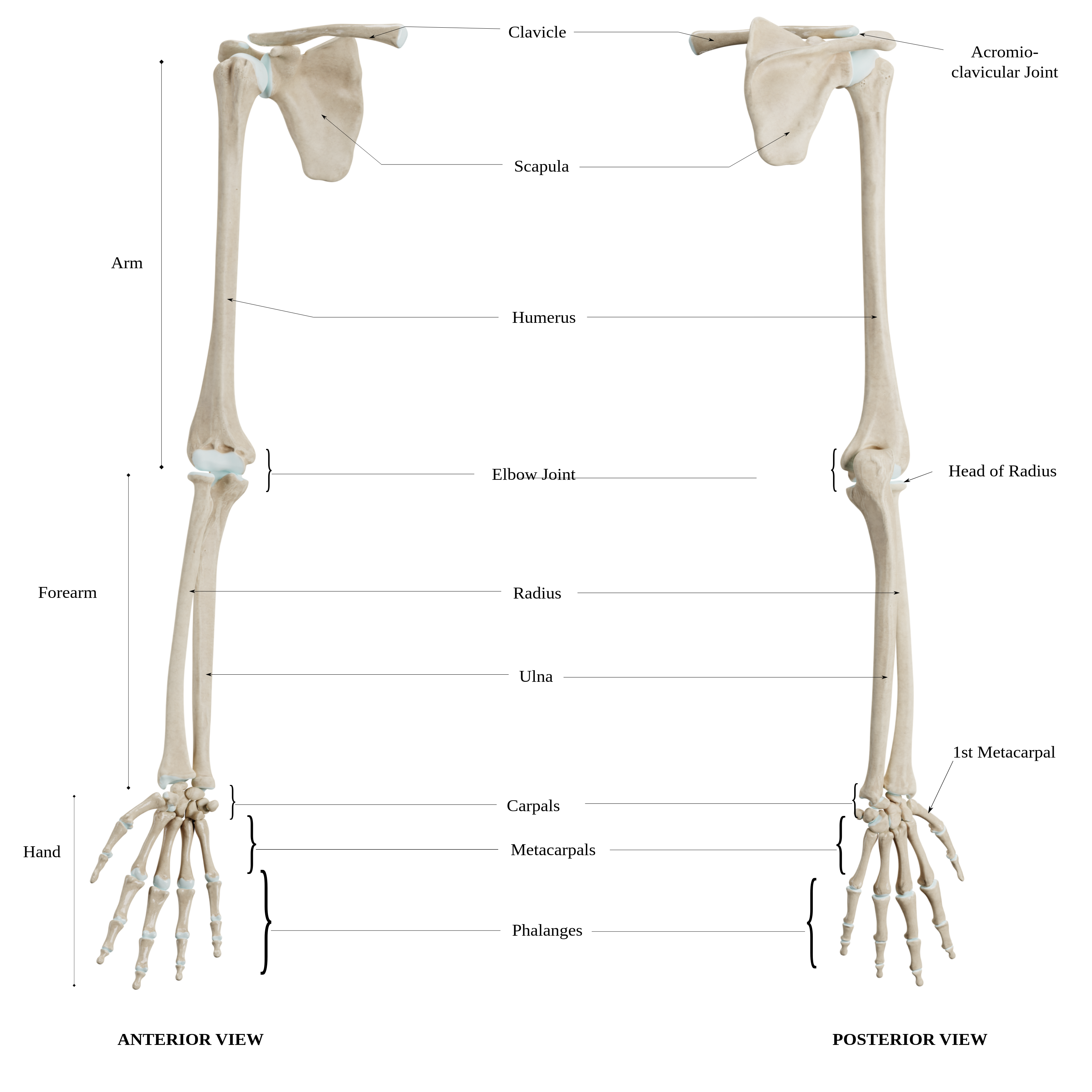
key arm bones
Humerus: The single bone of the upper arm, extending from the shoulder to the elbow.
Radius: Located on the thumb side of the forearm.
Ulna: Located on the pinky side of the forearm.
key arm muscles
Upper Arm: Primarily the biceps brachii (front) and triceps brachii (back).
Forearm: Contains 19 muscles (flexors and extensors) that control wrist and finger movement.
key arm nerves
Median Nerve: Controls muscles that bend the wrist and fingers.
Ulnar Nerve: Known as the "funny bone" nerve; it runs down the inside of the arm and controls hand muscles.
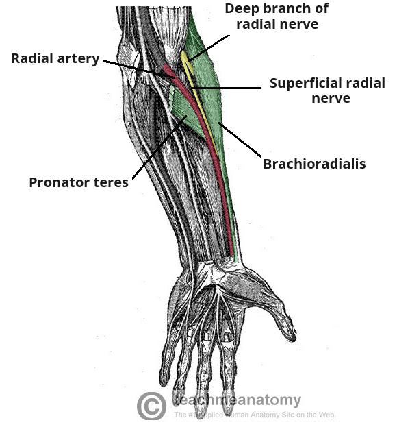
Radial Nerve: Controls muscles that straighten the elbow, wrist, and fingers.
key arm blood vessels
Brachial Artery: The main supply, which runs along the inside of the arm and divides into the radial and ulnar arteries near the elbow.
Veins: The superficial venous network includes the cephalic, basilic, and median cubital veins, along with deep brachial veins.
Humerus
the long, single bone of the upper arm extending from the shoulder to the elbow,
Radius
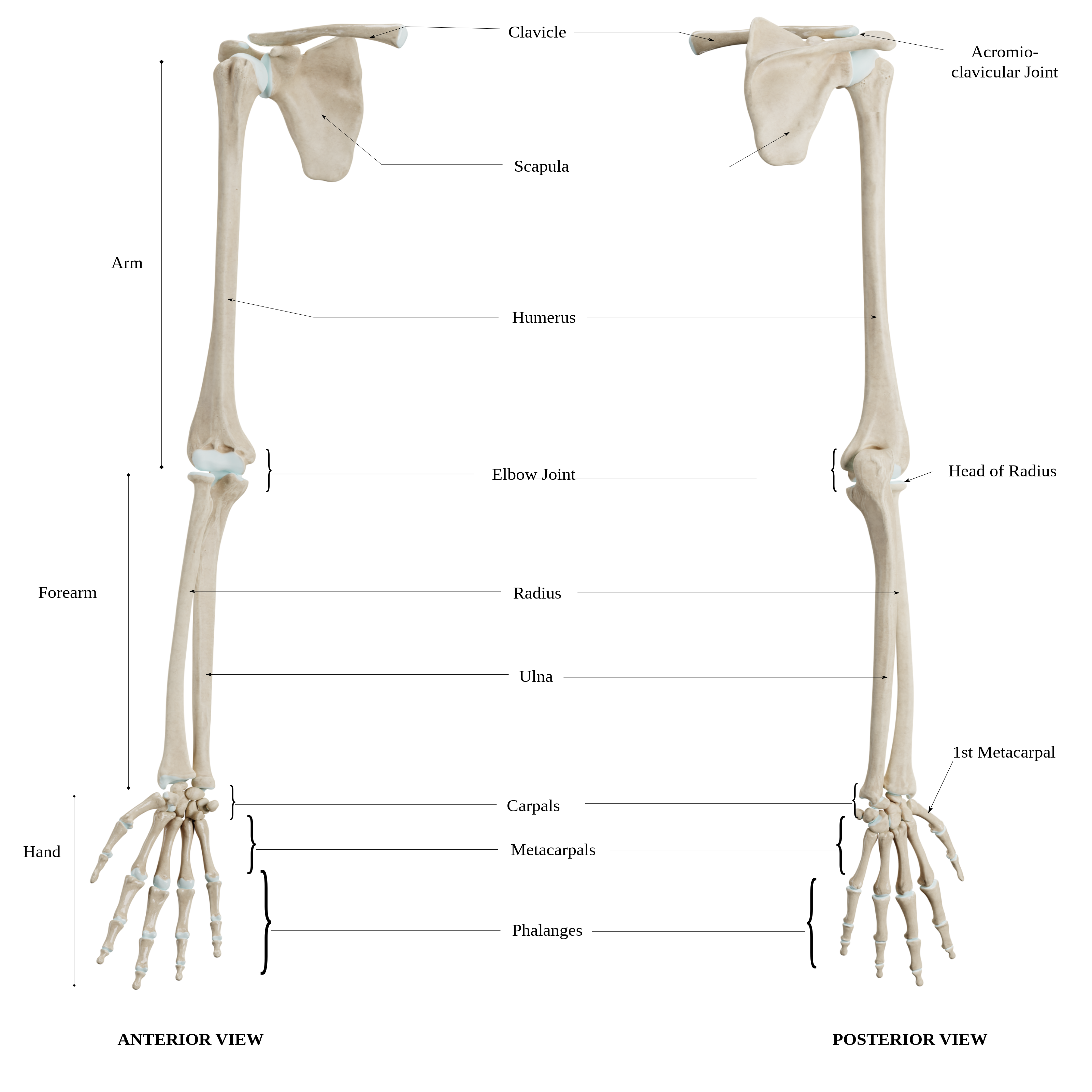
Located on the thumb side of the forearm.
generally thicker than the ulna
features a small head at the elbow and a wider, stronger base at the wrist,
which enables rotational movements like pronation(movement of the forearm that turns the palm to face downward or backward) and supination(movement of the forearm that turns the palm upward or forward, achieved by the radius rotating around the ulna).
running parallel to the ulna
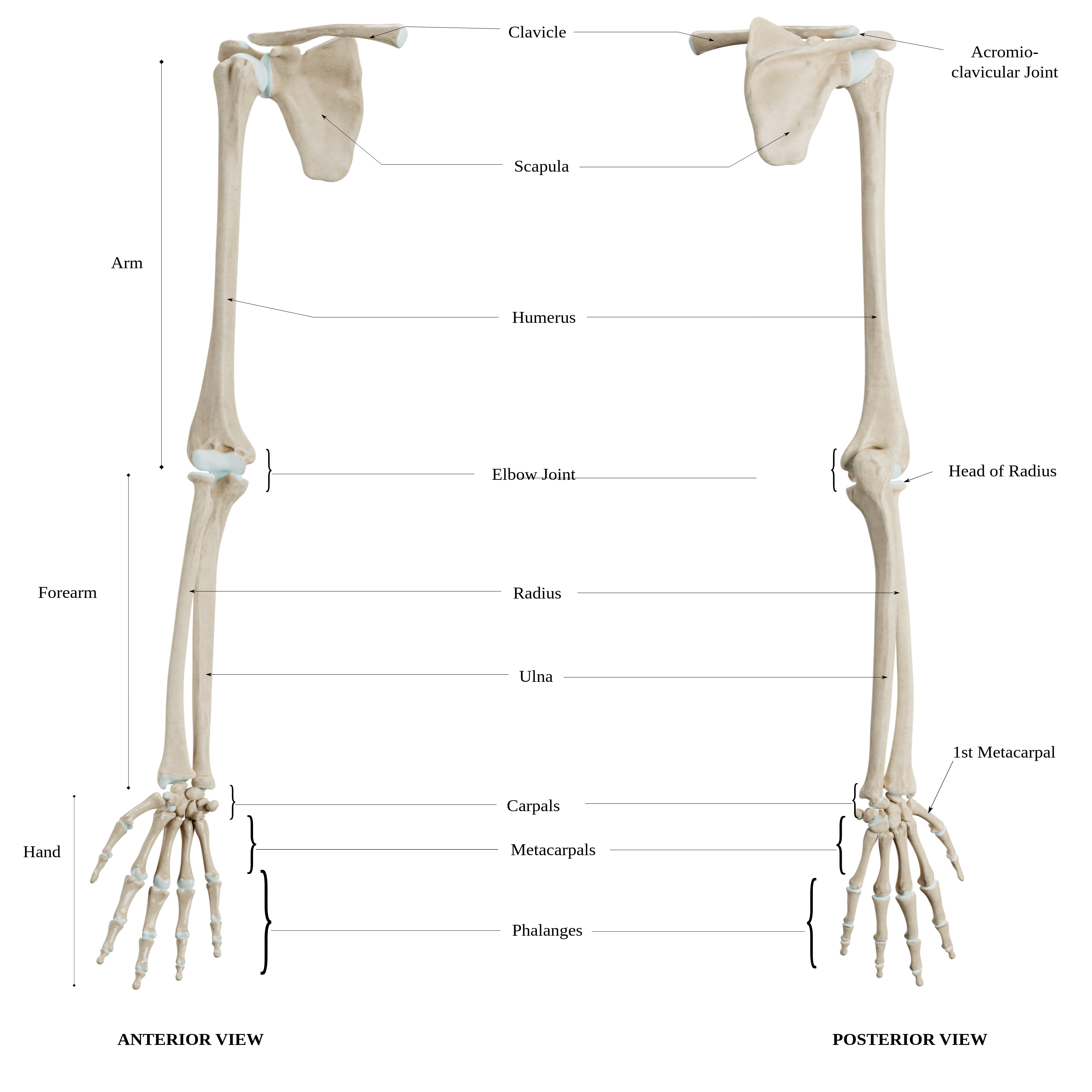
ulna
Located on the pinky side of the forearm.
longer, thinner bone
running parallel to the radius
It extends from the elbow to the wrist, forming the primary hinge joint for the elbow with the humerus. It stabilizes the forearm during movement.
Sites to avoid in venipuncture
Veins with hematomas or bruises.
Arms with IV lines, fistulas, or infections.
The arm on the side of a mastectomy, due to lymphedema risks.
Scarred or burned areas.
What to consider in the process of venipuncture
Tourniquet Placement: Apply 4–5 finger widths above the selected site.
Angle of Insertion: Typically 15 to 30 degrees, depending on vein depth.
Avoiding "Rolling" Veins: Ensure the skin is anchored properly to keep the vein stable during puncture.
vein choices in venipuncture
Median Cubital Vein (1st Choice):
Cephalic Vein (2nd Choice):
Basilic Vein (3rd Choice):
Alternate choices
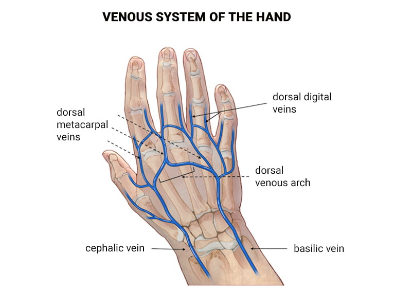
Dorsal Hand Veins (Metacarpal veins):
Forearm Veins:
Foot/Ankle Veins:
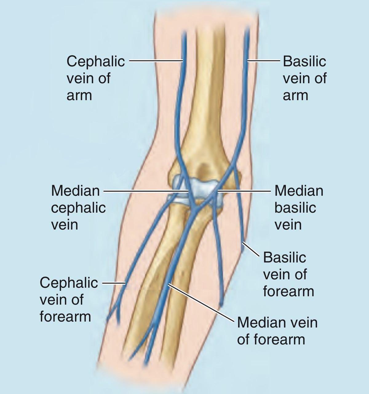
Median Cubital Vein
a prominent, superficial vein in the cubital fossa (inside elbow crease) that connects the cephalic and basilic veins. It is the preferred site for venipuncture and intravenous access due to its large size, superficial location, and relative stability. It runs obliquely across the elbow, separated from the brachial artery by the bicipital aponeurosis.
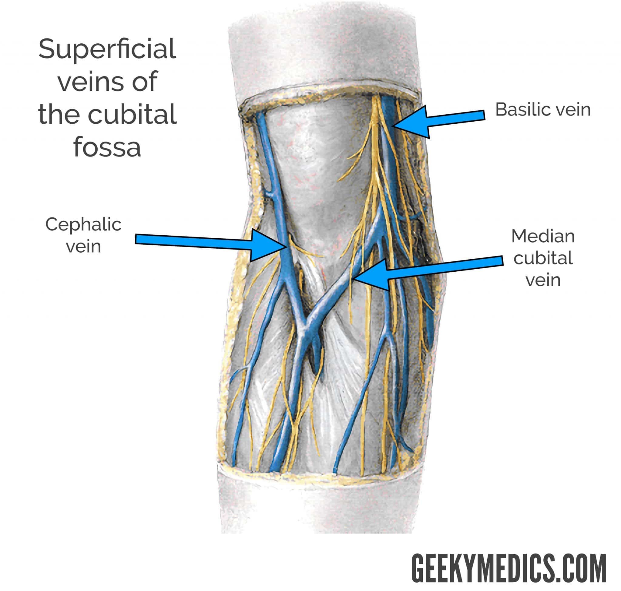
Cephalic Vein
a major superficial vein in the arm, running from the hand, along the radial (outer) side of the forearm and biceps, to the shoulder, where it empties into the axillary vein. It is commonly used for intravenous catheters, pacemaker lead insertion, and blood sampling due to its size and accessibility.
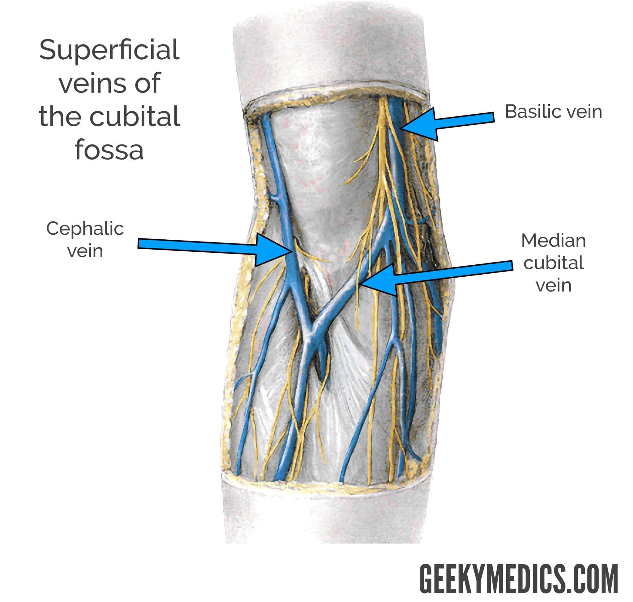
Basilic Vein
a major superficial vein in the upper limb, starting from the back of the hand and running up the inner (medial) side of the forearm and arm, visible under the skin, eventually piercing deep tissue in the upper arm to join the brachial veins and form the axillary vein, draining blood from the hand and forearm back towards the heart. It's a common site for venipuncture (drawing blood) but often a last resort due to its depth and proximity to nerves.
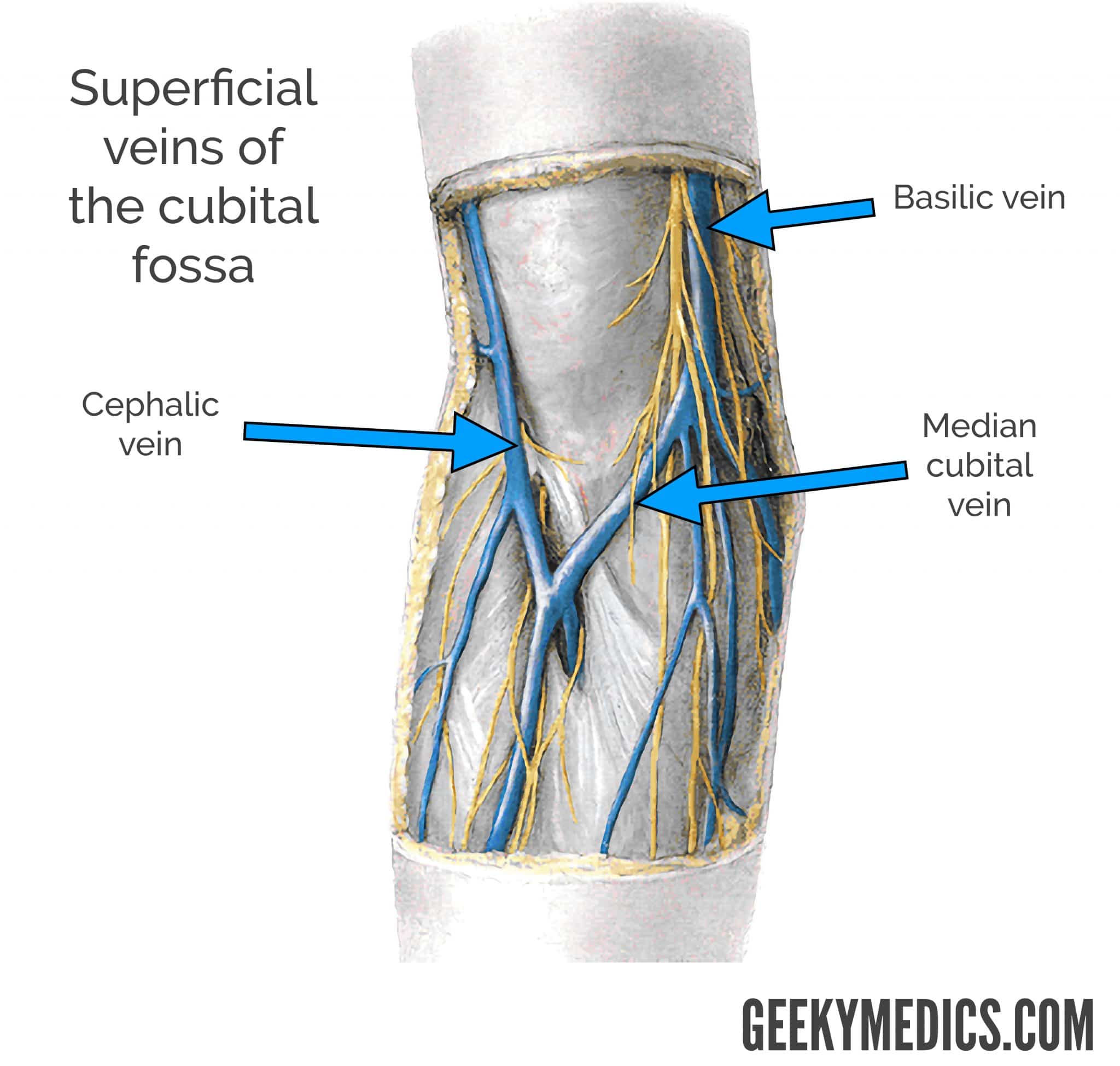
Dorsal Hand Veins
a prominent, superficial network of veins on the back of the hand that drain blood from the fingers and hand towards the forearm. Formed by dorsal metacarpal veins and dorsal digital veins, this network acts as the primary venous drainage for the hand, feeding into the cephalic vein (laterally) and basilic vein (medially).
Forearm Veins
superficial vessels, primarily the cephalic and basilic veins, that become prominent due to low body fat, muscle development from exercise, genetics, age (thinner skin, weaker valves), heat, or increased blood pressure,
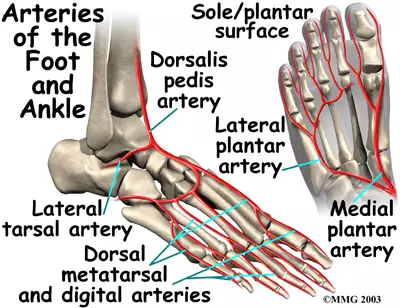
Foot/Ankle Veins
a last resort for phlebotomy in adults, often requiring explicit physician permission or performed only by specialized personnel used when upper extremity veins are unavailable, such as in cases of extensive scarring, burns, or when both arms are inaccessible due to casting or medical devices.
Great Saphenous Vein: Originates from the marginal vein at the foot and passes in front of the medial malleolus (ankle bone).
Dorsal Arch Veins: Veins on the top of the foot (dorsum of the foot) are sometimes used because they are easy to visualize and access.
Small Saphenous and Medial Marginal Veins: Located around the ank
Site selection for venipuncture prioritizes…
the antecubital fossa, specifically the median cubital vein, because it is large, superficial, well-anchored, and least likely to roll, offering a safer and less painful experience.
capillary blood obtained…
via fingerstick (adults/children >6 months) or heel stick (infants <6 months) is a minimally invasive, common procedure using a lancet to obtain small blood samples for diagnostic tests, including glucose monitoring, metabolic screening, and hematology. It offers an easier alternative to venous draws, allowing for rapid, in-home, or point-of-care testing.
Identify major nerves and nerve pathway on the arm
three main nerves median, ulnar, and radial
Blood samples are collected for…
for diagnosing diseases, monitoring treatment efficacy (e.g., drug levels), and performing therapeutic procedures like reducing excess red blood cells. Accurate, patient-identified samples ensure safety, allowing doctors to manage therapy, prevent toxicity, and assess patient health, with results influencing critical care decision
Phlebotomy collection methods include
venipuncture, capillary (dermal) puncture, and arterial sampling, used to obtain blood for diagnostic testing. The most common techniques, often performed at the elbow or back of the hand, are evacuated tube systems, syringe methods, and butterfly needles
Venipuncture Using a Butterfly
used for small, fragile veins (geriatric/pediatric patients) or difficult sticks to improve success and comfort. The process involves placing a tourniquet, sanitizing the site, and inserting the needle at a 15–30 degree angle, with the bevel up, until a flash of blood appears in the tubing. The wings are held for stabilization while tubes are attached to the holder, allowing blood to fill via vacuum, after which the tourniquet is released, the needle is withdrawn, and a safety device is activated.
Effective communication practices
Using patient‑appropriate vocabulary, confirming understanding, and clearly relaying critical information to clinicians; also using professional body language, eye contact, posture, and tone to convey empathy and competence
Order of draw with a butterfly needle
Blood Cultures
Light Blue (Coagulation/Sodium Citrate) - Note: Use discard tube here with butterfly!
Red/Gold/Tiger Top (Serum)
Green (Heparin)
Lavender/Pink (EDTA)
Gray (Sodium Fluoride/Oxalate)
If Light blue is first then a discard tube is used which isn't used for testing"
Tubes that must be completely full
Light Blue Tube (Sodium Citrate)
Used for coagulation tests (PT, PTT, INR, fibrinogen, D‑dimer).
MUST be 100% full to maintain the 9:1 blood‑to‑additive ratio.
Underfilling = invalid results → redraw.
2. Black Tube (ESR — Westergren method)
Also requires a specific blood‑to‑additive ratio.
Must be filled to the marked line.
List the process of conducting a normal venipuncture procedure for HOSA
Review requisition
Identify patient (name + DOB)
Explain procedure & get consent
Hand hygiene + gloves
Position arm
Apply tourniquet 3–4 in above site
Ask patient to make fist
Palpate & select vein
Release tourniquet
Clean site & let dry
Prepare ETS needle
Reapply tourniquet
Inspect needle
Ask patient to remake fist
Anchor skin
Warn patient
Insert needle bevel‑up at 15–30°
Ask patient to release fist
Collect tubes in correct order (blue → red/gold → green → lavender)
Remove tourniquet as last tube fills
Remove needle & activate safety
Apply gauze & mix tubes
Check site & bandage
Label tubes
Dispose sharps
Clean area
Thank patient
Remove gloves & hand hygiene
Evacuated blood collection tubes
sterile, closed-system glass or plastic vials with colored stoppers that use a premeasured vacuum to draw a specific volume of blood. These tubes often contain additives—such as anticoagulants, clot activators, or separation gels—designed to stabilize the specimen for laboratory analysis.
Ethylenediaminetetraacetic acid
EDTA) acts as a potent anticoagulant in hematology by chelating (binding) calcium ions (𝐶𝑎^2+) in the blood. Because calcium is essential for the coagulation cascade, its removal keeps the blood in a liquid state, preserving cell morphology for accurate CBC and smear analysis, usually in purple-top tubes.
Heparin
inhibits thrombin (Factor IIa) and Factor Xa, preventing blood clotting. It is primarily used in plasma collection for chemistry tests (e.g., blood gases, electrolytes)
Thrombin (Factor IIa)
a crucial enzyme in blood clotting, acting as the final activator that converts soluble fibrinogen into insoluble fibrin, forming the meshwork of a clot, and also activates platelets and other factors to amplify the clotting process.
Factor Xa inhibitors
a class of anticoagulant drugs (blood thinners) that prevent blood clots by blocking the activity of coagulation factor Xa, a crucial enzyme in the clotting cascade
Antithrombin III (AT III)
a vital plasma protein that acts as a natural anticoagulant by inhibiting key clotting factors, primarily thrombin and factor Xa, to control blood coagulation. A deficiency in AT III, often inherited, increases the risk of dangerous, recurring blood clots (thrombosis)
Sodium citrate
a reversible anticoagulant in coagulation testing by binding to (chelating) calcium ions in the blood, preventing the clotting cascade, and allowing laboratories to precisely reverse this effect by reintroducing calcium
Soudium Cirate Vs Ethylenediaminetetraacetic acid
Sodium citrate is used for coagulation studies (PT/INR, PTT) because its binding is reversible, while EDTA is preferred for hematology (CBC, blood films) and molecular testing because it preserves cell morphology and integrity better.
Sodium citrate is the standard for coagulation tests, as it keeps clotting factors (V and VIII) stable.
EDTA is the gold standard for complete blood counts (CBC), providing stable platelet counts and red cell morphology.
Reason for standardized tube filling order
to prevent cross-contamination of additives (like EDTA, heparin, or citrate) between tubes, which can cause inaccurate lab results, misdiagnosis, or invalid samples.
Light blue top tube contain and what tests is it used for
top tubes contain sodium citrate, which chelates calcium;
required for coagulation assays such as prothrombin time (PT) and partial thromboplastin time (PTT) and must be filled to the specified volume to maintain the citrate-to-blood ratio
coagulation (clotting) studies and hematology tests
prothrombin time (PT)
a blood test measuring how many seconds it takes for plasma to clot, evaluating the extrinsic and common coagulation pathways.
Partial Thromboplastin Time (PTT or aPTT)
a blood test measuring how many seconds it takes for a clot to form, usually taking 25 to 35 seconds for normal results.
Light blue top tube ratio and inversion count
ratio-9:1 ratio of blood to anticoagulant
inversion count-3-4 gentle inversions immediately after drawing to properly mix the anticoagulant with the blood,
ABO blood group system
classifies human blood into four types (A, B, AB, O) based on inherited A and B antigens on red blood cells.
ACESSION NUMBER
a number printed on all specimen labels and documents associated with a particular reason
advance directive
legal document that allows people to decide what kind of medical care they wish to have in the event they are unable to make those choices themselves
aerosolize
to disperse a substance through the air in such a way it might be inhaled
aliquot
a small amount of a larger specimen
American hospital association (AHA)
a non-profit organization concerned with providing information to the healthcare industry and the public regarding healthcare issues trends.
anaerobic bacteria
thrive in oxygen-free environments, often finding oxygen toxic
rely on fermentation, producing less energy (2 ATP), and exist in deep soil or gut environments.
Aerobic bacteria
require oxygen for survival and energy production (cellular respiration)
Aerobic bacteria produce more energy (30-38 ATP) and are found on surfaces,
aortic valve
the heart valve locating between the left ventricle and aorta
arterialize
convert (venous blood) into arterial blood by reoxygenation, especially in the lungs.
arteriovenous (AV) fistula
an abnormal, direct connection between an artery and a vein, allowing blood to bypass capillaries.
atrioventricular junction
the anatomical area separating the atrial and ventricular myocardia, containing the AV node and the nonbranching bundle of His. It facilitates, delays, and coordinates electrical signals between the atria and ventricles, acting as a secondary pacemaker (40–60 bpm).
atrial and ventricular myocardium
the muscular tissues of the heart's upper (atria) and lower (ventricles) chambers, respectively. Ventricular myocardium is significantly thicker, stronger, and more metabolically active to pump blood throughout the body, while thinner atrial myocardium serves to fill the ventricles.
atrioventricular (AV) node
a critical component of the cardiac electrical conduction system, located in the right atrium's Koch triangle. It functions as an electrical bridge between the atria and ventricles, introducing a brief, 0.09s delay to allow ventricular filling. It serves as a backup pacemaker (40–60 bpm).
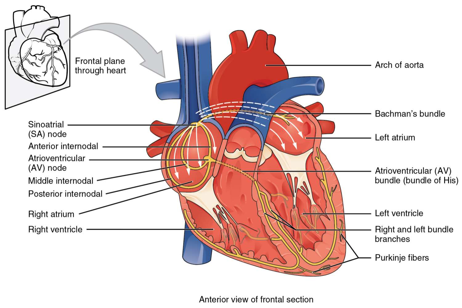
cardiac electrical conduction system function
a network of specialized muscle cells that generate and transmit electrical impulses, ensuring a rhythmic, coordinated contraction of the heart chambers (60–100 beats/min).
Sinoatrial (SA) Node:
Located in the right atrium, it acts as the heart's natural pacemaker, initiating each impulse.
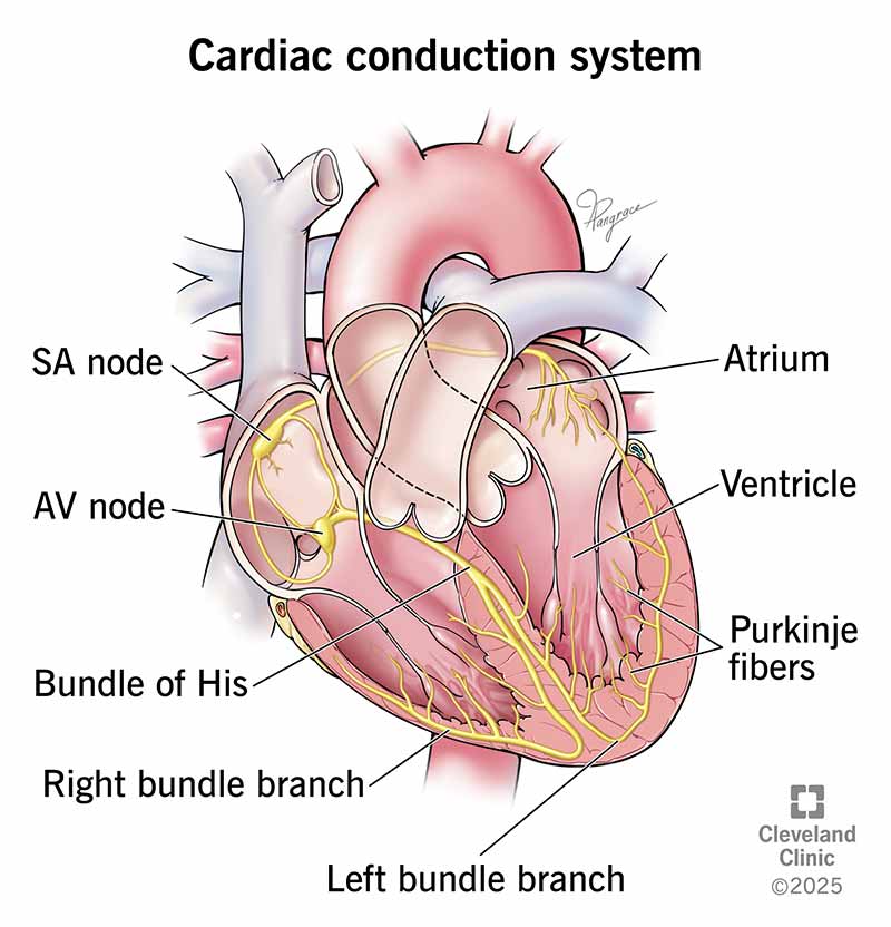
cardiac electrical conduction system list
Sinoatrial (SA) Node
Atrioventricular (AV) Node
Bundle of His (Atrioventricular Bundle)
Bundle Branches (Left and Right)
Purkinje Fibers
Bundle of His (Atrioventricular Bundle)
Receives the impulse from the AV node and passes it to the ventricles.
Bundle Branches (Left and Right)
key pathways in the heart's electrical system, splitting from the Bundle of His to carry electrical signals to the right and left ventricles, respectively, ensuring they contract in sync to pump blood effectively
Purkinje Fibers
specialized cardiac muscle cells located in the subendocardium of the ventricular walls, responsible for rapidly conducting electrical impulses (action potentials) throughout the ventricles. They ensure, efficient, and coordinated contraction of the heart, acting as the final, fastest, and most crucial component of the cardiac conduction system.

Lavender (purple) top tube contain, and what tests are they used for
contain the anticoagulant EDTA (Ethylenediaminetetraacetic acid), which prevents blood clotting by binding calcium ions
tests-Complete Blood Count (CBC), hemoglobin A1c, ESR, blood film, and HIV/hepatitis testing
Lavender (purple) top tube,ratio, and inversions
gently inverted 8 to 10 times immediately after blood collection
contain 1.5-2.2 mg EDTA per milliliter of blood.
Gold or tiger-top serum separator tube(SST) contain, and what tests are they used for and purpose
a clot activator (silica) and a gel barrier(a synthetic, thixotropic polymer gel.)
clinical chemistry tests (BMP, CMP), immunology, serology, and hormone testing. The gel enhances sample processing efficiency.
Chemistry Panels: Basic Metabolic Panel (BMP), Comprehensive Metabolic Panel (CMP).
Lipid Profiles: Cholesterol, HDL, LDL.
Liver & Kidney Function: LFTs, urea, electrolytes.
Endocrinology/Hormones: Thyroid panels, cortisol.
Serology/Immunology: Infectious disease screening.
Cardiac Markers: Troponin, creatine kinase.
Gold or tiger-top serum separator tube(SST) ratio, and inversions/important information
gentle inversion 5 times
Allow blood to clot for 30 minutes in an upright position.
Centrifuged for 10 to 15 minutes at 1,100–1,300 Relative Centrifugal Force (RCF) or g-force, or approximately 2,500-3,000 RPM.
Centrifuge within 2 hours of collection to prevent hemolysis and analyte degradation.
What tubes are centrifuged and why and which require time before
Don’t require prior time
Light Blue-Sodium citrate-Plasma for coagulation tests
Green (light or dark)-Lithium or sodium heparin-Plasma for chemistry
Mint Green (PST)-Heparin + gel-Plasma separated by gel
Require time for coagulation
Red (plain)-None/Clot activator- to get Serum
Red/Gray or Gold (SST)-Clot activator + gel-Serum separated by gel
Tiger-top (SST) -Clot activator + gel-Serum