SECTION 04: INTESTINES
 Call Kai
 Learn
 Practice Test
 Spaced Repetition
 Match
 Flashcards
 Knowt Play
1/22

There's no tags or description
Looks like no tags are added yet.
23 Terms
What are the two parts of the intestines, and what is their function?
Small intestine and large intestine.
Function: Further digest food and eventually eliminate waste from the body.

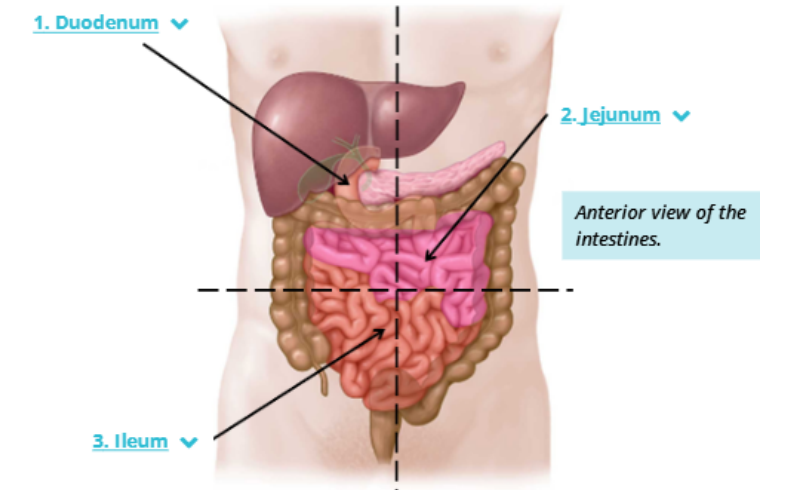
What are the 3 regions of the small intestine, their lengths, and locations?
1. Duodenum: (25 cm): It is C-shaped and encloses the head of the pancreas.
2. Jejunum: (2.5 m): Most of the jejunum lies in the left upper quadrant of the abdomen.
3. Ileum: (3.5 m): Most of the ileum lies in the right lower quadrant of the abdomen.
What are villi in the mucosa of the small intestine?
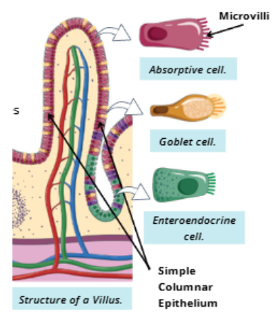
Villi: fingerlike projections that extend into the lumen of the small intestine.

What type of epithelium lines the villi in the mucosa of the small intestine, and what is the function of microvilli?
Epithelium: Simple columnar with absorptive cells.
Microvilli (brush border): Increase surface area for nutrient absorption.
What is the lamina propria of the mucosa in the small intestine, and what does it contain?
Location: Forms the core of each villus.
Contains: Blood capillaries and lymphatic capillaries.
Peyer's patches: Collections of lymphatic tissue, mainly found in the ileum, that help with immune defense.

What are intestinal glands in the mucosa (crypts of Lieberkühn), and what is their function?
Deep folds of the small intestine's mucosa between villi that secrete intestinal juices.

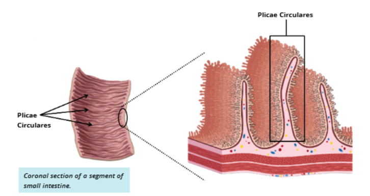
What are plicae circulares in the small intestine, and what is their function?
Permanent folds of the mucosa and submucosa in the small intestine that increase surface area for nutrient absorption.
Describe the submucosa of the duodenum
The duodenum's submucosa contains Brunner's glands, which secrete alkaline mucus to protect the small intestine from stomach acid.
These glands are absent in the jejunum and ileum.

Describe the muscularis externa of the small intestine.
Two layers of smooth muscle:
Inner: Circular muscle.
Outer: Longitudinal muscle.
Nerve plexus: Located between the two layers to control muscle contractions.

What is the outermost layer of the small intestine?
Serosa, the same outermost layer found in the stomach.

What are the 3 sections of the large intestine?
Cecum
Colon
Rectum
The large intestine is the final portion of the digestive tract, extending from the small intestine to the anal canal.

What is the cecum of the large intestine, and what structure extends from it?
The cecum is the first part of the large intestine and has an extension called the vermiform appendix.

What is the colon of the large intestine and the 4 sections and their locations?
The second portion of the large intestine is the colon
Ascending Colon: Extends from the cecum up the right side of the abdomen to join the transverse colon just below the liver.
Transverse Colon: Extends from below the liver, crossing the abdomen to join the descending colon just below the spleen.
Descending Colon: Extends down the left side of the abdomen from the splenic end of the transverse colon to the sigmoid colon.
Sigmoid Colon: The S-shaped terminal portion of the colon that leads into the rectum.

What is the rectum in the large intestine, and what is its function?
The last portion of the large intestine, extending from the sigmoid colon to the anal canal, that temporarily stores feces before defecation.

Describe the mucosa of the large intestine
No villi or plicae circulares.
Epithelium: Simple columnar with increasing goblet cells toward the anus.
Anal canal: Changes to stratified squamous epithelium.
Intestinal glands: Present, like in the small intestine.
Lymphatic tissue: Found in the lamina propria and submucosa.

What does the submucosa of the large intestine contain?
Blood vessels, lymphatics, glands, and nerve plexuses.

How does the muscularis externa of the large intestine differ from the small intestine?
The muscularis externa has a unique muscle arrangement depending on the region:
Cecum and colon
Rectum
Anal cana
The exact muscle arrangements are not specified in these notes.
Describe the muscularis externa of the cecum and colon.
Teniae coli: Three longitudinal muscle bands that contract.
Haustra: Sac-like pouches formed when the teniae coli bunch up the intestinal wall.

Describe the muscularis externa of the rectum.
three teniae coli merge to form the continuous longitudinal muscular layer

Describe the muscularis externa of the anal canal.
Anal Canal: the circular muscle layer thickens into the internal anal sphincter, which is involved with waste excretion.

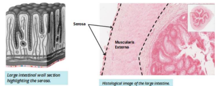
What is the outermost layer of the large intestine?
Either adventitia or serosa, depending on the region.
What is Crohn's disease, and what are its symptoms?
Inflammation of the digestive tract, mainly affecting the lower small intestine and large intestine.
Symptoms: Abdominal pain, diarrhea, fatigue, and malnutrition.

What is the anal canal, and what are its main structures?
The anal canal is the last portion of the digestive tract that leads into the external world.
Anal column: Longitudinal ridges in the canal.
Internal and external anal sphincters: Help with the excretion of waste during a bowel movement.
