ALFS, L5: Anatomy of the Lymphatic System
 Call Kai
 Learn
Learn Practice Test
 Spaced Repetition
 Match
Match Flashcards
 Knowt Play
1/41

Earn XP
Description and Tags
Given definition, answer w/ term
Name | Mastery | Learn | Test | Matching | Spaced | Call with Kai | Chat |
|---|
No analytics yet
Send a link to your students to track their progress
42 Terms
Cardiovascular and immune
List the 2 principal roles of the lymphatic system
Cardiovascular
1 of the 2 Roles of the Lymphatic System:
Maintains fluid balance and homeostasis via 2 distinct pressures that cause fluid to move between blood and tissues:
Hydrostatic pressure – push force that moves fluid _???___ from __???__ located in capillaries
Osmotic pressure – pull force generated by differences in the protein concentration between blood plasma and tissue fluid
Lymphatic system runs parallel to the venous cardiovascular system, drains interstitial fluids, and deposits back into the cardiovascular system
Immune
1 of the 2 Roles of the Lymphatic System:
Component of the surveillance system that defends the body
?? Creates excess fluid bluid, which would be bad, fill in reason
Enter the lymphatics
Lymphatic system
Function:
Returns excess fluid back into circulation
Surveillance of infection/abnormality
Anatomy:
Mirrors the venous system (e.g. lymphatic vessels are made of simple epithelial cells)
No intrinsic pump, the movement of lymph is driven by pressure changes caused by contraction of surrounding muscles
Has 2 main ducts ???? that prevent retrograde flow (??)
Lymph
Function: fluid balance & immune
Formation: ???
Edema
Definition: Abnormal accumulation of interstitial fluid
Examples:
Chronic heart disease – caused by increase in venous hydrostatic pressure
Loss of plasma protein – caused by decrease in blood osmotic pressure
Inflammation – caused by increased blood vessel permeability
Neoplastic obstruction of lymphatic vessels – caused by blockage of lymphatic drainage
Inflammation
Inflammatory response in local tissue, consequences include:
Edema
Extra-vasation of immune cells into interstitial fluid
Antigen presenting cells – Specialized antigen-presenting cells migrating ???
Antigen presenting cells (APC)
Function: Specialized antigen-presenting cells migrating ???
Examples: ???
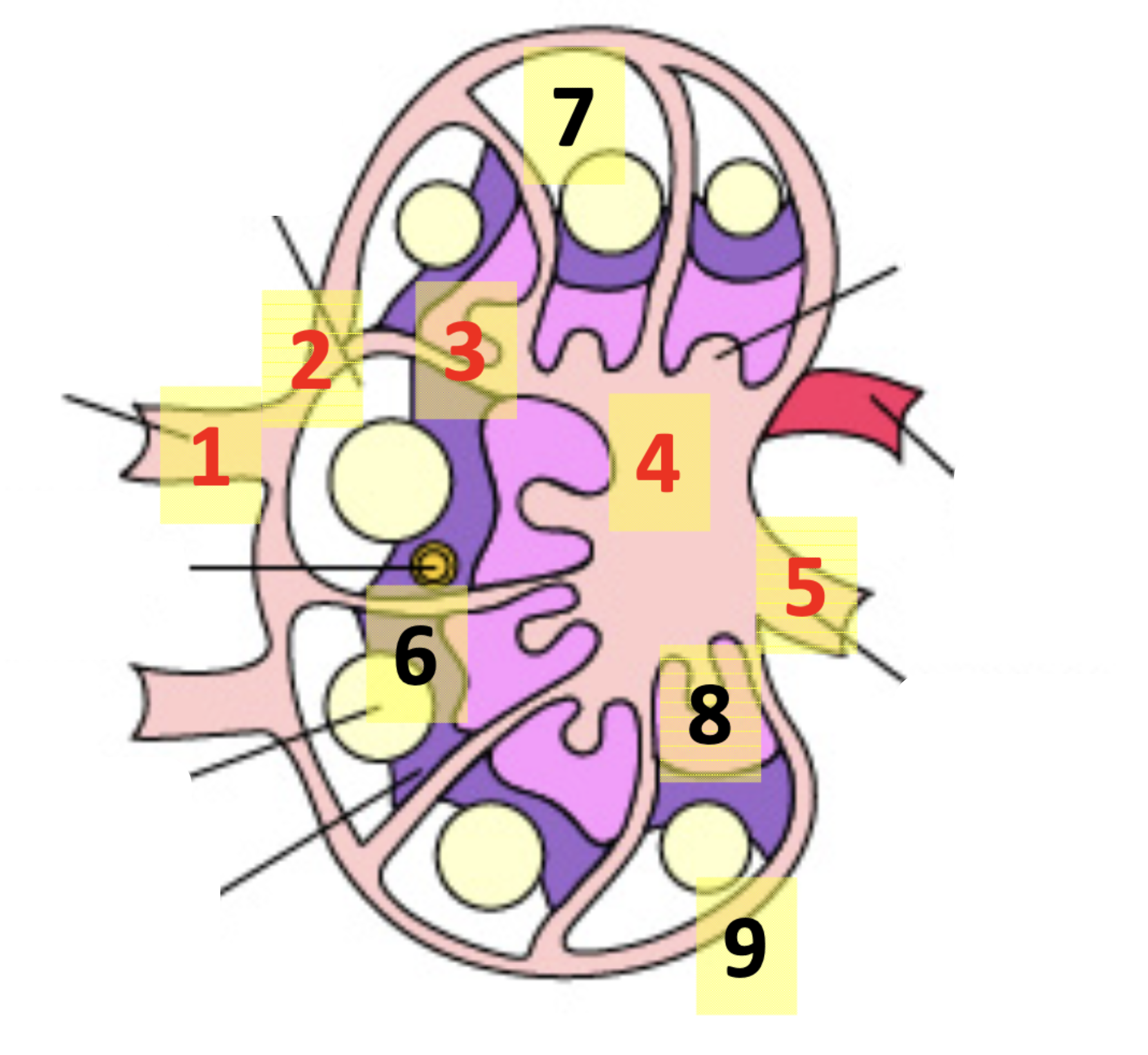
Afferent lymphatic vessels > subcapsular sinus > travecular sinuses > medullary sinus > efferent lymphatic vessel
List the main 5 steps in flow of lymph through a lymph node

T and B cells
While fluid is moving through the lymph node, they’re exposed to _______ and _______ cells
Afferent lymphatic vessels
Lymph Flow Through Lymph Node:
Diagram: #1

Subcapsular sinus
Lymph Flow Through Lymph Node:
Diagram: #2

Trabecular sinuses
Lymph Flow Through Lymph Node:
Diagram: #3

Medullary sinus
Lymph Flow Through Lymph Node:
Diagram: #4

Efferent lymphatic vessel
Lymph Flow Through Lymph Node:
Diagram: #5

T-cells in the paracortex
Lymph Flow Through Lymph Node:
Antigen presenting cells get trapped in trabecular sinuses and interact with #6

B cells located in the cortex
Lymph Flow Through Lymph Node:
#7 located in the cortex in lymphoid follicles – upon activation form germinal centers

Plasma cells in medullary cords
Lymph Flow Through Lymph Node:
_______ cells (antibody secreting B cells) located in _______ cords
Connective capsule
Lymph Node Histology
Diagram: A

Cortex
Lymph Node Histology
Diagram: B

Medulla
Lymph Node Histology
Diagram: C

Follicles
Lymph Node Histology
Diagram: D

Germinal center
Lymph Node Histology
Diagram: E

Antigen stimulation
Most B and T cells in lymph node are naive (unstimulated), and upon _______ stimulation proliferate, and cause enlargement of the lymph node
On a histological sample,
-inrease in ytoplasm
differences in how cytoplasm and nuclei pick up stain
put in picture
Primary lymphoid organs
Function: Site of production and early maturation of B and T cells:
Function:
Thymus – primary maturation of T cells
Bone marrow – production and primary maturation of B cells and production of T cells ??
Bursa of fabricius
Secondary lymphoid organs
Function:
Site for maintaining naive lymphocytes
Where lymphocytes are activated due to encountering & responding to antigens
Examples:
Spleen – screen lymph
Lymph nodes – screens blood
Peyer’s patches and mucosal associated lymphoid follicles (MALTs)
Tonsils
Spleen
Screening organ for blood, analogous to lymph in the lymph nodes
Arterioles > periarteriole lymphatic sheath > margina zone > red pulp > venus sinusoids > efferent veins
List the main 5 steps in flow of blood through the spleen

Arterioles
Blood Flow Through Spleen:
Diagram: #1

Perarteriole lymphatic shealth (PALS)
Blood Flow Through Spleen:
Diagram: #2

Marginal zone
Blood Flow Through Spleen:
Diagram: #3

Red pulp
Blood Flow Through Spleen:
Diagram: #4

Venus sinusoids
Blood Flow Through Spleen:
Diagram: #5

Efferent veins
Blood Flow Through Spleen:
Diagram: #6

T and B cells
While blood is moving through the spleen, they’re exposed to _______ and _______ cells
Thymus
Components of Lymphatic System:
where T cells mature
Bone marrow
Components of Lymphatic System:
Production of B and T cells
Maturation of B cells
Spleen
Components of Lymphatic System:
o
M cells
Take sample of gut
Pass it through a pseudo lymph node (aka pass it by T and B cells) and then out through an efferent lymphoid vessel
Problem is, it will identify many non-self particles that aren’t harmful to the host (aka food), ongoing area of research
M cell
function – specialized areas distributed throughout the small intestines, allows body to identify pathogens
locoated over the peyer’s patches
many pathogens have developed systems to hijack and attach thesmelves to M cell, and allow them to enter the GI tract ???
peyer’s patch - organized lymphatic tissue, APC, B and T cells, that activate the immune response
Peyer’s patches
Components of Lymphatic System:
Definition: Organized lymphoid follicles in distal ileum
Location: Lies underneath M cells
function: screens mucosal membrane of GI tract