DH 235 Clinical Science Midterm Study Terms & Definitions
 Call Kai
 Learn
 Practice Test
 Spaced Repetition
 Match
 Flashcards
 Knowt Play
1/178

There's no tags or description
Looks like no tags are added yet.
179 Terms
Disadvantages of Chlorhexidine
- Staining
- Taste
- Supra calc build-up
- Interferes with Fl toothpaste
What is Arestin?
Minocycline HCl 1mg

What depth of pockets would a clinician place Arestin?
5mm & above
How long does Arestin stay in the periodontal pocket?
21 days
POI after Arestin placement
- Advise pt to floss gently in the area for 10 days
Difference between rigid vs. flexible instruments
Rigid instruments offer limited tactile sensitivity while flexible instruments offer a lot of tactile information
Where is an area that is common to miss calculus?
In the mesial concavity of the maxillary 1st premolar
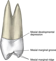
Where do you use the after five minis?
In narrow pockets deeper than 5mm

What is the length of the after five mini terminal shank?
3mm elongated terminal shank than traditional gracey

Why are the after five minis so useful?
They allow for deeper and easier insertion (thinner blade and elongated shank)
TRUE OR FALSE: The 15/16 resembles the 13/14 gracey but it is for mesial surfaces and not distal
True

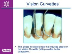
What is a special feature of the subzero vision curvette?
Has a (+) marking where the blade is at and it has a smaller curved blade
Two raised bands indicating 5mm and 10mm

Why is the smaller curved blade of the subzero vision curette useful?
It is useful for deep vertical defects for anterior segments
The use of files
Used for burnished calculus or large, tenacious pieces

Why is the working end of the file important?
The end of the file is serrated, which allows the operator to break apart the large deposits into smaller segments

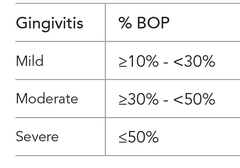
If a patient has less than 10% bleeding sites, are they considered to have incipient gingivitis?
YES, bleeding is an indicator of infection activity
When would a diamond tip coated instrument be useful?
In class 4 furcations because you can see. You do not want to use in areas you cannot see bc the diamond could abrade the root surface

Purpose of a periodontal file
An instrument used to prepare calculus deposits before removal with another instrument. Used to crush or roughen a heavy deposit so that it can be removed with a sickle or curet
What areas are files limited to?
Enamel surface or on outer surfaces of calculus deposits
Design features of a periodontal files
Has a series of cutting edges lined up on a base
Adaptation of periodontal files
The face of the working end should be flat against the calculus deposit so that the corners of the cutting edges do not gauge the root surface or soft tissue

Two-point contact for periodontal files
1. Adaptation of the working end to a calculus deposit
2. Resting the lower shank against the tooth
Periodontal hoes design
Single cutting edge -- right angle to shank
Shank bent at a variety of angles
Face of blade at 100 degrees to shank
Edge beveled at 45 degrees
Cutting edge = union of bevel and face
Periodontal hoes
Goldman fox 6 -- mesial/distal surfaces
Goldman fox 5 -- buccal/lingual surfaces

Disadvantages of periodontal hoes
Does not reach bottom of pocket or adapt to the curvature of roots
Advantages of periodontal hoes
Good for hard heavy deposits and distals of last molars
1/2 Barnhart USC/suter curet instrument design
Two cutting edges, less pronounced curvature, longer shank for posterior access, used for line angles

7/8 Younger-good curet instrument designs
Offset elliptical, similar to Nevi 4, smooth angle, ideal as universal --> posterior to anterior

Why do we need implants
Missing teeth = resorption of osseous

Bone graft -- autograft
Bone obtained from the patient from another area
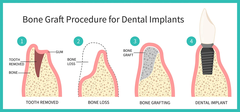
Bone graft -- allograft
Bone obtained from another human (cadaver bone)

Bone graft -- xenograft
bone obtained from another species (cow or bovine)

Bone graft -- alloplast
synthetic derivative of bone (beta-tricalcium phosphate)

How to qualify the success of an implant
No visible inflammation
No pain or tenderness
No mobility
Radiograph shows less than 2mm loss from initial surgery
No bleeding or suppuration
No damage to components
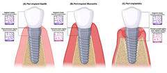
Peri-implant health
No inflammation
No BOP
No swelling or suppuration
Bone loss

Peri-implant mucositis
(also known as peri-implant gingivitis) plaque-induced gingivitis in tissues surrounding the implant

Peri-implantitis
periodontitis in tissues surrounding osseointegrated implant, resulting in bone loss
Probing depth >4mm-8mm
Bone loss >3mm+
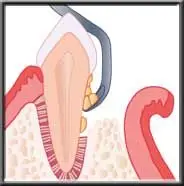
Gingival curettage
The scraping of the gingival wall of a periodontal pocket to remove inflammed soft tissue

Surgical curettage
"flap surgery"
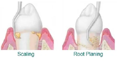
Hard tissue curettage or root curettage is another name for...
Root planing
Inadvertent curettage
Unintentional removal of pocket wall and junctional epithelium
Types of gingival curettage
Definitive and non-definitive
Definitive curettage procedure
Eliminates supra bony pockets (surgeon does this)
Non-definitive curettage procedure
Reduces inflammation to improve status of tissue before surgery (we do this)
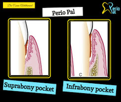
Suprabony pocket
- horizontal bone loss
- JE is located coronal to the crest of the alveolar bone

Infrabony pocket
It occurs when there is vertical bone loss.
JE is located apical to the crest of the alveolar bone (below the crest of bone).
Base of the pocket is located apical to the level of adjacent bone
Indications for definitive gingival curettage
1. soft tissue
2. spongy tissue
3. suprabony pockets
4. shallow pocket (4-6mm)
5. Edematous tissue
6. Recall visits (to maintain areas)
Aim of gingival curettage
To reduce pocket depth by enhancing gingival shrinkage and/or creating a new connective tissue attachment (long junctional epithelium)
Contraindications for definitive gingival curettage
1. fibrotic tissue
2. if aesthetics is a concern -- papilla may not grow back
3. Deep supra bony pockets = bottom of the pocket is coronal to alveolar bone (vertical loss)
4. Mucogingival involvements
5. pockets associated with frenum attachments -- after curettage, the tissue is thinned, therefore the frenum attachment can cause recession as it pulls on the thinned tissue
Rationale for non definitive curettage
Reduction of inflammation is needed before other pocket elimination techniques
The patient in whom more aggressive surgical techniques are contraindicated (age, systemic problems, financial problems, etc.)
Curettage technique
1. Scale, root place until root surface is clean, smooth & hard, as detected by a sharp explorer
2. Make sure patient is anesthetized
3. Select a sharp instrument, the curette is selected so that the cutting edge is against the tissue
4. Place cutting edge against tissue
5. Insert blade into sulcus to desired depth
6. Sweet horizontally, using short strokes
7. Use finger pressure against tissue with non-dominant hand
8. Make sure to turn instrument into interproximals
9. Clean out all the tissue with instrument or explorer
10. Flush with water
11. Use dressing as needed
Healing process after gingival curettage
- Clot formation
- Granulation tissue
- Restoration and epithelialization of sulcus occurs within 2-7 days
- Junctional epithelium restores in 5 days
- Clinically normal within 2 weeks
How does silver diamine fluoride work?
arrests tooth decay
acts as an antimicrobial -->
1. the silver acts as a stimulate dentin sclerosis and calcification/binds with collagen
2. the fluoride promotes the remineralization
3. the ammonia stabilizes the ingredients in solution

Various uses of SDF
- Desensitizer
- Caries arrest
- As a liner under restoration
- Indirect pulp cap material
- Around existing restorations
- Preventive
Application of SDF
1. Clean the site
2. Isolate and dry the site
3. Apply & soak
4. Blot dry
5. Fluoride varnish
6. No post-treatment patient restrictions

A single application of SDF arrests...
80% of caries
A second application of SDF arrests...
95% of caries
True or false: SDF stains sound enamel and dentin
False, it only stains decayed enamel or dentin
When SDF is applied on demineralized areas, _____________ occurs
staining
What does SMART stand for?
Silver Modified Atraumatic Restorative Treatment
Restorative protocols of SDF
If a restoration is placed at the same appointment, the restoration will likely gray
Self-curing restorative materials are used for better aesthetics
CDT Code for Advantage Arrest SDF -- Primary prevention/remineralization
D1355
CDT Code for Advantage Arrest SDF -- Caries arrest
D1354
CDT Code for Advantage Arrest SDF -- Desensitize
D9910
Define periodontics
The specialty of dentistry that studies the supporting structures of teeth, as well as diseases and conditions that affect them
Pathophysiology of Periodontal disease
chronic inflammatory disease mostly caused by bacteria
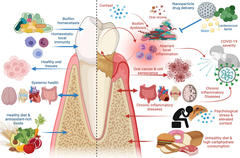
Multifactorial modifiers of periodontal disease
poor oral hygiene, genetics, smoking, uncontrolled diabetes, stress, age, etc.
Medical consequences of periodontal disease
Relationship to heart disease, diabetes, immune diseases
Beginning stages of periodontitis
virulent bacteria are introduced. periodontal pathogens are gram (-), they have LPS present in their cell walls (lipopolysaccharides)
Once the immune system is initiated in someone with periodontitis...
the host response is turned on, cytokines are released to the site by the responding neutrophils and macrophages
The release of...
TNF-a, IL-1, and MMP occurs during the pathogenesis of periodontitis

MMP stands for..
matrix metalloproteinases
IL-1 stands for
interleukin-1
Role of MMP
responsible for collagen break down
TNF-a is responsible for...
increased osteoclast activity
Red complex bacteria
P. gingivalis
T. forsythia
T. denticola

Periodontitis and diabetes
- Periodontitis has a higher prevalence among diabetics and is 3-5 times more severe
Proposed mechanism:
- pathogenesis of periodontitis releases certain cytokines
- HbA1c levels of more than 7% = twice the level of cytokines in GCF
- Glycemic control linked to progression of periodontal disease through poor wound healing

Periodontitis and smoking
- Increase in periodontal pathogens in deeper pockets (anaerobic)
- Altered neutrophil function: chemotaxis, phagocytosis, and oxidative burst
- Vasoconstriction and decrease in blood vessels
- Decrease of bone healing: altered osteoblast function -- from nicotine

Periodontitis and heart disease
- inflammation from oral bacteria may modify thrombus formation
- Pro-inflammatory bacteria attach to thrombus and its bursts

Genetics and periodontitis
- In the presence of periodontal pathogens - it is the balance of destructive and protective inflammatory mediators that determines disease progression
- Expressions for TNF-a, IL-1, MMPs, RANKL, osteoprotegerins, and TIMPs are genetically determined
TNF-alpha inhibitors
a chemical messenger produced by the immune system that regulates inflammation
major regulator of inflammatory responses and is involved in the pathogenesis of some inflammatory and autoimmune diseases
function of IL-1 in inflammation
interleukin 1 is a cytokine that introduces local and systemic responses that are characteristic of inflammation such as:
Fever
Pain
Vasodilation
Hypotension
RANKL
Receptor activator of nuclear factor-kB ligand is a protein that plays a key role in bone metabolism and the immune system
Osteoprotegerina
A protein that regulates bone remodeling. Prevents bone breakdown and increases bone strength by binding to the receptor activator of RANKL, which inactivates RANKL and prevents osteoclast development
What are the forms of measurement for periodontitis
Visual
Indices (plaque index and bleeding index)
Mobility
Furcation
Recession
CAL
Attached tissue - bio-type i.e keratinized
Radiographs
Purpose of mobility measurement
To measure tooth stability with respect to the underlying bone. Assess bone support and trauma from occlusion
Why do we measure furcations?
Because furcations harbor bacteria and are difficult to maintain
Periodontal biotype — keratinized attached tissue
Minimum of 1mm is needed, but ideally 2+mm
Keratinized attached tissue is needed to withstand day-to-day trauma

Periodontitis grading
aims to indicate the rate of periodontitis progression, responsiveness to standard therapy, and potential impact on systemic health
Diagnosis of periodontal disease — gingivitis
Biofilm-induced
Non-biofilm induced (i.e. hormones, medication-induced etc.)
Diagnosis of periodontal disease — periodontitis
Extent: localized, molar-incisor, or generalized (30% rule)
Severity: initial, moderate, severe (stages I-IV)
Form: chronic, grade
Algorithm for treatment planning — periodontally stable/healthy pt
- OHI
- Recommended 6 month prophy
- Recommended for annual/periodic periodontal evaluations

Algorithm for treatment planning — Localized/generalized gingivitis
- OHI
- Recommended for 4-6 months prophies and tissue evaluation
- Recommended for 6 month periodic periodontal evaluation
- Continue course until pt is periodontal stable/healthy, then use algorithm in that column
- Diagnosis: Generalized or localized, plaque induced gingivitis

Algorithm for treatment planning — Localized/generalized periodontitis
- OHI
- Localized or generalized scaling and root planing
- 4-6 week periodontal re-evaluation is needed
- Diagnosis: Generalized or localized, chronic/aggressive, slight/moderate/advanced perio & Stage I-IV and Grade A, B, or C

Algorithm for treatment planning — Localized/generalized periodontitis if pockets have decreased and osseous surgery is NOT indicated
Periodontal maintenance 3-4 months

Periodontal maintenance algorithm
- usually done every 3 months following initial SRP for 1 year, then modified to 3-4 months after annual re-assessment
- however, monitoring every perio maint is important
- If perio status is stable, you can graduate pt to perio maint for 4 months

Algorithm for treatment planning — Localized/generalized periodontitis if pockets remained or has residual deep pockets, surgery is indicated
- Osseous surgery: 1-3 teeth (localized) or 4 or more (quadrant) OR open flat surgery (without osseous recontouring)
- After surgery, 3 months of perio maintenance is recommended for at least 1 year. After 1 year, an annual re-assessment is recommended
- However, monitoring at every apt is critical
- if perio status is stable you can graduate pt to 4 months

Prophylaxis
Is done when there are no signs of periodontitis. Only done in the presence of health or gingivitis
Debridement
Is done prior to periodontal exam to clear heavy plaque and calculus that is preventing you from performing proper periodontal exam
Scaling and root planning
Done when there is attachment loss/bone loss in conjunction with a deeper pocket (4+mm). Although SRP can be initial treatment to address periodontitis, it can also be a definitive treatment. It is unlike prophylaxis in that both of these are "preventive" and NOT a "definitive" treatment