Maternity Exam 2
 Call Kai
 Learn
Learn Practice Test
 Spaced Repetition
 Match
Match Flashcards
 Knowt Play
1/65

There's no tags or description
Looks like no tags are added yet.
Name | Mastery | Learn | Test | Matching | Spaced | Call with Kai | Chat |
|---|
No analytics yet
Send a link to your students to track their progress
66 Terms
Postpartum Hemorrhage
Leading cause of maternal mortality & morbidity. Is life-threatening.
Blood loss over 500cc after vaginal birth
Blood loss over 1,000cc after C-section
OR blood loss with signs of hypovolemia within 24 hours after birth (all types)
SIGNS
Unresponsive to fundal massage or medication (indicates uterine atony)
10% decrease in HCT between admission & postpartum
The “4 T’s”
Early v. Delayed Postpartum Hemorrhage
Early: Occurs within the First 24 hours
Late: 24 hours to 6 weeks PP
4 T’s of Postpartum Hemorrhage (causes for PPH)
Tone: Uterine Atony → Vessels in uterus aren’t contracted, allowing bleeding
Trauma: Tearing/Trauma to Genital Track
Thrombin: Coagulation Disorders
Tissue: Retained Placenta → Placenta or part of placenta stuck in uterus. Causes uterus to not contract (subinvolution)
Client delivered 4 hours ago. Assessment data: BP 102/58, P96, R 22, T 98.2, Fundus firm at the umbilicus, lochia heavy rubra. Your first action should be:
A. Massage the uterus
B. Administer an oxytocic as ordered
C. Notify the provider
D. Assist the client to the bathroom
C. Notify the provider
Heavy rubra lochia indicates a tear causing PPH. Is not a uterine problem. Nursing interventions are exhausted, so provider must fix the tear.
Not B, because oxytocin is only used to make fundus firm, and the uterus is already firm
Uterine Atony
Uterus fails to contract despite being empty. Most common cause for PPH
Risk Factors
Overdistention: If pt has multiples, uterus can be over-distended and will not contract
Traumatic birth: Overstimulation of uterus
MgSO4: Magnesium Sulfate, used to soothe uterus to treat pre-eclampsia. Stops uterine contractions, increasing risk for hemorrhage
Precipitous Labor: Very fast birth
Augmented labor w/Oxytocin: “Pitocin”, used to contract uterus during pregnancy. If too much is used, receptors will become sensitized and Pitocin will not work during postpartum to help with uterine contractions.
Chorioamnionitis (infection): Will have maternal fever & tachycardia, fetal tachycardia, fundal tenderness, & foul-smelling discharge
Retained placental fragments: Cause uterus to become flaccid because it is not fully empty
High Parity: High number of pregnancies (G5+) or short-spaced pregnancies (2 babies in less than a year)
Hx or uterine atony
Treatment of Uterine Atony
Firm Fundal Massage
Manual expression of clots → Clots can get trapped in uterus, can be manually removed
Elimination of bladder distention → Make sure bladder is empty before fundal massage (if fundus is midline, then bladder is NOT distended)
IV infusion of oxytocin & Lactated Ringer’s or Normal Saline Solution (or IM if no IV access)
Can give alternatives
Bimanual compression and exploration for fragments (provider only)
Uses sterile technique with antibiotics after expulsion
Oxytocin Alternatives
Methergine (IM): Can use in pts w/HTN or elevated BP
Hemabate (IM): Contraindicates in pts w/asthma. Can cause bronchospasm
Prostaglandin E2 (Cytotec/Misoprostol) (Rectal or PO)
Tranexamic Acid (TXA)
Crystalloid solution & blood products
Oxygen (non-rebreather 10L) & Urinary Catheter (minimum of 30cc/hr), Labs
Bimanual Compression/Exploration for Atonic Uterus
Compressing of uterine front and back walls, held until uterus contracts. Provider uses hand to manually sweep clots out of uterus
Which symptom do you expect to observe in a postpartum client with a vaginal hematoma?
A. Bleeding
B. Pain
C. Redness
D. Warmth
B. Pain
Only symptom of hematoma (like a bruise)
Will have intense pain
Trauma to Genital Tract
Laceration to the cervix, vagina, & perineum
CAUSES
Precipitous birth→ any birth that occurs in less than 15mins at start of contractions or less than 3hrs at start of contractions
Congenital abnormalities
Contracted pelvis
Infant size (large baby)
Operative Births (forceps or vacuum assistance)
Abnormal fetal presentation/position
Previous scars from infection, injury, or operations
Vulvar, perineal, and vaginal varicosities (enlarged veins)
Treatment of Trauma to Genital Tract
Identifying site of bleeding (call provider to identify)
Venous
Arterial → Will have bright red discharge
Placenta → Will have spurts of discharge; uterus will go back and forth between boggy & firm
Control bleeding
Analgesia (pain relievers)
Ice
Diet
Stool softeners
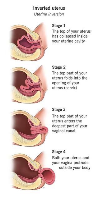
Uterine Inversion
Life-threatening complication where uterus flips inside out
NO PULLING ON CORD unless separation is certain
RISK FACTORS
Fundal pressure or improper fundal massage → Never apply fundal pressure if fundus is firm o
Short cord → Can pull the placenta out before its ready, pulling on uterus
Cord traction→ Pulling cord by force. Placenta is ready when cord is lengthening outside the vagina & will have spurts of blood
Uterine Atony
Fibroids
Multiples
Hx of placenta accreta/increta → Part of placenta grows inside uterus, instead on being attach to it.
Hx of Past inversion
SIGNS & SYMPTOMS
Hemorrhage (94% of cases)
Shock
Pain
TYPES
Partial or complete
TREATMENT
Lactated Ringer’s & blood products → treat shock; must have IV access
Fundal repositioning after placenta separation
Give MgSO4 & analgesia to relax uterus (discontinue after repositioning)
MgSO4 relaxes uterine muscle, uterus must be completely relaxed to put back in place
Oxytocic medications & bimanual compression
Antibiotic therapy
Coagulopathies Affecting Postpartum
Idopathic Thronbocytopenia Purpra (ITP)
von Willebrand Disease (vWD)
Disseminated Intravascular Coagulation (DIC)
A client is being monitored after a significant postpartum hemorrhage. Which of the following do you need to report to the provider?
A. Urine output of 200cc for last 8 hours
B. Weight decrease of 2 lbs since delivery
C. Drop in Hematocrit of 2% since admission
D. Pulse rate of 68 beats per minute
A. Urine output of 200cc for last 8 hours
25cc/hr is too low (should be at least 30cc/hr)
Not C bc hematocrit is only concerning if its >10% drop
Idiopathic Thrombocytopenia Purpura (ITP)
An autoimmune disorder where antiplatelet antibodies decrease lifespan of platelets
Can causes severe hemorrhage in C-section or in lacerations
Increased risk of uterine bleeding or vaginal hematomas
DIAGNOSTICS
Thrombocytopenia (decreased platelets)
Capillary Fragility (multiple bruises & petechiae/purpura, bloody gums)
Increased bleeding time (PT & PTT → takes longer for platelets to clot)
TREATMENTS
Control platelet stability with platelet transfusion
Glucocorticoids → Steroids help maintain vessel walls & decrease bleeding
Splenectomy → Done during pregancy. Removes spleen to raise platelet count by 75%
Danazol postpartum: Male sex hormone that may raise platelet count. Only give postpartum as causes tetragenic effects during pregnancy
von Willebrand Disease (vWD)
Genentic deficiency in von Willebrand factor, blood clotting proteins.
Most common clotting disorder in US (type of hemophilia)
SYMPTOMS
Nose bleeds
Bruising
Prolonged bleeding after procedures
Factor VIII Deficiency
TREATMENT
Desmopression & Factor VIII supplementation
Disseminated Intravascular Coagulation (DIC)
Overactivation of clotting & anti-clotting processes. An exaggeration of widespread clotting cascade
Will have initial hypercoagulability is followed by HYPOcoagulability & hemorrhage. Once all platelets are used up, there will be a decrease in fibrinogen & increase in PT & PTT
Pt must still have 2 IVs even when bleeding
CAUSES
Disease or Injury
Snake bites
Severe trauma
Extensive surgery
Acute hypotension
SYMPTOMS
Widespread internal and external bleeding
Gums, nose, petechiae around pressure sites and trauma sites
LAB VALUES
Low platelets
Low fibrinogen
Prolonged PT & PTT
Abnormal RBC morphology → RBCs trying to produced quickly, leading to immature, non-functional RBCs
TREATMENT
Removal of underlying cause → Address uterine atony or hemorrhage
Volume replacement & blood component therapy → For every cc of blood loss, must be replaced with 3cc of crystalloids or LR
Optimize O2 & perfusion status
NURSING ASSESSMENT
Vital Signs → RED FLAGS: decreased BP & increased HR
2 IV lines
Observe signs of bleeding
Observe adverse blood reactions
Monitor urinary OP → Insert cath, maintain 30cc/hr
O2 administration
Maintain quiet/non-stress environment (pt will be admitted to ICU)
A client who is diagnosed with DIC is to receive a blood transfusion. All actions are critical for the nurse to perform except..?
A. Look up patient’s blood type in the chart
B. Check the patients arm bracelet
C. Check the blood type on the infusion bag
D. Obtain an infusion bag of dextrose and water
E. Document the time the infusion begins
D. Obtain an infusion bag of dextrose and water
Blood products can only be mixed with Normal Saline Solution
Retained Placenta
2 Types
Non-Adherent Retained Placenta
Adherent Retained Placenta
RISK FACTORS
Pt w/scars on uterus → scars cause placenta to have less fluffy tissue to implant onto.
TREATMENTS
Non-Adherent
Manual separation & removal
IV analgesia
Still at risk for PPH & infection
Adherent
Attempts to remove may not be successful → May result in lacerations of uterus
Blood replacement
Possible hysterectomy (removal of uterus)
Non-adherent Retained Placenta
Partial separation from uterus. Part of placenta is still in uterus.
Caused by mismanagement during the 3rd stage of labor or abnormal adherence
Adherent Retained Placenta
Placenta grows into uterus.
TYPES
Placenta Accreta
Placenta Increta
Placenta Perceta

A G5P3105 is undergoing repeat C/S. The physician declares the patient has a placenta accreta. Which of the following is consistent with this diagnosis
A. Blood pressure 160/110
B. Blood loss of 2000 cc
C. Jaundice skin color
D. Shortened Prothrombin time
B. Blood loss of 2000 cc
Adherence placenta due to blood loss
Sub-Involution
Delayed return of enlarged uterus to normal size & function
@ 6 weeks uterus should be back to normal. If not, then there is subinvolution
RISK FACTORS
Retained Placental Fragments
Pelvic Infection
SIGNS & SYMPTOMS
Prolonged, irregular, or excessive vaginal bleeding
Enlarged uterus by exam
Boggy Uterus
TREATMENTS
Methergine 0.2mg q4h for 2-3 days
Antibiotic therapy
Dilation & curettage → removes retained placental parts w/ instruments
Review of PPH Assessment
Check for uterine atony/bogginess
Massage if atonic
Empty bladder if displaced
Possible bimanual uterine compression/exploration by MD
Assess vaginal canal & cervix for lacerations
Assess bleeding from non-genital areas
Check venous puncture sites
Lab studies → INR, PTT, platelet count, & fibrinogen
Restore Blood Volume
3cc crystalloid for every 1cc of blood lost
10L oxygen by non-rebreather mask
Anticipate meds and potential surgical intervention
Nursing Discharge Instructions After PPH
Same as normal postpartum, but emphasize potential for fatigue/exhaustion related ot low blood volume
Increase iron & protein intake
Increase fluid intake
Limit physical activties/need for support system
Call MD if…
Soaking pad after 1 hr or less
Clots larger than a plum
Fever/chills
Dizziness/fainting
Postpartum Infections
Any infection of the genital tract in the first 28 days after birth or abortion.
Or temperature >100.4F orally after the first 24 hours on 2 separate occasions within the first 10 days postpartum
Low-grade fever is common in the first 24 hours postpartum. Is normal & resolves on its own
COMMON INFECTIONS
Metritis (Most common) → Infection of endometrial lining
Wound infections (C-section or lacerations)
Cystitis → Infection of bladder
Mastitis → Breast Infection, occurs after 1st week
RISK FACTORS
General: Hx of throbosis, UTI, mastitis, DM, Alcholism, drug abuse, anemia, or malnutrition
OB Factors: C-section, operative vaginal delivery, Prolonged ROM (18-24hrs), Chorioamnionitis, Prolonged labor, catheterization, Epidural anesthesia (can’t feel urge to void, requiring a Foley), retained fragments, PPH, lacerations, & hematomas
You should suspect puerperal infection when a client exhibits which of the following?
A. Temperature of 100.2
B. White blood cell count of 14,500 cells/mm3
C. Diaphoresis during the night
D. Malodorous lochial discharge
D. Malodorous lochial discharge
Foul smelling lochia
Endometritis/ Metritis
Infection of Uterine cavity or muscle
Most Common PP infection
Elective C-section have lower rates (have more time to prep and minimize infection risk)
SYMPTOMS
Tachycardia
Jagged Temp elevation → temps shift quickly. 2 spikes in 24 hours (concerning)
Uterine tenderness
Prolonged afterbirth pains
Subinvolution
Scant, odorous lochia or heavy, foul-smelling bloody, seropurulent lochia (yellow-green discharge)
Elevated WBCs after birth
Increased RBC sedimentation rate (ESR → indicates inflammation)
NURSING INTERVENTIONS
Get blood cultures before giving antibiotics
Give broad-spectrum antibiotics → usually IV until afebrile, pain-free for 24-48 hrs, & no fundal tenderness
Analgesia (NSAIDs are best)
Wound Infections
NURSING ASSESSMENTS
Vital Signs q4h
Wound Assessment → MD might need to reopen wound & repack
RED FLAGS: Redness, irritation, warmth, tenderness, oozing, foul smell, poor healing, splitting
NURSING INTERVENTIONS
Open & cleanse wound
Antibiotic therapy
PATIENT SELF CARE
Hydration, rest, & good nutrition
Increase roughage (fiber) in diet
Clean perineum from front to back
Report increases in temp, drainage, or pain
Cystitis
Infection of bladder
RISK FACTORS
Abdominal distention
Dehydration
Poor hygiene
Catherization
Pelvic exams
Epidural
Genital tract injury
Hx of UTI
C-section
SIGNS & SYMPTOMS
Urinary frequency, urgency, & buring
Suprapubic discomfort
CVA tenderness if pyelonephritis (assessed by percusing kidneys)
Fever
Tachycardia
N&V
NURSING INTERVENTIONS
Give antibiotics & analgesics as ordered (NSAIDs & urinary analgesics)
Increase fluid intake
encourage frequency voiding
Routine peri-care (change pad each time they use bathroom)
Mastitis
Infection of breast tissue
Typically from organism on maternal skin or infants mouth
Occurs 2 days to 2 weeks PP
BEST PREVENTION: Proper latch & complete emptying of breast, should keep feeding on effected breast. Best remedy is to empty breast completely
NURSING ASSESSMENT
Breast pain, swelling, & tenderness
Temp elevation
Axillary Adenopathy (swelling in axillary lymphnodes)
Usually unilateral but can affect both breasts
Possible purulent drainage
TREATMENT
Antibiotics
Continuation of pump/feed/expressing
Incision & drainage (I & D) if abscess is present
Analgesics as ordered
A breastfeeding patient is diagnosed with mastitis. Which of the following nursing interventions is appropriate?
A. Advise the woman to apply ice packs to her breasts.
B. Encourage the woman to breastfeed frequently
C. Inform the woman that she should wean immediately
D. Direct the woman to notify her pediatrician as soon as possible
B. Encourage the woman to breastfeed frequently
Best remedy for mastitis is to completely empty breasts
Thrombosis/ Thromboembolic Disease
Formation of blood clots inside blood vessels caused by inflammation or partial obstruction of vessel
Seen in pelvis or legs
RISK FACTORS
Stasis & hypercoagulation (pregnancy is a hypercoagulable state)
C-section, multiparity, Hx of thrombosis, obesity, tobacco, immobility
COMPLICATIONS
Superficial Venous Thrombosis (SVT)
Deep Vein Thrombosis (DVT)
Pulmonary Embolism (PE)
SIGNS & SYMPTOMS
Tachycardia
Edema, warmth, redness, & enlarged, hard veins over thrombis
Superficial Venous Thrombosis (SVT)
Clot in vein that is just below the skin. Superficial, close to skin.
Most common in PP
Usually harmless, very superficial
SYMPTOMS
Calf pain
Tenderness
Swelling
NURSING INTERVENTIONS
Analgesics (NSAIDS)
Rest & Elevation
Heat
Use compression socks prophylactically
Deep vein Thrombosis (DVT)
Extends from foot to iliofemoral region
NURSING INTERVENTION
Measure thigh circumference in affected leg
Assess cap refill & pedal pulse (affected leg will be diminished)
Anticoagulation therapy (heparin IV for 3-5 days)
Strict bedrest
Elevation of leg
Analgesia
Compression sock ONLY WHEN symptoms go away and used prophylactically
Compression socks can cause even more clotting if DVT is still present
Assess for symptoms of PE (will see wet cough)
SYMPTOMS
Reduced peripheral pulses on affected side
Will have unilateral swelling, pain, & redness
DO NOT perform Homan’s Sign → Shape flexion of pt calf to access pain (can dislodge DVT)
DIAGNOSTICS
Real-time & color Doppler ultrasound
Pulmonary Embolism (PE)
Complication of DVT where clot dislodges and is carries to pulmonary artery, occludes the vessel, and obstructs blood flow to lungs
IS AN EMERGENCY
A postpartum woman is diagnosed with deep vein thrombosis. For which additional complication is this client at high risk?
A. Hemorrhage
B. Stroke
C. Endometritis
D. Hematoma
B. Stroke
thrombus can dislodge and become an embolus, which will travel to occlude a vessel. Causing a stroke or PE
Baby Blues
Short term mood swings, sadness, or fatigue that occurs 1-2 days after delivery through 2 weeks PP (should be gone after 2 weeks)
IS NORMAL - affects 85% of PP women
NURSING INTERVENTIONS
Encourage communication of needs & fears
Praise mom on feeding, assessing infant needs, etc
Encourage rest when baby sleeps
Continue to monitor mother & infant bonding
Educate on what signs are not normal and who to contact if postpartum depression occurs
Postpartum Depression
Can occur at anytime in the first year PP
Greatest risk: during 1st month post delivery
SYMPTOMS
Insomnia (can’t sleep even when given the chance)
Crying & prolonged sadness
Obsessive thoughts/not being able to care for baby
Decline in personal hygiene
Suicidal ideation or harming baby/self
NURSING INTERVENTIONS
Edinburgh Postnatal Depression Screen: Test that assesses severity of PPD.
If 10 or greater, set up w/help
If pt is having suicidal ideation, set up help regardless of score
Early assessment & education
Home visitation
Stress rest
Give contacts for mental health care
Give information on meds & complementary treatments (yoga, massage, etc)
Postpartum Psychosis
Can occur at any time within the year of birth
Nursing Assessment
Signs of agitation/confusion
Irrational statements or behaviors
Delusions (false sensory perception) or hallucinations
Insomnia & hyperactivity
NURSING INTERVENTIONS
Education & assessment of family → Never leave baby alone with woman
Provide emergency contact info
Home visitation if available
If mother is having delusions, should be brought to ER asap
A postpartum woman is diagnosed with postpartum psychosis. Which of the following is essential to be included in family teaching?
A. The woman should never be left alone with her infant
B. Symptoms rarely last more than a week
C. Clinical response to medications is usually poor.
D. The woman must have her vitals assessed every two days
A. The woman should never be left alone with her infant
The Fiver P’s
Factors that affect birth
Passageway
Passenger
Power
Position
Psyche
Passageway
The birth canal
Includes bony pelvis & soft tissue
Passenger
Include the baby and the placenta. The fetal movement through the birth canal.
Movement depends on head size, presentation, lie, attitude, & position
Placenta Previa
Placenta comes out before baby, needs immediate C-section
Fetal Head Size
Ability to devliery depends on baby’s head size. The larger the head, the more dilated the cervix needs to be
Sutures
Membrane filled gaps between cranial bones
Include sagital, lambdoidal, coronal, & frontal suture
Fontanel
Where sutures intersect
Most important: anterior & posterior fontanels
Palpation after ROM reveals fetal presentation, position, & attitude
The more pushing during labor, the more molding of the infant’s head, due to pressure
Fetal Presentation
Types
Cephalic → Head first
Breech → Sacrum first
Shoulder → scapula first (transverse lie)
Never deliver transverse lies because of increased risk of umbilical cord prolapse (baby's head should come out first)
Molding
Overlap/sliding of bones to change the shape of the head
Fetal Lie
Relationship of the baby’s spine to the mother’s spine
Longitudinal/Vertical: Mother and baby’s spine are align. Cephalic or breech presentation
Transverse/Horizontal: Mother and baby’s spines are not aligned. Can be at any angle. (shoulder is the presenting part)
Biparietal Diameter (BPD)
Largest diameter of baby’s head
Important indicator of fetal size
Fetal Position
Relationship of the presenting part to the 4 quadrants of the mother’s pelvis. Noted by 3 letter abbreviation
First letter: location of presenting part in right or left side of mom’s pelvis (is posterior fontanel facing the right or left)
Middle letter: the specific presenting part of fetus (occiput or sacrum)
Third letter: location of presenting part in relation to anterior, posterior or transverse portion of maternal pelvis ( is back facing anterior or posterior)
EX: ROA → Right, occiput, anterior
What is the best position for a fetus to be in before delivery
LOA & ROA
Occiput anteriors are easier to deliver than Occiput posterior
If mother has lower back pain, indicates fetus is OP
Fetal Station
Relationship of the presenting part to maternal ischial spines (smallest part of pelvis)
Measured in centimeters above or below the ischial spines
Negative Station
Station -3 to -1. The fetus is higher up
0 Station
The “Point of Engagement”. When the baby’s head descends into the ischial spine
The infant should be at least in this station before active labor
Positive Station
+1 to +3, the more positive the number, lower the fetus’s head is to the pelvic bone
The lower the better
Pt will have the urge to push at station +3 (should not encourage pushing UNTIL urge is felt
Engagement
Term used to indicated that the largest part of presenting part has passed through the maternal pelvic brim.
Largest part of baby or placenta has passed the pelvic brim
Usually corresponds to station 0
If fetus is still high (has - station) even after hours of dilation/active labor, then should consider C-section
What part tells us where we are in labor?
The cervix. Dilation is more important than effacement or station. If not at least 10cm dilated, cannot start pushing.
Effacement
Ability of cervix to get thinner and shorter
Measure in percentage
Cervix needs to thin out for baby to be delivered
Cephalopelvic Dysproportion (CPD)
When pelvic bone is too small for infant to pass. Will have negative station after hours of labor. Will need C-section
Bony Pelvis
Separated by the brim/inlet into two parts
False pelvis → Upper iliac crest & pelvic rim (width of hips)
True pelvis → Pelvic inlet (width of pelvic opening)
Soft Tissues
Corpus (Uterine Body)
Upper uterine segment → contracts during labor
Physiologic retraction ring → tells us that upper is more muscular & lower thins out
Lower uterine segment → Thins out (cervix)
Fetal Attitude
Posting of the fetal joints and the relationship of the fetal parts to one another
Flexion or extension
Chin must be tucked in (flexion) so that head circumference decreases, making it easier to deliver head
Power
Power is needed to expel the fetus & placenta
Primary Power: Involuntary contraction that start at beginning of labor
Measured by frequency, duration, & intensity
Secondary Power: Voluntary bearing down efforts by woman
Pushing only after 10 cm dilated