ansc314 - vitamin E & K
 Call Kai
 Learn
 Practice Test
 Spaced Repetition
 Match
 Flashcards
 Knowt Play
1/33

There's no tags or description
Looks like no tags are added yet.
34 Terms
vitamin E
abundant in vegetable oil, nuts & seeds
tocopherol
tokos - childbirth/offspring
pherein - to bring forth
ol - alcohol
crucial to break lipid peroxidation & protection cell membrane
vitamin E structure
tocopherols & tocotrienols (active forms of vit E)
group of chromanol compounds
play crucial role in breaking lipid peroxidation & protecting cell membrane function

tocopherols
a form of tocochromanols
4 forms: 𝛼, ꞵ, γ, δ
diff in # of position of methyl groups on chromanol head group

tocochromanols
made of:
head - chromanol ring system (antioxidant portion)
tail - hydrophobic prenyl side chain
2 groups
tocopherols & tocotrienols
differ in saturation

vitamin E functions
antioxidant (most important)
stop lipid peroxidation as molecular oxidant
cooperate w/ enzymatic antioxidants
immune enhancer
adequate vit E status plays an important role immune function
cows w/ depleted vit E status have impaired neutrophil function
lipid peroxidation
Lipid peroxidation
free radicals oxidize lipid membranes, producing lipid radicals & and H2O
aerobic environment (L)
can oxidize 1 free oxygen molecule, giving rise to a lipid peroxyl radical (LOO•)
highly reactive continue to oxidize PUFAs to produce lipid hydroperoxide + additional
Lipid peroxidation chain reaction continues in the cell membrane leading to cellular dysfunction
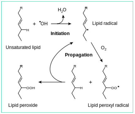
radical
unsaturated (more DB) FA ↑ prone to oxidation (BAD)
lipids prone to oxidation
vit E helps prevent oxidization of FA

free radical oxidation
vit E donates e- to protect cell membranes
reduces free radicals that dmg FA in phospholipid bilayers
free radials have unpaired e- → unstable
dmg (oxidize) FA in phospho. bilayer
vit E reduce free rad → stable → no dmg

vitamin E pathway
vit E 1st line of defense
vit E reduce free radical → stable
vit C reduce oxidized vit E
glutathione peroxidases reduce vit C
all so vit E can keep reducing phospholipid bilayer
glutathione peroxidase
superoxide dismutase
GSSG – glutathione
GSH – reduced glutathione

vitamin E & selenium
selenium spares vit E & delays onset of deficiency symptoms
vit E protects against / delays onset of several forms of selenium deficiency symptoms
↑ dietary PUFA ↑ vit E requirements
↑ PUFA → ↑ C=C bonds attacked by OH radical
vit E & selenium protects against FA tissue peroxidation
vit E 1st line of defense in cellular & subcellular membranes
selenium 2nd line of defense as part of glutathione peroxidase
vitamin E sources
wheat germ (most concentrated)
almonds
olive & sunflower oils (𝛼-tocopherol)
soybean & corn oils: (gama γ - tocopherol
gras lipid fraction
synthetic forms: all-rac-𝛼-TOH
DL mixture (rac - racemic)
½ potency of natural d-𝛼-TOH
natural vit E

vitamin E absorption, transport, storage
high affinity receptor for LDL demonstrated to function as mechanism of delivery of vit E → cells

vitamin E roles
coronary heart disease
vit E inhibits platelet aggregation → reduce risk of CHD
disease resistance
vit E & selenium (glutathione peroxidase) protect leukocytes & macrophages during phagocytosis of bacteria
chicks: large doses of vit E protect against E. coli infections
pigs: vit E ↑ resistance to dysentery
calves: 125 IU of vit E daily maximizes their immune response
vitamin E requirements
humans - fairly low
animals
1 mg vit E per 1 g of polyunsaturated FA in diet
adding essential FA ↑ need for vit E supplementation
vit E requirements ↑ if PUFA are ↑ & vice versa
vitamin deficiency symptoms
stiff lamb disease - nutritional myopathy in sheep & cattle (antioxidant role)
cardiac disease in young
encephalomalacia (crazy chick disease)
affects 204 wk-old chicks, severe muscle incoordination, vascular lesions in brain
RBC fragility - prone to hemolysis
erythrocyte destruction
testicular degeneration in most male animals
impaired fetal development → intrauterine death
encephalomalacia
softening of the brain
stiff lamb disease
most common vit E deficiency symptom
nutritional muscular dystrophy
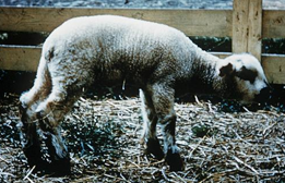
nutritional muscular dystrophy
disease caused by deficiency of selenium & vit E in dietary intake
soils that contain ↓ lvl selenium produce forages & grains are deficient in selenium

vitamin K
fat soluble vitamin
stable to heat, but oxidized by light & UV radiation
name derived “Koagulation factor” Danish

vitamin K forms
phylloquinone
menaquinone
menadione
phylloquinone
vitamin K1
synthesized by plants

menaquinone
vitamin K2
synthesized by gut/rumen
microbes (bacteria-protozoa)
soluble by cutting it off

menadione
vitamin K3
synthetic form
no hydrophobic side chain
relatively water soluble & ↑ efficiently absorbed on ↓ diets
body can’t store it → need ↑ of it
make ↑ water soluble to be ↑ easily absorbed
fat soluble is going to just be stored & long process to absorb it

vitamin K sources
rumen bacteria
↓ gut bacteria
alfalfa
kale
cauliflower
green tea
beef lier
green, leafy veggies
vitamin K absorption, transport, storage

vit K blood clotting

vit K blood clotting

blood clotting pathway
carboxylation of glutamyl residues in prothrombin allows binding Ca+ for further conversion → thrombin
vit K is cofactor for carboxylase to add CO2 on 1 AA on 1 protein (glutamic acid)
(γ) gama-carboxyglutamate
vit K → prothrombin → thrombin
vit K activates prothrombin
antagonists
anticoagulants
inhibitors of blood clotting system
vitamin K deficiency
poor blood clotting & anemia
hemorrhagic disease of newborn
uncommon in humans and animals
antagonists
warfarin
dicoumarol
block Vit K-dep carboxylase reaction
story line: warfarin
Warfarin
used to prevent & treat deep-vein thrombosis & pulmonary embolism to prevent stroke in patients who have atrial fibrillation, valvular heart disease, or a prosthetic heart valve
antagonism
1920s
Sweet Clover Disease (moldy clover)
Death by internal bleeding
1940
Active chemical isolated from blood
Coumarin → Dicoumarol (mold)
Patent to WI Alumni Research Foundation
1945
Wisconsin Alumni Research Foundation (WARF) funded development of rat poison
Warfarin (coumarin derivative)
1950’s
Developed to prevent thrombosis
Given to Dwight D. Eisenhower, 1955
Dose affected by vitamin K levels