Microbio
 Call Kai
 Learn
Learn Practice Test
 Spaced Repetition
 Match
Match Flashcards
 Knowt Play
1/193

There's no tags or description
Looks like no tags are added yet.
Name | Mastery | Learn | Test | Matching | Spaced | Call with Kai | Chat |
|---|
No analytics yet
Send a link to your students to track their progress
194 Terms
What is a microbe
A living organism you need a microscope to see (the general definition)— microbial cells range from millimeters (rare) to 0.2 micrometers (typical in bacteria and fungi)
viruses can be 10 times smaller—
some microbes consist of a single cell(such as bacteria)
Fungi are multicellular— why there microbes: because there is a microscopic form/aspect to their life cycle as they form spores which is how they reproduce karyogamy etc
every microbe has the capacity in its genome (genome is its genetic blue print) to reproduce its own kind—definition of microbe
Types of microbes
Various types: Cyanobacteria are microbes and can make oxygen: are 20 microns
2nd image is bacteria trying to get into stomata
3rd is eukaryotic protist: they are bigger than other
D is a bacteria
E is a mutualistic relationship: its a lichen is a fungus and symbiote
F is a virus and its very tiny compared to bacteria: can be around 50 nanometers

What is a microbe
The simple definition of a microbe leaves us with contradictions
Examples of microbes
Supersize microbial cells: Thiomargarita namibiensis: A giant sulfur eating bacterium can grow up to a millimeter wide and can be seen with the naked eye—breaks the rule that we need a microscope to see it size of fruitfly head
Microbial communities: Biofilms: Microbes living in complex coordinated communities like dental plaque or slime on a rock acting together—
bacteria are single celled microorganisms: they reproduce but they can also communicate and build communities called biofilms
can be single or multispecies biofilms and are a community of organisms collaborating together through division of labor:
produce protect matrix—some involved in metabolism providing food for biofilm: so every microbe or bacteria can participate in different aspects—form a division of labor this is called biofilm
Viruses: Mimivirus: Massive viruses rivaling small bacteria—however they can’t reproduce on their own without hijacking a host cell—infect amoebas (single-celled organism that can change its shape by stretching parts of its body outward)
Microbes include members of the three domains of life
Basically microbes are found across all three domains of life:
Bacteria and Archaea are prokaryotes with are simpler cells with no nucleus
Eukarya: Organisms with complex cells with a nucleus includes microbes like protists and microscopic fungi like yeasts along with algae
bacteria use to be grouped with archaea than we found out they’re different through a common ancestor becoming their own domain of life are still prokaryotes (archaea)

Microbial genomes are sequenced
A genome is the total genetic information in an organism’s chromosomal DNA (The DNA that does this is inside the chromosome but DNA is found everywhere)
(Basically a genome is a set of instructions or DNA with all the genetic information needing for the microbe to rebuild itself function survive and reproduce)
First method of DNA sequencing was made by Fred Sanger—1980 nobel prize in chemistry winner
in 1995 scientists completed first genome sequence of a cellular microbe the bacterium called Haemophilus influenzae
Metagenome: A collection of sequences taken directly from the environment
its very quick to get bacteria genome—back than you had to plate them and separate and than plot genome—hard to do not every bacteria is growable—captures everythinig there that might not be culturable
Microbes shape human history
Microbes affect food availability: They destroy crops but preserve food
Microbes preserve and create: Bread, Wine, Cheese, Chocolate
Microbial disease also changes history
Bubonic plague, smallpox in Americas, Irish Potato Famine (caused mass migration to new world to find place were they didn’t need potatoes), Covid-19
Microbial Disease Devastates Human Populations
More soldiers have died in microbial infections than of wounds in battle
British nurse Florence Nightingale discovered how significant disease was in warfare she lived from (1820 to 1910)
She founded the science of medical statistics which shows the deaths of soldiers due to various causes- she made the “polar area chart”—found more soldiers died from bacteria than from battle
Polar area chart is a circular statistical graphic looks like a pie chart but the radius changes based off the size of the data—used in Crimean war to prove infectious disease kill soldiers
Image attached is polar area chart—heres people we thought died from bullet wound but this chunk actually died from the bacteria

Discovery of Microbes
A light microscope was invented in the 1600s and the quality kept improving ( it used visible light and glass lenses to magnify things)
in the mid 1600s Robert Hooke made the first compound microscope and observed small eukaryotes
in 1676 Van Leeunwenhoek was the first to observe single cell organisms and discovered bacteria
Do microbes have parents—spontaneous generation
spontaneous generation: The theory that living creatures could arise without parents
Francesco Redi in the 1660s showed that maggots in decaying meat were the offspring of flies—disproved that maggots can just appear out of nowhere through a spontaneous generation
Lazzaro Spallanzani in the 1760s showed that a sealed flask of meat broth sterilized by boiling failed to grow microbes—Showed that microbial life can’t just spontaneously generate and needs a good environment to grow
if you seal flask after boiling microbes to death, no more will get in or spawn out of no where
Louis Pasteur in the 1860s
He discovered the microbial basis of fermentation
He devised swan-neck flasks
Showed that after boiling the contents remain free of microbial growth despite access to air
the father of modern day microbiology: showed that just because theirs oxygen you won’t get microbes: got around criticism there’s no oxygen
s curve excludes dust and microbes but if you turn it microbes can enter and reproduce so its not about the oxygen but microbes reproducing

Germ theory of Disease by Pasteur continued
Pasteur’s theory was that transmission of germs causes disease
What all scientific Theories do
Explain many known observations: For example transmission of rabies
Provide framework for understanding: Where do diseases come from
Can be tested further: Do germs cause anthrax
A scientific theory is NOT a guess its based on facts
we didn’t know why we got sick at first so this helps us understand and break the transmission cycle
Medical Microbiology
Germ theory of disease: Many diseases are caused by microbes
Robert Koch who is a German physician is the founder of the scientific method of microbiology
He applied his methods to numerous lethal diseases around the world
one of the founders of scientific method so we use his method to classify diseases
growth of microbes in pure culture
Working with Anthrax, Koch demonstrated an important principle of epidemiology: The chain of infection or transmission of a disease
To prove a particular bacterium caused a specific disease, pure culture of microorganisms were needed—we needed a matrix to spread and grow different microbes but we needed a way to get broth into it to grow them
So basically scientists figured out how to grow microbes by themselves in lab as pure cultures
People who figured out how to get that broth and oxygen in to grow microbes
Angelina and Walther Hesse: Figured how to use agar to make a solid growth medium instead of just a liquid broth- helps separate different types of microbes
found out awther is best to use to grow microbes
Julius Petri: Invented the double dish container: Called the petri dish
basically we needed to learn how to grow microbes ourself
Koch’s postulates
Criteria for establishing a causative link between an infectious agent and a disease
1.Microbe is always present in a diseased host and absent in a healthy host
The microbe is grown in a pure culture and No other microbe is present
Introduce pure microbe into healthy host: Individual becomes sick
Same microbe reisolated from now sick individual (basically catch it again from sick host)
koch’s postulates defining the causative agent of a disease
This is an example of Koch’s postulate
First the healthy rabbit has no microbes for the disease but found in the sick rabbit that has the disease
Scientists take microbe from sick rabbit grow it by itself in petri dish
They inject the pure microbe into a healthy rabbit and it gets sick with the exact same disease
They take a sample from the newly sick rabbit and isolate the same exact strain of microbe from it—which shows the microbe caused the illness

Immunization prevents Disease
In the eighteenth century, smallpox infected a large fraction of the European population
Mary montagu introduced smallpox inoculation in Europe 1717
Edward Jenner than infected patients on purpose with matter from cowpox lesions— practice of cowpox inoculations was called vaccination (Vacca meant cow)
inoculation and vaccinations are almost the same: Innocculation is introducing infectious material to a healthy person to make them immune
vaccination is the type of inoculation made by edward jenner using cowpox to protect people against small pox
Small pox vaccination
Different people got vaccinated

Immunization prevents Disease
Louis Pasteur developed the first vaccines based on attenuated (weakened) strains
Fowl cholera and rabies were used
Immunization: It is the stimulation of an immune response by deliberate inoculation with an attenuated pathogen
Antiseptics
In 1847 Ignaz Semmelweis ordered doctors to wash their hands with chlorine which is an antiseptic agent: This caused mortality rates to fall
In 1865 Joseph Lister developed carbolic acid to treat wounds and clean surgical instruments
In the twentieth century, aseptic surgery was developed
Environments completely microbe-free
Antibiotics
Alexander Fleming discovered that penicillium mold generated a substance that kills bacteria in 1929
In 1941 Howard Florey and Ernst Chain purified Penicillin: The first commercial antibiotic to save human lives
The discovery of viruses
In 1892 Dmitri Ivanovsky studied the tobacco mosaic disease
The agent of transmission could pass through a porcelain filter that blocked all know microbes
Martinus Beijernick: The agent of tobacco mosaic disease is not a bacterium, it passes through a filter that retains bacteria
The filterable agent was purified and crystallized by Wendell Stanley (the scientist):
This proved it was Tobacco mosaic virus (TMV)
Tobacco mosaic virus
Capsid proteins are protein building blocks that wrap around and protect a virus’s genetic material

Microbial ecology
Microbes cycle the many minerals essential for all life, including the global N2 and much of the O2 of the world
less than 0.1% of microbial species can be cultured in the lab
The remainder make up the majority of Earth’s entire biosphere
Only the outer skin of Earth supports complex multicellular life
Microbes Support Natural Ecosystems
Sergei Winogradsky (1856–1953) A Russian scientist who was among the first to study microbes in natural habitats
Discovered lithotrophs - organisms that eat inorganic materials like rocks instead of organic food
Developed enrichment cultures - grow microbes by giving nutrients
Built the Winogradsky column - Sealed glass column naturally seperating and growing layers of different microbes with different metabolisms based on Light, oxygen and nutrients.
A wetland model ecosystem containing regions of enrichment for microbes of diverse metabolism

The winogradsky coloum
Cyanobacteria at the type, than purple,green sulfur bacteria. Sulfate reducing bacteria at bottom.

Geochemical cycling
This is showing the global nitrogen cycle
The problem is N2 makes up most air but plants and animals can’t use it unless converted to NH4+ and NO3- by bacteria
Winogradsky discovered bacteria drive geochemical cycling
Nitrogen fixation: Soil bacteria and bacterial symbionts living on plant roots (like legumes) convert atmospheric nitrogen gas into ammonium NH4+
Nitrification & Transformation: Other groups of bacteria convert ammonium into nitrite NO2- and then into nitrate NO3-
A way to remember: Nitrification is when you add oxygen to the mix
Plants absorb these nitrogen compounds from soil to grow and animals eat the plants driving the food web
Industrial fixation: Humans manufacture commercial fertilizers to add reactive nitrogen into the soil.
Bacterial Endosymbiosis with Plants and Animals
Endosymbionts are microbes livings symbiotically inside a larger organism
Rhizobia are enodymbiotic bacteria that induce roots of legumes (like beans or peas) to form special nodules to facilitate bacterial nitrogen fixation
(Nitrogen fixation happens inside nodules) and plant provides nutrients to bacteria
Endosymbiotic microbes also make essential nutritional contributions to host animals that host can’t produce on own
Colonic bacteria such as Escherichia coli and Bacteroides species grow as biofilm and help digest food synthesize vitamins and protect against harmful pathogens
Endosymbiont Theory-Lynn Margulis
Explored how Eukaryotes arose
Their DNA is similar to archaea’s
Mitochondrial, chloroplast DNA: Similar to bacterial DNA
Led to endosymbiont theory
Mitochondria use to be the bacteria
Chloroplasts were the cyanobacteria
they were infected or eaten by other species
ended up living together inside through endosymbiosis
Eukaryotes Evolved through Endosymbiosis
The five kingdom system was modified dramatically by Lynn Margulis
She said eukaryotic organelles such as mitochondria and chloroplasts:
evolved through endosymbiosis from
prokaryotic cells that were engulfed by pre-eukaryotes
so basically the prokaryotic cells got eaten by pre eukaryotes leading to eukaryotic organelles evolving
Highly controversial endosymbiosis theory
because it implied polyphyletic ancestry of living species and not the long held assumption that:
species evolve only by divergence from a common ancestor (monophyletic ancestry)
The microbial Family tree
Archaea are not bacteria
They have a similar size and shape but very different biochemistry
For example:
Different membranes
Archaeal ribosomes are similar to eukaryotic ribosomes: More similar to eukaryotic than bacterial ribosomes
Many archaea live in harsh environments
16s rRNA gene sequence
is found in all creatures including Archaea
Archaea are a separate domain: Uses 16S rRNA gene as molecular clock to map out microbial family trees:
Shows archaea are separate domain of life
Archaea Differ from Bacteria and Eukaryotes
Carl Woese was studying recently discovered prokaryotes that:
- Live in hot springs
- Produce methane
Analysis of the 16S rRNA revealed that these prokaryotes were a distinct form of life - He called them archaea
Cell Biology Techniques
Electron microscopy: The observations of cell composition you see eukaryotic organelles and membrane
Ultracentrifuge: Separation of cell components
Study of biochemistry of organelles
n Fluorescence microscopy: Identification of cell components and Subcellular location of individual proteins
Genetics and DNA Revolution
Molecular genetics depends on bacteria especially E.Coli
Concept of “ gene ” proposed for bacteria
Rosalind Franklin- discovered the double helix along with James Watson, Francis Crick, Maurice Wilkins
Genetic code, Transcription, translation: Show us how how life works at the molecular level
Tools used for genetic Engineering
Restriction enzymes: Molecular scissors used to cut DNA at specific sequences
Recombinant DNA: Combining DNA from different sources
Cloning: Making identical copies of genes or organisms.
PCR reaction: A technique to rapidly copy and amplify DNA segments.
E. coli has best understood genome
n Molecular genetics depends on bacteria ¨ Concept of “ gene ” proposed for bacteria
¨ DNA structure- Rosalind Franklin- discovered the double helix; James Watson, Francis Crick, Maurice Wilkins
¨ Genetic code
¨ Transcription, translation
¨ Restriction enzymes
¨ Recombinant DNA ¨ Cloning ¨ PCR reaction ¨ E. coli has best understood genome
Start of lectures 3 and 4
3 and 4
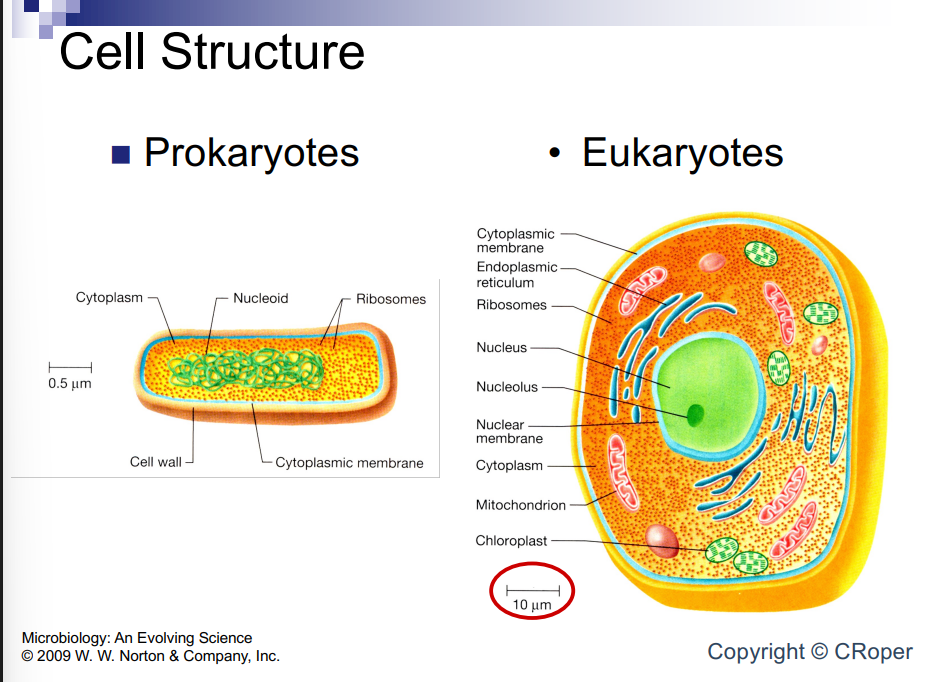
Structural differences between Prokaryotes and Eukaryotes
Prokaryotes have no defined organelles
¨DNA is not bounded by a membrane
Prokaryotes are generally much smaller in size
¨Equivalent to a mitochondrion in eukaryotes
Eukaryotes are the biggest and have a nucleus, than prokaryotic cell and virus is the smallest
Eukaryotes have order and function to each part
DNA not bounded by nuclear membrane in prokaryote like the eukaryotic has

Cell structure
In eukaryote there is membrane around chromosomes
Prokaryotes don’t have this
Ribosomes are out in the cytoplasm in prokaryote and have dna genes coding for messenger RNA allowing transcription and translation to change very fast
Know prokaryote organelles have no membrane bound organelles, condensed form nucleoid and parts. Condensed because theres a single circular chromosome, DNA is bunched up and tightly packed
Prokaryote Size
Prokaryote Sizes Microbiology:
Small size of Prokaryotes affects biological properties
Large surface area/volume ratio means more rapid transport rates
¨ Excretion of waste products
¨ Import of nutrient
Bacteria like cyanobacteria are big and live in ocean and are photosynthetic
large surface area means large transport rate

Bacteria Cell Shapes
Many have rod shape (Bacilli) so you can’t identify them just from how they look
can come in
Cocci = spheres – in bunches
Bacilli = sticks, rods – alone
Vibrios = bent rods
Spiral
Irregular
in chains

Morphogenesis: change in shape
Monomorphic: one shape; observed in most pure cultures of bacteria
• Pleomorphic: multiple shapes
- Changes during growth
Cells become smaller as they age -
Response to environmental cues
Sporulation (nutrient limitation)
Sometimes they can change shape if they are under nutrient or environment stress
The Bacterial Cell
Cytoplasm surrounded by cell envelope
¨Cytoplasm contains DNA in the nucleoid- a system of looped coils of DNA
heres the chromosome and most bacterial genomes are circular
surrounded to cell envelope which are specific to bacteria as only bacteria have cell envelopes which helps with making antibiotics targeting this
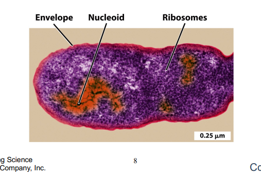
The Bacterial Nucleoid
Single loop of double-stranded DNA- most bacteria have a circular chromosome made of one continuous double-stranded DNA molecule
¨ Single molecule of DNA ¨
~4x10^6 bp (base pairs) in most bacteria comparadly humans only have 3 million so they have far smaller and more compact genomes
¨ Compacted via supercoiling (instead of using histone proteins bacteria twist and coil the DNA tightly on itself in a compact shape)
Attached to cell envelope
¨ No membrane separates DNA from cytoplasm allowing transcription and translation to happen simultaneously
Replicates once for each cell division: Both cells get a complete clone copy of each other
Dna origin (oriC) is the start of DNA replication and guides cell division during binary fission
the two origins move toward opposite sides of the cell. helping organize the cell so that when it splits via binary fission, each daughter cell gets one complete chromosome.
• Cell envelope-
Very important to bacteria and target for antibiotics
envelope is made of different layers
we have cell membrane aka inner membrane, than cell wall which is rigid and forms its structure
Gram-Negative: Has a three-layer setup: Inner membrane than Cell wall than Outer membrane.
Gram-Positive (Not Gram-Negative): Has a two-layer setup: Inner membrane Cell wall (with no outer membrane)
if not gram negative same but no outer membrane
Comprised of the cell membrane + the cell wall + the outer membrane (if there is one)
• Keep in mind the cell envelope is different between Gram+ and Gram- bacteria
Helps us identify and classify bacteria in clinical setting when thinking of clinical solutions to infection

Cell Membranes
It is a phospholipid bilayer
it seperates cytoplasm from outer word and controls what goes into cell
and collection of membranes dictates what goes in and out through different transporters
dictated by the type of proteins that bacteria have
they can help anchor membrane to envelope and sense what’s going on
and can be transporters and channels for things like glucose to come into and waste to go out of cell
ATPase makes ATP for the cell
Made of lipid bilayer
¨ Double layer of phospholipids: two layers of fat molecules (phospholipids) arranged with their hydrophilic (water-loving) heads facing outward and hydrophobic (water-fearing) tails facing inward
Separate cytoplasm from outside world
Permeability barrier
Proteins embedded in membrane
¨ Anchor membranes to envelope They help link the membrane to other layers of the cell envelope.
¨ Sense the outside world detect changes in the external environment (like nutrients, toxins, or temperature).
¨ Transport materials into cell
Phospholipid Bilayer
Hyddrophobic inside and hydrophilic outside and it has fatty acids and glycerol
Forms general structure of biological membranes
Composed of fatty acids (hydrophobic) and glycerolphosphate (hydrophilic

Cell Transport
Alot of transport the cell allows like oxygen and water that can passively move in and out through transporters
Small or uncharged molecules (like oxygen and water) can slip right through the lipid bilayer or use unassisted channels to move along their concentration gradient (from high concentration to low concentration)
passive transport doesn’t cost ATP
some dictated by type of transporter/protein some specific to type of sugar
alot are active need ATP energy or through proton motor force and is cheap energy from electron transport chain: When the cell needs to pull in nutrients when there is already a high concentration of them inside, it has to force them against their gradient (from low to high concentration)
active transport needs energy either from ATP or PMF
Bacteria stores them in cell wall and they are a good source of energy to drive motor force of ATPsynthase
Transporters pass material into/out of cell
¨Passive transport follows gradient of material
¨Active transport of some sugars
PMF drives ATP synthase to make ATP
Pumps use energy
¨ATP or PMF ¨Move material against their gradient
Cell Transport. cont
Atp snthase is collection of proteins in cell to make atp
inexpensive biproduct of electron transport chain with protons to drive ATP chain and they have energetic potential turning motor of ATP synthase to make ATP
ATP made through ATP motor force and synthase with biproduct of glycolysis and electron transport chain
Proton pumps push protons out of cell
¨Generates proton-motive force (PMF)
Osmotic force tries to push protons back into cell
electrical force tries to push protons back into cell
PMF is used to create ATP ¨ATP synthase uses PMF for energy
Bacterial Cell Walls
Rigid structural component that lies just outside the cytoplasmic membrane Enables bacteria to withstand turgor pressure due to dissolved solutes in cell
2 major types: 1)Gram positive (thick) and 2)Gram negative (thin)
The cell wall is the rigid layer on top of the membrane and helps dictate the shape and the size of bacterial cell and is specific to bacteria so others don’t have it
can withstand terminal pressure of too much salt or water coming in and maintaining osmotic pressure, etc
gram positive or negative named after scientist gram: gram negative have thin cell wall gram positive have thick cell wall made of peptidoglycan
Peptidoglycan- polymer that makes up the bacterial cell wall
Peptidoglycan synthesis is unique to bacteria
Makes up a large % of the cell mass (≈ 1%)
Often a target for antibiotics (penicillin)
Peptidoglycan makes up the cell wall and is easily accessed and is a target for antibiotics
the process of building cell wall can be interrupted by penicillin which is a cell wall inhibitor which stops cell wall from being made which kills the bacteria
The Cell Wall Is a Single Molecule
The cell wall confers shape and rigidity to the cell, and helps it withstand turgor pressure (internal water pressure trying to make cell swell and explode, rigid cell wall stops cell from bursting).
The bacterial cell wall, or the sacculus, consists of a single interlinked molecule.The cell wall is made of a repeating polymer called peptidoglycan (sugar chains cross-linked by short amino acid peptides)
You can extract a cell wall or sacculus also called and image it with electron microscope and image is sacculus
repeat polymer of peptido glycan and is a hotdog shaped cell single linked molecule and interrupting this can cause cell to lice

The Bacterial Cell Wall
Sacculus made of peptidoglycan (murein)
¨Sugar chains wrapped in circles around cell
“glyco” = “sweet” The wall is built from long repeating chains of two sugar units (specifically, N-acetylglucosamine [NAG] and N-acetylmuramic acid [NAM])
¨Sugar chains linked to each other by short polymers of amino acids
amino acid = “peptide”
peptido glycan is peptides and sugars linked together to make this long chain becoming peptidoglycan
so the wall is called peptidoglycan and its made of NAG and NAM
Monomeric units of peptidoglycan (murein) in bacteria
Peptido = peptide links
Four amino acids are linked together by peptide bonds.
Glycan (Sugar component) = NAM+ NAG disaccharide
NAG: N-Acetylglucosamine
NAM: N-Acetylmuramic acid
Monomers are made of NAG and NAM and have peptides that are tiny proteins and are amino acids linked to each other linked to sugar chain
a monomer is a single, small molecule that can chemically bond with other similar molecules to form a larger chain, which is called a polymer
Differences between Gram negative and Gram positive peptidoglycan crosslinks
Direct cross-linking
Thin layer Gram -
Peptide interbridge
Thick layer Gram +
in gram negatives We have sugar backbone and peptides and in gram negative layer is thin and linked to one another and is very close to each other giving thin layer of peptidoglycan
with gram positives we have sugarback bone and peptides but instead of being linked theres a peptide interbridge linking peptides creating thicker peptidoglycan layer

The Gram-Positive Envelope
Capsule (not all species): Called a Polysaccharide: A slippery, protective outer layer made of sugars (polysaccharides) found in some bacteria: helps them hide from immune system
S Layer (not all species): ¨Made of protein: Rigid armor like for structural support
Thick cell wall in gram positive: gives the cell high structural strength
¨9-amino acid crosslinks in peptidoglycan:Gram-positive cell walls use specific peptide cross-links (such as 9-amino acid bridges): use ¨Teichoic acids for strength in gram positive
Thin periplasm in gram positive (space between the plasma membrane and the cell wall)
Thick blue cell wall and there is a space between cell membrane and outer membrane which is were all those protons are stored in periplasm
sometimes they have capsules on outside with sugar
2 layers cell wall and membrane

Gram Negative Cell Envelope
A thin layer of peptidoglycan
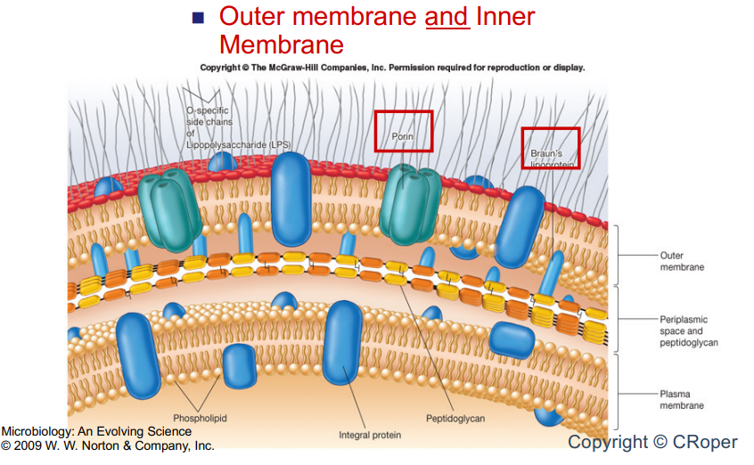
Outer membrane and Inner Membrane
The Gram-Negative Envelope
Capsule (not all species) ¨Polysaccharide
Outer Membrane ¨Lipopolysaccharide: In outer leaflet only: acts like endotoxin triggering strong immune responses
Thin cell wall : 4-amino acid crosslinks in peptidoglycan
Thick periplasm
Plasma membrane
inner and outermembrane and thin cell wall and can have extra sugar decorations on outside helping build biofilms and causing disease and virolence and bacteria interactions
lipopolysaccharides which is unique to gram negatives
The difference
Link makes it thicker
in gram negative you have thin peptidoglycan layer

Lipopolysaccharide
Outer membrane made of lipids and sugar (Lipopolysaccharides)
sugar part on outside and no other organism makes it
our eukaryotic systems have evolved to think that’s foreign triggering immune response

The Gram Stain
Developed by Hans Christian Gram in 1884
The most widely-used stain in microbiology
Differential Stain: Does not stain all cells equally, Based on differences in composition of cell wall
Gram Positive: blue Gram Negative: pink
We have rod shaped bacteria from patient sample and this will decide what type of antibiotics to give
use differential stain it doesn’t stain equally based on differences of chemistry of peptidoglycan thin vs thick
it stains to gram positive cells but nothing happens to gram negative until later on in the reaction
How to stain
Get pure culture and grow it make sure its nice and not dead
1.Put some in microscope slide to see and add methanol to stick cells to surface and air dry and here they are all stuck (why cells don’t die from this: its such a short time that ethanol doesnt kill them )
2.than stain them with crystal violet stain which is super blue and pipet the stain and let it sit for one minute and is time specific now everything is blue
3.Add iodine which will stain At this point, both Gram-positive and Gram-negative cells still look purple.
than wash with ethanol for 20 seconds and stains are removed from gram negative cells gram positive stay dark purple. colorless cells are practically invisible under a standard light microscope so add safranin to tell
gram positive cells remain dark purple so add safranin and counterstain to be able to tell the difference of what’s positive and negative. positive stay purple and negative turn pink or red.

Outer Coverings
Glycocalyx: network of polysaccharides extending from cell surface, ¨ survival, attachment, virulence
Capsule: well organized glycocalyx; difficult to remove
S-layers – Gram positive bacteria ¨ layers of protein or glycoprotein ¨ Common among Archaea: basically protein armor: S-layer is a rigid, crystalline sheet made entirely of interlocking proteins or glycoproteins
Glycocalyx are sugars coating the bacteria and is sticky helping them stick to surfaces and survive
can be called glycocalyx, capsule, and gram positive bacteria have S-layers
all of these are coverings of the cell
not all have this and others can produce/not produce as it takes a lot of energy to make this layer
remember some have outer slimy coverings on outside
The Bacterial Nucleoid
Single loop of double-stranded DNA, Single molecule of DNA ¨~4x10⁶ bp in many bacteria ¨Compacted via supercoiling
Attached to cell envelope ¨No membrane separates DNA from cytoplasm
Replicates once for each cell division
No nuclear membrane and is a condensed and organized
is bidirectional DNA replicaqtion 2 circles orginating from one
bp= base pairs
Nucleoid organization
Lice cell you disrupt DNA and a bunch of it spills out
The nucleoid forms about 50 loops of chromosome called domains
Within each domain, the DNA is supercoiled and partly compacted by DNA-binding proteins

Gene Expression
RNA Polymerase transcribes DNA to mRNA
Ribosome translates RNA to Protein
¨Processes occur simultaneously
Enabled by polymerase transcribing it into mRNA
MRNA transcribed from DNA
can happen simultaneously transcription and translations so are coupled and ribosomes can hop onto that RNA before its even transcribed
From one you can get many

RNA polymerase
In bacteria, making mRNA (transcription) and building protein (translation) happen at the same time in the same
Important: RNA Polymerase (Light Blue): Reads the DNA template and builds a growing single strand of RNA (Nascent RNA
Secondary Channel (ATP, GTP, CTP, UTP): Allows free ribonucleotides (the building blocks of RNA) to enter the enzyme to build the strand
Ribosome (Pink / Red): While the RNA strand is still being made by RNA polymerase, a ribosome latches onto it and immediately starts reading it to make a protein
30S & 50S Subunits: The two bacterial ribosomal subunits that form the functional ribosome
Transfer RNA (tRNA): Delivers incoming amino acids corresponding to the genetic code on the mRNA
Peptide Chain & Exit Channel: As amino acids link together, the growing polypeptide chain feeds out through the exit channel
Correctly Folded Protein: Once completed, the chain folds into a functional 3D protein

Lecture 4
Start
Cell Division
Cell elongates as it grows ¨ Adds new wall at cell equator
DNA replicates to make 2 chromosomes ¨ DNA replicates bidirectionally ¨ Can begin next replication before cell divides
Cell undergoes septation ¨ Usually at equator ¨ Each daughter has same shape
Binary fision turns one cell into 2 identical cells
during that time dna is tethered to cell envelope
this is were dna replication begins and his hooked by the inner cell wall (cell envelope)
theres a collection of proteins 2 of them starting dna replication (ori site )
ropel zones go around and hit termination side: cell grows and expands as origins of replication detatch slightly
each of 2 circles go into daughter cells
bacteria does this quickly and transcript and transcribe at same time
sometimes next round can start before even completing first cycle
in prokaryotes replication begins at ori sight and Enzymes pull the double-stranded DNA apart, creating two "replication forks"
each strand in parental dna used as template to build new matching strain
replisome perform replication function: (contains multiple copies of DNA polymerase (the enzyme that builds the new DNA) along with other helper enzymes, copying both directions at the same time.)
each contains 2 dna polymerase, enzymes etc
2 copies of dna origin replication attatch to envelope and move along expanding cell
2 progency helices get made
replication happens on 2 ends until the termination site: They keep copying until they meet at the exact opposite side of the circle, known as the termination site.
this causes septum to grow inwards to sides of cell constricting and sealing off called septation
inward growth eventually meets in the middle, sealing off the two halves completely. leading to 2 daughter cells
Bacterial Cytoskeleton
Shape-determining proteins
- FtsZ = forms a “Z-ring” in spherical cells
- MreB = forms a coil inside rod-shaped cells
- CreS “crescentin” = forms a polymer along the inner side of crescent-shaped bacteria
actin contributes to cytoskeleton
recent discoveries show they do have cytoskeletons improtant to cell processes
different proteins determine shape
Ftsz forms one ring along center of cell and makes spherical cells so whole cell is spherice
MReB forms a coil inside rod shaped cells and runs along the length of cell and is rodshaped so MreB and FtsZ combined make a rod shaped
CreS crescentin makes a crescent shape and is only on one side of cell causing bend so they have ftsz mreb and crescentin present here

Specialized Structures-Cell Attachment
Fimbriae and pili attach cells to surfaces ¨ Thin filaments of protein “pilin” that extend from cell surface
¨ Conjugation – sex pili ¨ Essential for bacterial pathogenicity
¨ Receptors for viruses
• Stalks attach cells to surfaces – Extension of cell cytoplasm
jewlery of what bacteria were
has to do with attachment helping bacteria to attach to cells
most bacteria attatch to some bacteria in their life cycle
fimbriae are hairlike structures on ends of cell
fimbriae are shorter and are sticky and made of pillin
specialized pili going beyond attatchement and transfer of dna between bacteria is conjugation which is their sex pili
Dna is transfered from one to another mediating dna transfer-horizontal dna transfer
how bacteria exchange genes very quickly can go from brother to brother etc and not just from mother to son
bacteria can do that when ever they want and can pick up genes from environment or from other bacteria which could be related to antibiotics etc gives them fitness edge
this all happens through sex pili
specialized structure are essential for bacterial pathogenicity
stalk enables bacterium to attatch to surfaces with strong hydrodynamic flow such as rivers and can attatch to a rock for example-strongest discovered moleculare glu
Flagellar Ultrastructure:
3 main components:
-Filament: flagellin
-Hook: hook protein
-Basal Body: consists of rings
short things enable attatchment and they use flagellin to swim to were they want to be
prokaryotes have flagellin but different structure than eukaryotes
flagellum is made of flagellin and all this is important in this
the hook protein helps with torque of system
basal body has rings that turn inside and rotate flagellum filament
all powered by proton motor force which is where the bacteria harbor protons in periplasmis space and use them for energy to create atp
or energy to drive motor
inexpensive energy lying around and use it to power flagellum motor
Flagella and Motility
Monotrichous (Polar) Flagella ¨Single flagellum attached to one end n Lophotrichous Flagella ¨A group (tuft) of flagella is attached to one or both ends n Peritrichous Flagella ¨Flagella are arranged on all sides
were are focusing on peritrichous and ecoli have this
Movement of Polar Flagella
n Reversible ¨ CCW rotation leads to forward swimming ¨ CW leads to reversal of direction n Unidirectional ¨ CW leads to forward movement ¨ Cell stops, reorients ¨ CW rotation commences, leading to forward movement
flagella aqueously enable movement
some flagella are reversible in the way they rotate so they can go forward or backwards
if a reversible is going ccw its forward
if clockwise its going backwards
some flagella can only rotate in one direction so clockwise if cell wants to reorient and move backwards it can stop rotating and stop and reorient itself and thats called a biased random walk
polar i unidirectional flagella
can send signal to flagella when it sees food it needs to go to or if theres a toxin and it needs to stop rotating
the flagellum is enabled by sensors coupled to flagella through signal conduction cascade
Movement of Peritrichous Flagella
n Counterclockwise (CCW) rotation of multiple flagella leads to formation of a bundle ¨ Smooth swimming—more or less in a straight line ¨ Swim forward n Clockwise (CW) rotation leads to disaggregation ¨ Tumble—allows reorientation
how ecoli move
lots of flagella are directing body
they form a braid and its bundeld enable ccw rotation
if they need to stop moving the tumble flagella are pushed apart and bundle is disssaggregated and rotation of flaggella is stopped as they are pushed apart
flagella is just floating around until ready to go forward again and than they bundle form
called biased random walk and is based on chemotactic receptors enabling bacteria to sense theres something like a vitamin or amino acid they want
enables them to counterclockwise rotate with bundle
if off course they disbundle and float around but are biased to that attractant
they will remake bundle when they are in position and go forward
so when they disbundle they float until they are in position to go to attractant
Chemotaxis
n Runs + tumbles cause “random walk” ¨Receptors detect attractant concentrations n Sugars, amino acids ¨Attractant concentration increases and prolongs run n Biased random walk n Net movement of bacteria toward attractants
your taxing or moving to a chemical attractant or away chemotaxis receptors enable movement to attractant
Chemotaxis
n The ability of organisms to move towards or away from specific chemicals n Random cell movement in absence of attractant n Attractant causes directed movement up-gradient towards the attractant – prolonged runs
Motility and Chemotaxis
n Movement in response to a chemical gradient n Attractants: Cells move up a gradient of these molecules (nutrients) ¨ Suppress tumbles, so runs become longer n Repellents: Cells move down a gradient of these molecules (wastes or toxic chemicals)
can also run away from something like antibiotic using same bundles and disaggregation tactic
Flagellum motor video notes
biomechanical motor is in flagellur motor powering flagellum in prokaryotes
hydrogen ions/protons in outer layer in high concentration and low protons on inside
energy can be made using this high protons on outside low protons on inside used as gradient by motor to turn itself
protein comes and binds to motor making it turn clockwise to stop
lectures 5 and 6
Bacterial Culture, Growth,
and Developmen
Introduction
The adage “To eat well is to live well” is
as true for microbes as it is for humans.
n Over eons, bacteria have evolved
ingenious strategies to find, acquire, and
metabolize a wide assortment of food
sources.
n - This owes to the remarkable plasticity
of microbial genomes.
Microbiology: An Evolving Science
© 2009 W. W. Norton & Company, Inc. Copyri
they have ability to change genomes very quickly allowing microbes to inhabit every aspect in the planet
Importance of Bacteria to Our Lives
Greatest metabolic diversity of all life forms: major agents in the recycling of C, N, S, Fe, and other elements in the environment n Provide the genetic source and cellular scaffold for recombinant DNA biotechnology
microbes have the greatest metabolic diversity out of every lifeform
major recycling component in planet for nutrients and plants and animals can use them
Bacteria are scafold for DNA microtechnolgy which means taking gene and making it generate a protein, etc based on micrbiology and biochem we have learned
Microbial Nutrition
Essential nutrients are those that must be supplied from the environment. § Macronutrients - Major elements in cell macromolecules - C, O, H, N, P, S -
Cations necessary for protein function § Micronutrients - Trace elements necessary for enzyme function
Macro nutrients are carbon based and you need a lot of it
micronutrients you need in smaller amounts for nutrients

How Microbes Build Biomass
All of Earth’s life-forms are based on carbon, which they acquire in different ways.
- Autotrophs fix carbon dioxide and assemble into organic molecules (mainly sugars).
- Heterotrophs use preformed organic molecules
If you expose bacteria to alot of copper they will die so you can spray copper on plant itll kill bacteria
all of earths lifeforms are carbon based and get it in different ways. Autotrophs fix carbon from atmosphere that’s not usable and convert it to glucose
bacteria can also use energy from inorganic minirals to drive autotrophy: taking co2 from environment converting it into usable enrgy like sugar
heterotrophs acquire it from premade organic forms of energy
How Microbes Obtain Energy
§ All organisms require an energy source. ̵
Phototrophs obtain energy from chemical reactions triggered by light. ̵ Chemotrophs obtain energy from oxidationreduction reactions. ̵
Lithotrophs use inorganic molecules as a source of electrons.
̵ Organotrophs use organic molecules.
they convert carbon back to energy: photo trophs get energy from sun or light
chemotrophs get it from oxidation reduction reactions in envrionment
lithotrophs use inorganic molecules as source of energy and break down inorganic compounds
Organic trophs use organic molecules as energy
How Microbes Obtain Energy Part 2
§ In short, microbes are classified based on their carbon and energy acquisition as follows: - Autotrophs - Photoautotrophs - Chemolithoautotrophs (or lithotrophs) - Heterotrophs - Photoheterotrophs - Chemoheterotrophs (or organotrophs)
classified by how they get carbon and their energy:
autrophs can be photoautotrophs or chemolithoauto trophs
lithotrophs take inorganic compounds sas energy and degrade them into energy and use that to fix CO2
Hetertrophs can be photoheterotrophs or chemoheterotrophs: Can also have sunlight as energy to help them break stuff down and can also use redux reactions as metabolisms to
these metabolisms allow them to colonize extreme and non extreme environment
The Carbon Cycle
Driven by glucose production becoming polymerase to polysachhrides that heterotrophs break down back into glucose
you need autrophs to take co2 convert to organic and hetrrotrophs break it down and produce co2 as biproduct going back to autotrophs

Energy Is Stored for Later Use
A membrane potential is generated when chemical energy is used to pump protons outside of the cell. § The H+ gradient plus the charge difference form an electrochemical potential called the proton motive force. § The potential energy stored can be used to transport nutrients, drive flagellar rotation, and make ATP by the F1FO ATP synthase
energy can be used for later use and they do these by creating membrane potential that is storage of protons in periplasm stored in gradient creating electrochemical potential called proton motor force
when you have more of something going down its gradient to create energy to convert electrochemical energy through proton motor force
bacteria funnels energy through and make ATP synthase: funnel through damn to create electricity and energy
they store protons and funnel them through protein complexes like ATP synthase produce atp through phosphorisation

Nutrient Uptake
n Passive diffusion ¨Some gases pass freely through membranes n O2, CO2 ¨Follows gradient of material n Forms of Transporters ¨Facilitated diffusion n Transporters pass material into/out of cell n Follows gradient of material n Does not require energy ¨Active Transport n Requires energy
gases and things they need to live can pass through membranes and depends on gradient
Different forms of transporters dictate where proton can live and what membrane can bring in and send out
transporters can be through facilitated diffusion and follow gradient of other compound facilitated diffusion means its faccilated by something but doenst use energy like phosphate
Active transport is higher level and are dedicated transport but need energy
Facilitated diffusion
n Cannot move a molecule against a concentration gradient n Does not use cellular energy, relies on diffusion across a gradient n Example-glycerol transporter
cannot move molecule against a concentration gradient
does not use cellular energy and relies on diffusion
Three Classes of Active Transport Systems
Simple transport: Simplest form driven by protons can

Nutrient Uptake—Simple Active Transport
n Symport and Antiport ¨ Gradient of one molecule transports another ¨ Transports material against its gradient - In symport, the two molecules travel in the same direction. - In antiport, the actively transported molecule moves in the direction opposite to the driving ion. Symport: Gradient of pumps in same direction Antiport: Gradient of pumps in opposite direction
symport 2 molecules go in same direction and antiport mean they go in opposite direction through same transport
source of energy for simple active transport from proton transport chain
Nutrient Uptake— Active Transport
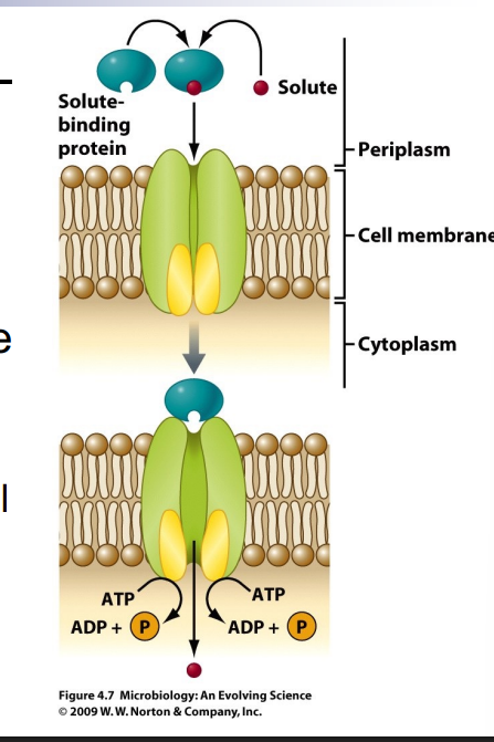
n ABC Transporters n ATP-binding cassette superfamily ¨Use ATP energy to pass material into cell ¨Transport material against gradient
solute binding protein binds to solute binds to transporter causing change using ATP energy transport things against gradient requires a lot of energy
Nutrient Uptake—Active Transport 2
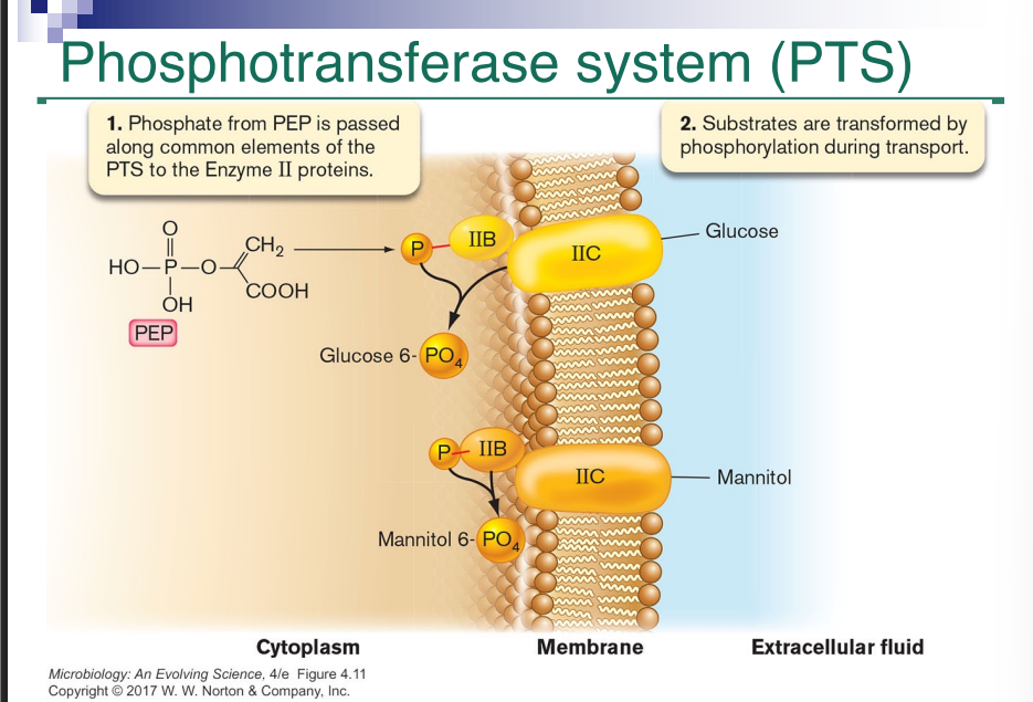
n Phosphotransferase System (PTS) ¨ Uses energy from phosphoenolpyruvate (PEP) to pass material into cell ¨ Modifies material as it enters cell n Gradient is maintained, pushing material into cell glucose enters cell and is phosphorylated. As a result, gradient of pushes more glucose inside. (glucose-6-phosphate) cannot pass out of cell
It capitalizes on fact it can maintain concentration gradient and uses energy
uses phoptransport system it doesn’t use ATP it uses phosphate and phosphoenolpyruvate
glucose becomes glucose 6 phosphate and cannot pass out of cell so inside of cell is different than outer glucose so it can’t leave
Nutrient Uptake—Active Transport 3
PEP transfers through a series of proteins
mannitol has a different enzyme 2 complex they pass phosphate through enzyme 1 and HDR than to enzyme to becoming glucose 6 phosphate
Video:Phosphotransport system use PEP energty to transport glucose into cell PEP is intermediate of glycolosys with high energy phosphate group
transfered to enzyme 1 transfered to HPR histidine protien than transfered to ENZyme 2: A to 2B to glucose from 2C
glucose converted into glucose 6 phosphate included mannos and mannitol can be transported
enzyme 2 is substrate specific
mannitol and mannos use unique enzyme 2 to transport Rewatch take notes for better understanding
each has own 2c complex
Culturing and Counting Bacteria
Microbes in nature exist in complex, multispecies communities, but for detailed studies they must be grown separately in pure culture. § After 120 years of trying, we have succeeded in culturing less than 1% of the microorganisms around us. - The vast majority has yet to be tamed.
We culture bacteria and manipulate them to isolate and study them
we can only culture 1 percent of all bacteria
sometimes they need things from other microbes, etc
we don’t know enough to grow all types of bacteria
Bacteria Are Grown in Culture Media
Bacteria are grown in culture media, which are of two main types: 1. Liquid or broth - Useful for studying the growth characteristics of a pure culture 2. Solid (usually gelled with agar) - Useful for trying to separate mixed cultures from clinical specimens or natural environments
passing something through ecoli
media can be liquid or broth we know what’s in them and use them to grow pure culture
in solid media we like to use that to separate bacteria and get them into pure culture
Bacteria Are Grown in Culture Media 2
We can be interested in a specific type of bacteria so we have to get that one away to study it in pure culture

Dilution Streaking and Spread Plates
How to get pure culture
§ Pure colonies are isolated via two main techniques: 1. Dilution streaking - A loop is dragged across the surface of an agar plate. 2. Spread plate - Tenfold serial dilutions are performed on a liquid culture. - A small amount of each dilution is then plated.
diluting them across solid matrix to grow them into new colonies we can consider pure could have been a mixed colony etc, so you have to do all steps
subculture trhough streaking or spreading into another plate
Dilution Streaking and Spread Plates 2
dilution Streaking is using metal with loop pass through plane to sterilize it in Bunsen burner
than go back through colony take little peace and streak it unto plate by moving loop back and forth across petri dish if you let it grow itll look like a smear can’t see individual colonies because theyre so concentrated so its not useful
to fix we dilute them so take loop put it back to flame to sterilize and pass it once or twice through first pass to pick up a few cells spreading them across plate
2nd pass we start to get single colonies but they are close and touching
do it 3rd time, sterilize in flame, pass 1 or 2 more times and spread on clean part of dish again so 3 steps
we finally get single colonies so we put Petri dish in incubator and we see individual colonies
the rule of thumb is that one colony represents being from a single cell spread of plate they are clonal so every cell grew from one cell so they are all brothers
all came from same cell from binary fission
some exceptions for bacteria that live in chains or clumps so separating is tricky because they are colony forming unit can be single cell or group of cells

Streaking video
inoculating loop is sterilized under flame which collects drop of liquid from bacteria culture
drop of liquid streaked across petri dish and organisms rub off loop: is sterilized and spotted again to be spreaded in another spot of dish
single cell multiples in colonies and pure culture obtained by touching single colony with loop
that colony loop dipped in the broth again
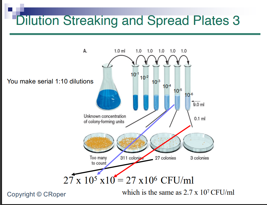
Dilution Streaking and Spread Plates 3
tells you number of living cells experiment biased towards living cells
take original flask, setup series of tubes with blank media and put 9mls take 1mL out of flask to create 1 to 10 dilution over and over so multiply by 10 every time
so 10 to 100 to 1000 to 1:100,000 to 1:1 million
don’t want two little or too many colonies between 10 and 50 is reliable range which is something you can count
311 is too many because you might miss some while counting we plated out a tenth of a milliliter from tube
so we have flask we take out 1mL than mix it around in other flask and mix out out over and over now we have a lot of tubes
different amounts and dilution gives you different amounts 1 to 10^5 is the ratio to use here it gets you 27 which is ideal

Spread plating again
Dilution streaking vs spread plating
Dilution streaking is a metal or plastic loop held against agar and you streak the surface
spread plating is moving a colony or liquid from one place to another so we are trying to spread liquid around the plate
you can use a slightly smaller volume like 100 microliters: use L shaped machine that spreads the liquid for you so it spreads sit instantly
we wanna know how much bacteria is in flask
when you start: put 9 ml of liquid media into tubes or 10 ml this becomes 10^-1 dilution
do that again 10^-2,10^-3, etc and you are diluting out bacteria
than put 100 microliters on petri dishes and seperate colonies we want a number 10 to 50, we got 27
so do 27 to 10%5 times 10
on exam: Info given certain dilution number of colonies you plated it out for X microliters so do math so you have X number