Structure and Function of the Liver in domestic animals
 Call Kai
 Learn
 Practice Test
 Spaced Repetition
 Match
 Flashcards
 Knowt Play
1/39

There's no tags or description
Looks like no tags are added yet.
40 Terms
Location and topography of the liver
Most cranial organ in the abdominal cavity
Located between the diaphragm and the stomach.
Oriented transversely
Parietal (Cranial) and visceral (Caudal) surfaces
From 7th to 9th ICS / 7th to 10th ribs – on the left.
From 7th to 11th/12th ICS / 7th to 12/13th ribs – on the Right.
8 or 9th ICS lower – Contact with the body (abdominal)wall

Parietal (Cranial) and visceral(Caudal) surfaces

Function of the Liver: Metabolic Regulation
Carbohydrate Metabolism, Lipid Metabolism and Protein Metabolism
Function of the liver: Detoxification
Metabolism of Toxins, Detoxifies endogenous and exogenous compounds (e.g., drugs, environmental toxins).
Function of the liver: Drug metabolism
metabolizes drugs primarily through enzymatic reactions, affecting their bioavailability, efficacy, and potential toxicity
Function of the liver: bile production and excretion
Produces bile, essential for the emulsification and digestion of fats.
Bile is stored in the gallbladder (in species that have one) and released into the small intestine to aid digestion.
Function of the liver: protein synthesis
synthesis of proteins such as albumin and coagulation factors
Function of the liver: Storage of nutrients and vitamins
Glycogen Storage, Stores fat-soluble vitamins (A, D, E, K) and vitamin B12, minerals like iron and copper
Function of the liver: Immune functions
important role in innate and adaptive immune response via different cells like Kupffer cells
In ruminants the liver is key in processing volatile fatty acids from ruminal fermentation
Lobation of the mammalian liver: Unique to the species (with same basic principles)
Right lobe, Left lobe, Quadrate lobe, Caudate lobe
Processes (extended parts): Caudate process, Papillary process

Borders – Applies to all species
Dorsal border: Esophageal notch – On the left
Notch for the caudal vena cava – on the right
Ventral border: Fissures/notches
Gall bladder – Between right and quadrate lobe
Falciform and round ligament – Between left and quadrate lobes
Hilus and root - Applies to all species
Opening (hilus) through which blood vessels and related structures (Collectively - root) enters the liver.
Root – Hepatic portal vein, hepatic artery, bile duct, lymphatic vessels, nerves.
Lobation of the liver in Dogs

Liver: The coronary ligament
Attachment to the diaphragm(semicircular /curved)
The triangular ligaments
Left and right
Attachment to the diaphragm
Liver: The falciform ligament
Median attachment to the diaphragm and abdominal floor
Remnant of the ventral mesentery/ventral connecting peritoneum
Liver: The round ligament (teres ligament)
Remnant of the fetal umbilical vein
Anatomy of ligaments of attachments of the liver

The liver: Sympathetic Innervation
originates from the Thoracolumbar(T7–T12) spinal cord Travel via the splanchnic nerves.
preganglionic fibers synapse in the right celiac ganglion, which is part of the celiac plexus.
The postganglionic fibers travel along the hepatic artery
Liver: Parasympathetic innervation
Originate from the dorsal motor nucleus of the Vagus n. in the medulla oblongata of the brainstem.
Ventral Vagal Trunk - Plexuses
Motor and Sensory
The liver of the cat
Lobation is same as dog
Left (L&M), Right (L&M), Caudate with caudate and papillary processes (both Larger)
Smaller liver, completely enclosed within the ribcage
Left side more cranial (deeper), Deeper renal impression
Anatomy of the cat liver

The Ruminant Liver
Mainly right to the median plane and rotated 90 degrees
Liver lies obliquely in the abdominal part of the thoracic cage: left lobe is cranioventral, right is caudo-dorsal.
Anatomy of the ruminant liver

The Porcine Liver
Well-lobated as in the dog.
Parietal surface fits into dome of diaphragm; visceral surface contacts stomach
Only domestic mammal in which liver has no contact with the right kidney → no renal impression in caudate lobe
Anatomy of the pig liver

The Equine Liver
Contact with diaphragm (parietal surface of liver).
Asymmetrical in shape; right lobe atrophies with age ( pressure from right dorsal colon)
Most cranial (left side)part at level of lower part of 6th or 7th rib.
Most caudal, and also the most dorsal (right Side) part at level of 16th and 17th ribs ventral to the right kidney.
The most ventral part does not reach the abdominal floor - at level of 7th or 8th rib.
Abdominal view - Equine Liver

Diaphragmatic surface of the equine liver.
1, left lateral lobe;
2 & 3, left medial lobe;
4, quadrate lobe;
5, right lobe; asterisks, hepatic veins near their entrance into the caudal vena cava (not shown).

The Equine Liver: Visceral surface
Stomach, duodenal and colic impressions; related to pancreas dorsally.
Esophageal impression (notch) on dorsal part of left lobe.
Caudal vena cava passes through the craniodorsal surface on the median plane of liver.
Renal impression on dorsal part of caudate process
The dog biliary system: Cystic duct
Arises from the neck of the gallbladder
Called cystic duct Until last hepatic duct opens into it
Dog biliary system: hepatic ducts
From each liver lone and larger processes
Dog biliary system: Common bile duct
From last hepatic duct To the small intestine
Dog biliary system: Major duodenal papilla
Opening of the common bile duct into the duodenum
Anatomy of the biliary system in the dog

The biliary system: storage and synthesis
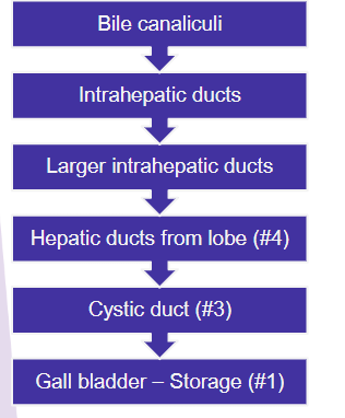
Biliary system: storage and release
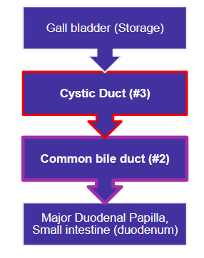
Major Duodenal papilla: Opening of the common bile duct into the duodenum.
Dog: opens via single opening but bile duct and pancreatic duct opens separately.
Cat: The pancreatic duct joins the common bile duct.
The biliary system horse: No gall bladder
The bile duct has a wide lumen; opens along with major pancreatic duct into the hepaticopancreatic ampulla[major duodenal papilla] of the distal and convex part of the duodenal sigmoid flexure.
Oblique passage of duct through wall acts as a valve