A&P lab lecture 1
 Call Kai
 Learn
Learn Practice Test
 Spaced Repetition
 Match
Match Flashcards
 Knowt Play
1/53

There's no tags or description
Looks like no tags are added yet.
Name | Mastery | Learn | Test | Matching | Spaced | Call with Kai | Chat |
|---|
No analytics yet
Send a link to your students to track their progress
54 Terms
What are the four abdominopelvic quadrants?
Right Upper (RUQ), Right Lower (RLQ), Left Upper (LUQ), Left Lower (LLQ).

What are the nine abdominopelvic regions?
Right hypochondriac, right lumbar, right inguinal, left hypochondriac, left lumbar, left inguinal, epigastric, umbilical, hypogastric (pubic).

Described the lines of the abdominopelvic regions.
The vertical lines are midclavicular lines.
The horizontal lines are made from the bottom of the 10th rib and at the iliac crest
Which plane divides the body into anterior and posterior portions?
Coronal (frontal) plane.
Which plane divides the body into equal left and right sections?
Sagittal plane.
Parasagittal vs midsagittal?
Parasagittal: Plane dividing the body into left and right halves, but not directly at the midline
Midsagittal: left and right at the midline.
Which plane divides the body into superior and inferior portions?
Transverse (horizontal) plane.
Cranial vs. Caudal?
Cranial = toward head
Caudal = toward feet.
Medial vs. Lateral?
Medial = toward midline
Lateral = away from midline.
Dorsal vs. Ventral?
Dorsal = back
Ventral = front/abdomen.
Describe flexion vs extension.
Flexion is toward the body except for the legs because the legs turn while in development.
Extension is away from the body except for the legs.
Abduction vs adduction?
Abduction is moving away from midline.
Adduction is moving toward the midline.
Proximal vs distal?
Proximal is toward the base of a limb.
Distal is away from the base of a limb.
Must be of a limb and must be comparing multiple things.
Which cavities are part of the ventral cavity?
Thoracic and abdominopelvic cavity (abdominal and pelvic)
What is in the thoracic cavity?
Heart and lungs including the associated trachea, bronchial tubes, esophagus, great vessels.
What separates the thoracic and abdominal cavities?
The diaphragm.
Which organs are in the pelvic cavity?
Bladder, reproductive organs.
Which structures are in the abdominal cavity?
Digestive organs & associated structures:
-Small intestines/ Large intestines, appendix
-Stomach
-Liver
-Pancreas
-Spleen
What surrounds the abdominal organs?
Peritoneum
What are the dorsal body cavities?
The cranial cavity and the vertebral cavity.
What surrounds the lung?
Pleura. Parietal pleura is the outer layer and the visceral pleura is the inner layer. The pleural cavity contains pleural (serous) fluid that acts as a lubricant.

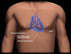
What surrounds the heart?
Pericardium. The parietal pericardium is the outer layer. The visceral pericardium is the inner layer. The pericardial cavity contains serous fluid for lubricant
What is the mesentery
The mesentery is a fold of the peritoneum that attaches the small intestine to the posterior abdominal wall. It provides support and contains blood vessels, nerves, and lymphatics that supply the intestines.

What is the border of the pelvic cavity?
Pelvis

Which imaging gives the best view?
MRI>CT>X-ray
How do X-rays and CT scans create an image?
By using ionizing radiation. CT scan uses more. Denser structures like bones reflect the radiation causing a white image.
How does an MRI make an image?
Magnetic resonance (protons bouncing around). Used for soft tissue and ligaments. Bone appears dark.
Visual, function, & example of simple squamous epithelium?
1 layer of flat cells
Rapid diffusion/filtration
Alveoli, capillaries.

Visual, function, & example of simple cuboidal epithelium?
1 layer of cube shaped cells
Secretion & absorption
Kidney tubules.

Visual, function, & example of simple columnar epithelium?
1 layer of tall column cells. Can be ciliated or nonciliated.
Absorption/secretion
Digestive tract lining.

What is pseudostratified columnar epithelium known for?
Appears layered, but all cells touch basement membrane; ciliated in trachea.

Difference between keratinized and nonkeratinized stratified squamous epithelium?
Keratinized = skin
Nonkeratinized = moist surfaces (esophagus, mouth).
Where is transitional epithelium found?
Bladder, ureters (stretchable).
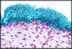
Describe epithelium.
Anything above the basement membrane and is connected to a cavity has to have polarity and is called epithelium
Simple vs stratified?
Simple = one layer
Stratified = multiple layers
What are the layers of the stratified squamous epithelium (keratinized)
Layers of the skin: Can Lucy Get Some Bacon
Stratum corneum
Stratum lucidum (only in palms and soles)
Stratum granulosum
Stratum spinosum
Stratum basale

What do dermal papilla make?
Ridges between the epidermis and dermis that make fingerprints
What is lamina propria?
The lamina propria is a thin layer of connective tissue that lies directly beneath the epithelial lining of various organs and body systems, such as the gastrointestinal tract, respiratory tract, and urinary tract.

What are the three types of loose connective tissue?
Areolar, adipose, reticular
Dense regular connective tissue is found where?
Tendons and ligaments.
What type of cartilage forms the fetal skeleton and trachea rings?
Hyaline cartilage.
What cartilage provides shock absorption in intervertebral discs?
Fibrocartilage.
What cartilage provides flexibility in the ear?
Elastic cartilage.
What are the two types of bone tissue?
Compact bone and spongy bone.
What are the main components of blood (fluid connective tissue)?
Erythrocytes, leukocytes, platelets, plasma.
What are the 2 layers of the dermis
Papillary (areolar tissue) and reticular (dense irregular connective tissue)
Describe the papillary layer
Consists of areolar tissue. It contains the capillaries, lymphatic vessels, and sensory neurons that supply the surface of the skin. The papillary layer gets its name from the dermal papillae that project between the epidermal ridges.
Describe the dermal layer
lies deep to the papillary layer. It consists of an interwoven meshwork of dense irregular connective tissue containing both collagen and elastic fibers.
Which dermal layer provides strength and elasticity?
Reticular layer.
What are the main parts of a neuron?
Cell body, dendrites, axon.
What are supporting cells in nervous tissue called?
Glial cells.
What muscle tissue is striated and voluntary?
Skeletal muscle.
What muscle tissue is striated, branched, and has intercalated discs?
Cardiac muscle.
What muscle tissue is non-striated and involuntary?
Smooth muscle.