RESP 210 quiz 4 study guide
 Call Kai
 Learn
 Practice Test
 Spaced Repetition
 Match
 Flashcards
 Knowt Play
1/48

There's no tags or description
Looks like no tags are added yet.
49 Terms
Patient Transport: think patient stability
team
what do they need to maintain
risks
Do not go on a transport unless
ALL members of the team are present
Be sure that patient is able to maintain
hemodynamic stability during the transport
If your patient is
hemodynamically unstable (2 – 3 + pressors)
and medically complicated
make sure the field trip benefits outweigh the risks
Patient Transport: Know when transporting a very unstable/high-PEEP patient may be unsafe.
If very high PEEP, be cautious about the transport
use Kelly clamp between transition from ventilator to
AMBU bag
or transport circuit
don’t lose PEEP!
Patient Transport: battery
Sufficient battery – bring backup
Patient Transport: airway/emergency equipment
equipment
Stethoscope
Self-inflating AMBU
Emergency airway supplies
O2 tanks
transport vent
Portable vent circuit
PEEP capacity
Sufficient battery – bring backup
Vent w/alarms, pressure monitoring, Vt capacity *
Make sure vent at destination
Patient Transport: monitoring
Pulse ox
Heart rate monitor, ideally with vascular pressure capacity
CO2 monitor (won’t always see but is recommended for pt safety)
Do NOT get comfortable at any time during a patient transport
Watch your patient and the monitoring screens at all times
on the way to the destination
at the destination
and the return back
Bring portable suction if you don’t think your patient can make it to the destination without coughing up material that could compromise safety
Be present and involved every time pt moves from bed to bed or onto the vennt set up at your location. This is a high-risk time
For special transports (iNO, continuous neb, trache etc.), bring specialty supplies
Patient Transport: backup plan
Bring back up oxygen and batteries in case things go wrong/get delayed
Be able to calculate whether an E-cylinder contains enough oxygen for transport, including an appropriate reserve.
(Tank pressure - reserve x 0.28) / tank flow in LPM
500 PSI common reserve
dont use if it mentions how much is left
Know jet nebulizer vs. Aerogen on the ventilator:
location
flow, VT, pressure
alarms
medications left
pulm deposition
Jet nebs
Place on the inspiratory limb near the Wye
cheaper
add flow 6 – 10 LPM depending on the device
can change Vt, MV, pressures, triggering, alarms and FiO₂;
↑
PIP
delivered Vt
MV
May
set off vent alarms
make it more difficult for pt to trigger a breath
alter the intended pattern of ventilation
Leaves substantial medication behind in the cup
lower less consistent pulm deposition
Aerogen does not add flow
Place on dry side of the humidifier
more expensvie
Produces aerosol by vibrating medication through a microscopic mesh
Does not alter delivered Vt, pressures, or impact patient triggering
Can remain inline
no circuit disconnection needed for tx
Minimal medication remains in the reservoir
Can deliver scheduled or continuous nebulization
can be used for non-bronchodilator meds, like:
inhaled epoprostenol
TXA
heparin
higher more consistent pulm deposition
Know basic MDI administration through a ventilator circuit and how inspiratory flow/Ti affect delivery.
If pt has important home medication in MDI form, may need to give through a vent circuit
Shake MDI first (prime if necessary) + remove HME
Set MDI spacer in inspiratory limb of the circuit
Temporarily ↓ inspiratory flow
Temporarily ↑ Ti
Coordinate MDI with precise start of inspiration
Give one puff at a time
Suctioning: indications
Patient’s inability to generate acough
Deterioration of ABG or SpO2 values
Decreased volume in pressure ventilation
Increased PIP in volume ventilation
Changes in monitored flow-volume graphics
Suctioning: monitoring
Before, during, and after suctioning:
BS, RR, and pattern
O2 saturation and FiO2
Pulse, BP, ECG if available
Cough effort
Sputum (color, consistency, etc.)
ICP if indicated
Suctioning: complications
Cardiac dysrhythmias
Hypoxemia or hypoxia
Airway trauma/bleeding
Atelectasis or reduction of lung volume
Increased intracranial pressure
Bronchospasm
Loss of PEEP/derecruitment
Do we preooxygenate before suctioning?
yes @100% FIO2
Understand the issues of decompensation when suctioning patient on high PEEP
Loss of PEEP/derecruitment
Know what SpO₂ measures
SpO2 is a useful early warning system to detect hypoxemia in critically illpatients – keep in mind that SpO2 ≠ SaO2
Hypoxemic events in the ICU are associated with
apnea
airway obstruction
equipment failure
disconnection
sometimes incorrect gas flow settings
How pulse oximetry works
To differentiate oxygenated Hb from deoxygenated Hb, two wavelengths of light are passed through the sampling site. (660 nm and 940 nm)
Deoxygenated Hb absorbs more of the
660 nm (red light)
Oxygenated Hb absorbs more of the
940 nm light (infrared)
Pulse rate is determined from cyclic changes in light absorption:
Systole: arterial blood volume ↑ → absorption ↑ → transmitted light ↓
Pulse oximetry provides continuous, non-invasive measurements of arterial oxygen saturation
Sensor commonly placed on:
finger
earlobe
forehead
bridge of nose
•Side note: In NICU, location matters.
Preductal vs postductal location
The basic relationship between SpO₂/SaO₂ and PaO₂ on the oxyhemoglobin dissociation curve
SaO2 varies with PaO2 in a ‘sigmoidal’ or S-shaped manner
When SaO2 is > 90%, PaO2 goes
up without much bump in SaO2
Below 80% SaO2, the PaO2
falls more dramatically
Although 97% is considered a normal adult SaO2, 90% or higher is acceptable for most adult patients
Know when pulse oximetry can lie or become unreliable
poor perfusion
motion
shivering, tremors, seizures, or patient movement
probe/site problems
incorrect fit, or inappropriate site
low saturations
ambient light
Nail polish, artificial nails, or tattoo ink
Does the SpO₂ fit the patient?
Check the pleth and pulse agreement
reposition or warm the site
confirm with an ABG when clinically necessary
Know COHb vs. MetHb: CO can give a falsely reassuring SpO₂; significant MetHb tends toward ~85%;
Carboxyhemoglobin (COHb)
A standard pulse oximeter cannot distinguish COHb from O₂Hb.
COHb may therefore produce a falsely reassuring SpO₂.
A normal SpO₂ does not rule out carbon monoxide poisoning.
Methemoglobin (MetHb)
MetHb absorbs 660-nm red light and 940-nm infrared light nearly equally
As MetHb increases, the red-to- infrared absorption ratio approaches
As MetHb becomes significant, the displayed SpO₂ trends toward approximately 85% and no longer reliably reflects the true arterial saturation
suspected dyshemoglobinemia requires co-oximetry.
Suspect COHb or MetHb? Order a blood gas with co-oximetry to directly measure O₂Hb, HHb, COHb, and MetHb
Review right and left shifts of the oxyhemoglobin dissociation curve and what happens to Hb-O₂ affinity.
right shift
↓
pH
↑
CO2
2-3 DPG
temp
affinity for Hb
left shift
↑
pH
↓
CO2
2-3 DPG
temp
affinity for Hb
Know the difference between capnometry, capnography, and a capnogram
Capnometry
measurement of exhaled CO2
(numerically without a waveform)
Capnography
continuous display of exhaled CO2 concentrations as a graphic waveform
Capnogram
graphic waveform of CO2 against time
Major clinical uses of capnography—including ET tube verification, ventilation/CO₂ elimination, V/Q assessment, perfusion, and detecting rebreathing.
Monitoring V/Q relationships and response to therapy
Verify ET tube placement
Monitor adequacy of pulmonary and coronary blood flow
Screening for pulmonary embolism
Detection of CO2 rebreathing and effects of sedation/neuromuscular blockade
Monitoring CO2 elimination
Optimizing mechanical ventilation

Know the four phases of a normal capnogram, including where PETCO₂/ETCO₂ is measured, and recognize the normal waveform.
Phase 1 - the initial gas exhaled is from the conducting airways, which contain low levels of CO2 (mostly dead space).
Phase 2 - alveolar gas containing CO2 mixes with gas in the anatomical airways and the CO2 concentration increases.
Phase 3 - the curve plateaus as alveolar gas is exhaled (this phase is often referred to as the alveolar plateau). Note that the concentration of CO2 at the endof the alveolar phase (just before inspiration begins) is called the end-tidal PCO2 or PETCO2.
Phase 4 - Inspiration, the concentration falls to zero.
Capnogram: Phase 1
Phase 1 - the initial gas exhaled is from the conducting airways, which contain low levels of CO2 (mostly dead space).
Capnogram: Phase 2
Phase 2 - alveolar gas containing CO2 mixes with gas in the anatomical airways and the CO2 concentration increases.
Capnogram: Phase 3
where is PETCO₂/ETCO₂ measured?
Phase 3 - the curve plateaus as alveolar gas is exhaled (this phase is often referred to as the alveolar plateau). Note that the concentration of CO2 at the endof the alveolar phase (just before inspiration begins) is called the end-tidal PCO2 or PETCO2.
Capnogram: Phase 4
Phase 4 - Inspiration, the concentration falls to zero.
Know normal PETCO₂ (~35–38 mmHg) and its relationship to PaCO₂ (normally ~2–5 mmHg lower); understand why an increased PaCO₂–PETCO₂ gradient suggests increased dead space/V-Q problems such as pulmonary embolism.
The normal end-tidal value is approximately:
35- 38 mmHg
or around 5% PETCO2.
Trends are more important than value.
PETCO2 normal =
~ 3 to 5mmHg less than PaCO2
*Why would you have a wide gradient with your PaCO2 and your PETCO2? What could be the problem?
*Pulmonary embolism
Will have
low PETCO2
high PaCO2.
*Other dead space issues
Be able to interpret changes in the capnogram/PETCO₂: sudden drop to zero
A sudden drop in CO2 to ZERO always indicates a problem such as:
Kinked ET-tube
CO2 analyzer defective
Disconnection
Ventilator defective
Patient stopped breathing, circulatory arrest
Condensation affecting sensor
Embolism
Sudden decrease in blood pressure
Be able to interpret changes in the capnogram/PETCO₂: sudden decrease, or sudden increase—and connect them to likely causes such as disconnection/airway problems, poor perfusion or compressions, ROSC, and changes in CO₂ production/metabolism.
A sudden decrease in CO2 but not to 0 may be caused by:
Leakage in the respiratory system
(low airway pressure)
Obstruction (high airway pressure)
Poor compressions
Be able to interpret changes in the capnogram/PETCO₂: s sudden increase—and connect them to likely causes such as disconnection/airway problems, poor perfusion or compressions, ROSC, and changes in CO₂ production/metabolism.
A sudden increase in PETCO2 can be caused by:
Injection of sodium bicarbonate
Sudden release of tourniquet
Sudden increase in blood pressure
Achieved ROSC
Production of CO2 is determined by the metabolic rate
*Factors that INCREASE metabolic rate and CO2 production:
- Fever, sepsis, hyperthyroidism, seizures
*Factors that DECREASE metabolic rate and CO2 production:
- Hypothermia, starvation, sedation
Know how to read the basic pressure, flow, and volume vs. time waveforms and recognize what the patient/ventilator is doing.
y axis = flow
x axis = time
a-b = inspiration
b-c = expiration
d= peak insp flow
e= PEFR

Know how to read the basic flow waveforms

Be able to recognize air trapping/auto-PEEP and inadequate expiratory time, including what the flow waveform should do before the next breath begins.
not enough time to exhale

type of disease from graph?

Increase Ti on pt A, will you get more vol?

Use graphics to identify airway obstruction, changes in compliance, patient triggering/active exhalation, flow demand, and patient–ventilator synchrony.

type of breath?
mandatory breath

type of breath?
spontaneous breath
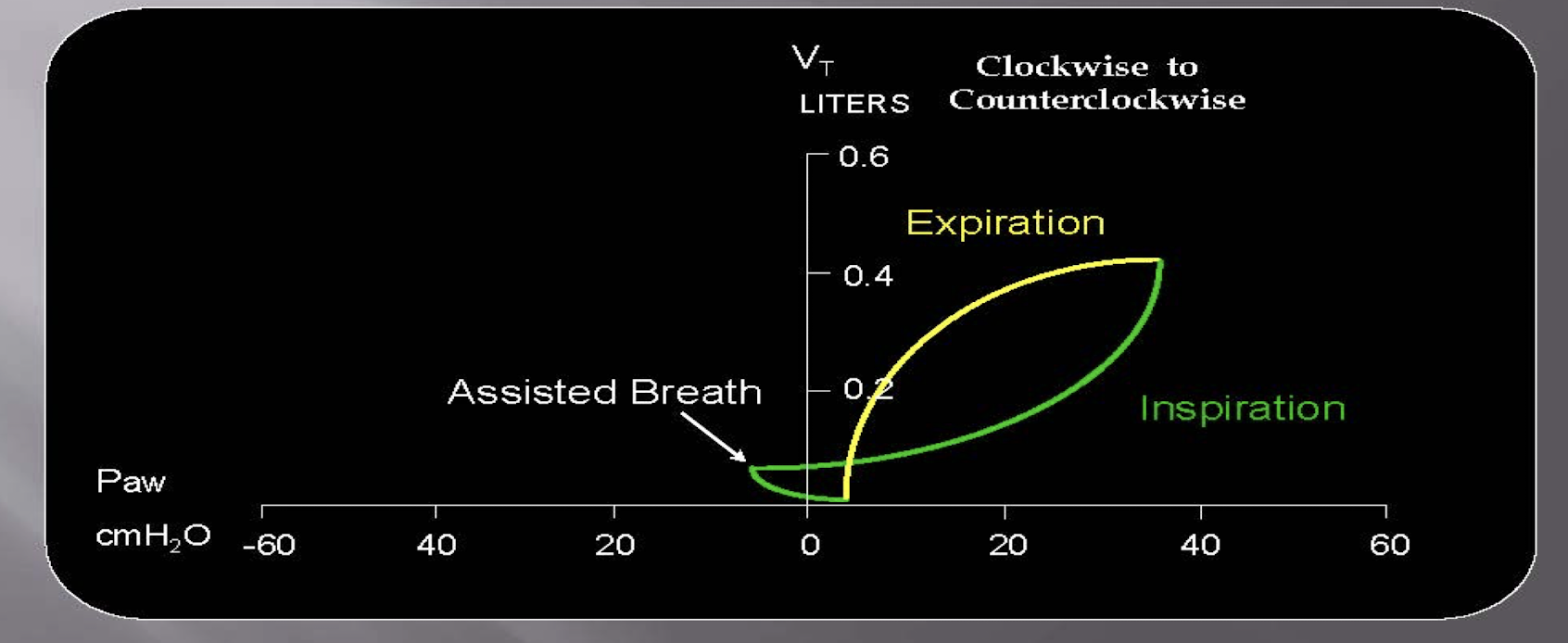
type of breath?
assisster breath
analyze graph: ACVC
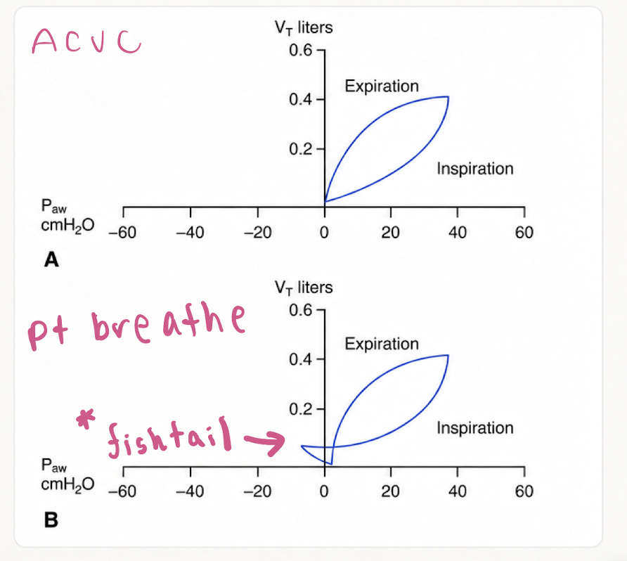
static compliance line

Understand how changes in Pplat, PEEP, Vt and compliance relate to one another. Know Cstat = Vte/(Pplat − PEEP)
PATIENT A
Vte 500 ml
PEEP 5 cmH2O
Pplat 20 cmH2O
PATIENT B
Vte 500 ml
PEEP 5 cmH2O
Pplat 30 cmH2O
Without using a calculator, tell me who has the best static compliance?
patient A
lower pPlat
overdistention graph

zone & inflection points on graphics

Be able to identify P-V loop compliance issues in volume mode

Be able to identify P-V loop compliance issues in pressure mode

Be able to identify LIP and place for optimal PEEP; be able to identify HIP and identify the dangers of that point; know how to make changes to the vent to solve overdistention or inadequate PEEP based on P-V loop

Be able to look at a graphic and determine the likely ventilator mode/problem and appropriate correction—not just name the waveform.
Is the patient getting enough time to exhale?
→ Air trapping / auto-PEEP
Is it taking too long to get air out?
→ Airway obstruction
Did our treatment make a difference?
→ Response to bronchodilator therapy
Is the patient comfortable with the breath?
→ Inspiratory flow demand & patient–ventilator synchrony
What is the patient doing?
→ Triggering effort & active exhalation