Cases 2018

0.0(0)
Studied by 0 people
call kaiCall Kai
learnLearn
examPractice Test
spaced repetitionSpaced Repetition
heart puzzleMatch
flashcardsFlashcards
GameKnowt Play
Card Sorting

1/12

flashcard set

Earn XP

Description and Tags

ok man.

13 Terms

1
New cards
<p>Case #481</p><p><span>A 22-year-old male noticed painless, ulcerating lesions with crusting and mild swelling on his back and face. He had travelled to the rain forest of Peru five months earlier, where he experienced multiple insect bites all over his body. A biopsy specimen from a lesion on his back was sectioned and stained with hematoxylin and eosin (H&amp;E) (Figure </span><strong>A</strong><span>).&nbsp;A smear of a scraping from the biopsy site was also made and stained with Giemsa stain (Figure </span><strong>B</strong><span>). Both figures show what was observed on the slides.</span></p>

Case #481

A 22-year-old male noticed painless, ulcerating lesions with crusting and mild swelling on his back and face. He had travelled to the rain forest of Peru five months earlier, where he experienced multiple insect bites all over his body. A biopsy specimen from a lesion on his back was sectioned and stained with hematoxylin and eosin (H&E) (Figure A). A smear of a scraping from the biopsy site was also made and stained with Giemsa stain (Figure B). Both figures show what was observed on the slides.

Leishmania sp.

2
New cards
<p>Case #480</p><p><span>A man from New England returned from a trip to Africa with fever, chills, sweats, and fatigue. He sought medical attention from his health care provider.&nbsp; A blood smear was ordered, prepared, stained with Wright-Giemsa and examined. A diagnosis of </span><em>Babesia</em><span> was made.&nbsp; The slides and a blood specimen were sent to the CDC for confirmation.&nbsp; Figures </span><strong>A-D</strong><span> show what was observed at 1000x oil magnification on one of the stained blood smears.&nbsp;</span></p>

Case #480

A man from New England returned from a trip to Africa with fever, chills, sweats, and fatigue. He sought medical attention from his health care provider.  A blood smear was ordered, prepared, stained with Wright-Giemsa and examined. A diagnosis of Babesia was made.  The slides and a blood specimen were sent to the CDC for confirmation.  Figures A-D show what was observed at 1000x oil magnification on one of the stained blood smears. 

Plasmodium falciparum

3
New cards
<p>Case #477</p><p><span>A 58-year-old male, with recent travel to Asia, was seen at an out-patient clinic for abdominal discomfort that lasted for two days. A stool sample was collected in 10% formalin and zinc PVA (Zn-PVA) and processed for ova and parasites (O&amp;P) examination. A presumptive worm egg was recovered from the stool and was processed in the pathology laboratory for identification. An image of the hematoxylin and eosin (H&amp;E) stained slide is shown in Figure </span><strong>A</strong><span>.</span></p>

Case #477

A 58-year-old male, with recent travel to Asia, was seen at an out-patient clinic for abdominal discomfort that lasted for two days. A stool sample was collected in 10% formalin and zinc PVA (Zn-PVA) and processed for ova and parasites (O&P) examination. A presumptive worm egg was recovered from the stool and was processed in the pathology laboratory for identification. An image of the hematoxylin and eosin (H&E) stained slide is shown in Figure A.

Artifact (plant seed)

4
New cards
<p>Case #478</p><p><span>A 4-year-old boy returned from a summer vacation in Tanzania with his parents with symptoms of fever and chills. He was evaluated at a clinic in St. Louis, Missouri where blood smears were ordered, stained with Giemsa and examined at 1000x magnification with oil. Images captured from the stained thin smear were sent to the DPDx Team for identification. Figures </span><strong>A</strong><span> – </span><strong>F</strong><span> show what was observed in some of the images submitted. What is your diagnosis?</span></p>

Case #478

A 4-year-old boy returned from a summer vacation in Tanzania with his parents with symptoms of fever and chills. He was evaluated at a clinic in St. Louis, Missouri where blood smears were ordered, stained with Giemsa and examined at 1000x magnification with oil. Images captured from the stained thin smear were sent to the DPDx Team for identification. Figures A – F show what was observed in some of the images submitted. What is your diagnosis?

Plasmodium malarie

5
New cards
<p>Case #473</p><p><span>A 67-year-old female presented with a 2-3 day history of lower back pain. She also reported a burning sensation all over her body.&nbsp;The patient’s history included&nbsp;emphysema of the lungs, history of smoking, pancreatitis, and cholelithiasis. She underwent endoscopic retrograde cholangio-pancreatography (ERCP) at which time a worm was detected in the common bile duct and removed. The worm was damaged during retrieval but images were taken of what was obtained and sent to the DPDx Team for diagnostic assistance. Figures </span><strong>A </strong><span>and </span><strong>B</strong><span> shows the portion of the worm recovered.</span></p>

Case #473

A 67-year-old female presented with a 2-3 day history of lower back pain. She also reported a burning sensation all over her body. The patient’s history included emphysema of the lungs, history of smoking, pancreatitis, and cholelithiasis. She underwent endoscopic retrograde cholangio-pancreatography (ERCP) at which time a worm was detected in the common bile duct and removed. The worm was damaged during retrieval but images were taken of what was obtained and sent to the DPDx Team for diagnostic assistance. Figures A and B shows the portion of the worm recovered.

Hookworm (Anclystoma duodenale)

6
New cards
<p>Case #474</p><p><span>A 27 year-old male presented to his health care provider, after a 10-day work related trip to Ethiopia, with lymphadenopathy of the head and neck, intermittent fevers, and malaise. Thick and thin blood smears were ordered, stained with Giemsa, and examined. Figures </span><strong>A</strong><span> – </span><strong>C</strong><span> show what was found on a stained thick smear in very low numbers.</span></p>

Case #474

A 27 year-old male presented to his health care provider, after a 10-day work related trip to Ethiopia, with lymphadenopathy of the head and neck, intermittent fevers, and malaise. Thick and thin blood smears were ordered, stained with Giemsa, and examined. Figures A – C show what was found on a stained thick smear in very low numbers.

Artifact (fungi)

7
New cards
<p>Case #472</p><p><span>A 22 year old man diagnosed with gastroenteritis underwent subsequent gastrointestinal endoscopic examination. The gastroenterologist observed a worm which he extracted from the duodenum and sent to the laboratory for identification. Stool samples were also processed for ova and parasites (O&amp;P) examination. Images of the worm and objects seen in a wet mount preparation from a formalin-ethyl acetate (FEA) concentration of the stool samples were captured by the laboratory technologist and sent to DPDx for identification. Travel history was not provided. The objects in Figures </span><strong>C</strong><span> – </span><strong>E</strong><span> measured 130 µm in length by 65 µm in width on average. The adult worm measured approximately 70 mm by 18 mm.</span></p>

Case #472

A 22 year old man diagnosed with gastroenteritis underwent subsequent gastrointestinal endoscopic examination. The gastroenterologist observed a worm which he extracted from the duodenum and sent to the laboratory for identification. Stool samples were also processed for ova and parasites (O&P) examination. Images of the worm and objects seen in a wet mount preparation from a formalin-ethyl acetate (FEA) concentration of the stool samples were captured by the laboratory technologist and sent to DPDx for identification. Travel history was not provided. The objects in Figures C – E measured 130 µm in length by 65 µm in width on average. The adult worm measured approximately 70 mm by 18 mm.

Fasciolopsis buski

8
New cards
<p>Case #470</p><p><span>A 51 year-old immigrant man from Honduras was prescribed steroids at a health care facility for asthma exacerbation. After feeling better, he continued to self-medicate with injectable steroids every three months obtained via mail order from Honduras for approximately one year. He returned to the health care facility with symptoms including abdominal pain, worsening fatigue, shortening of breath and bright red rectal bleeding. Figures </span><strong>A</strong><span>–</span><strong>E</strong><span> are from hematoxylin-and-eosin stained sections of biopsies taken from the terminal ileum to the rectum (do not let the “sugar skull” in Fig </span><strong>A</strong><span> distract you!). Figures </span><strong>F</strong><span>–</span><strong>H</strong><span> are from a stained (presumably Papanicolaou) cytospin smear of a sputum specimen.</span></p>

Case #470

A 51 year-old immigrant man from Honduras was prescribed steroids at a health care facility for asthma exacerbation. After feeling better, he continued to self-medicate with injectable steroids every three months obtained via mail order from Honduras for approximately one year. He returned to the health care facility with symptoms including abdominal pain, worsening fatigue, shortening of breath and bright red rectal bleeding. Figures A–E are from hematoxylin-and-eosin stained sections of biopsies taken from the terminal ileum to the rectum (do not let the “sugar skull” in Fig A distract you!). Figures F–H are from a stained (presumably Papanicolaou) cytospin smear of a sputum specimen.

Strongyloides stercoralis
9
New cards
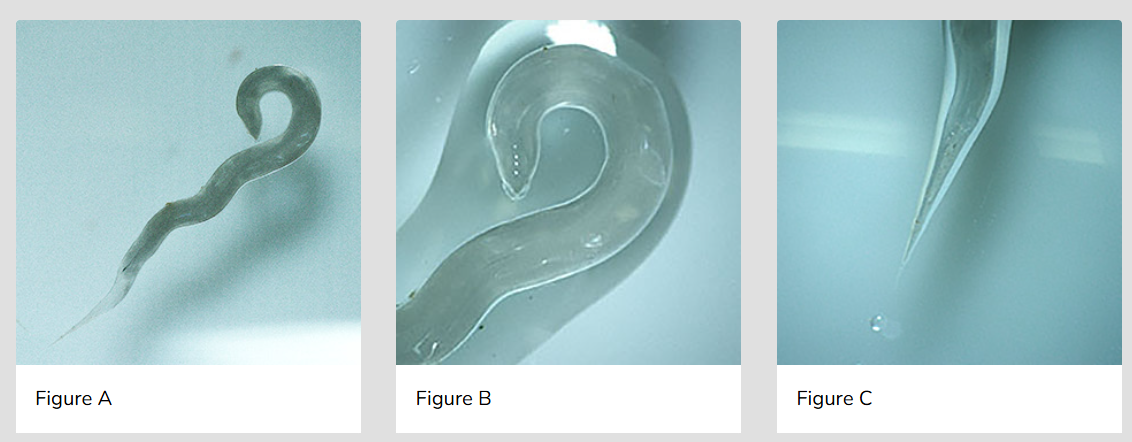
<p>Case #468</p><p><span>The object shown in Figures </span><strong>A</strong><span>–</span><strong>C</strong><span> was found in a stool specimen collected in SAF (sodium acetate-acetic acid- formalin) from a 43-year-old woman born and raised in Manitoba, Canada with no recent travel reported. Estimated length of the object was 8 millimeters.</span></p>

Case #468

The object shown in Figures A–C was found in a stool specimen collected in SAF (sodium acetate-acetic acid- formalin) from a 43-year-old woman born and raised in Manitoba, Canada with no recent travel reported. Estimated length of the object was 8 millimeters.

Enterobius vermicularis (pinworm)

10
New cards
<p>Case #465</p><p><span>A 74-year-old man, with a prior history of urothelial carcinoma, high grade pT1 (invasive, into lamina propria), was seen at a medical facility for follow-up care. Travel history for the patient was not available. A urinary cytology specimen was examined by a pathologist who observed structures thought to be parasitic in nature. Digital images were captured and sent to the CDC DPDx Team for diagnostic assistance. Figures </span><strong>A</strong><span> and </span><strong>B</strong><span> represent the structures that were observed.</span></p>

Case #465

A 74-year-old man, with a prior history of urothelial carcinoma, high grade pT1 (invasive, into lamina propria), was seen at a medical facility for follow-up care. Travel history for the patient was not available. A urinary cytology specimen was examined by a pathologist who observed structures thought to be parasitic in nature. Digital images were captured and sent to the CDC DPDx Team for diagnostic assistance. Figures A and B represent the structures that were observed.

Artifact (crystals)

11
New cards
<p>Case #464</p><p><span>A 45-year-old man from Paraguay went on a safari in eastern Zambia. He reported multiple insect bites at one location and noticed that some bites had developed into sores while still in country. One week after returning, he developed fever, headache, and enlarged lymph nodes and sought medical attention. A rapid test for malaria was negative but a blood smear was still ordered and stained with Wright-Giemsa. Figures </span><strong>A</strong><span>– </span><strong>D</strong><span> show what was observed on the blood smear at 1000x oil.</span></p>

Case #464

A 45-year-old man from Paraguay went on a safari in eastern Zambia. He reported multiple insect bites at one location and noticed that some bites had developed into sores while still in country. One week after returning, he developed fever, headache, and enlarged lymph nodes and sought medical attention. A rapid test for malaria was negative but a blood smear was still ordered and stained with Wright-Giemsa. Figures A– D show what was observed on the blood smear at 1000x oil.

Trypanosoma brucei

12
New cards
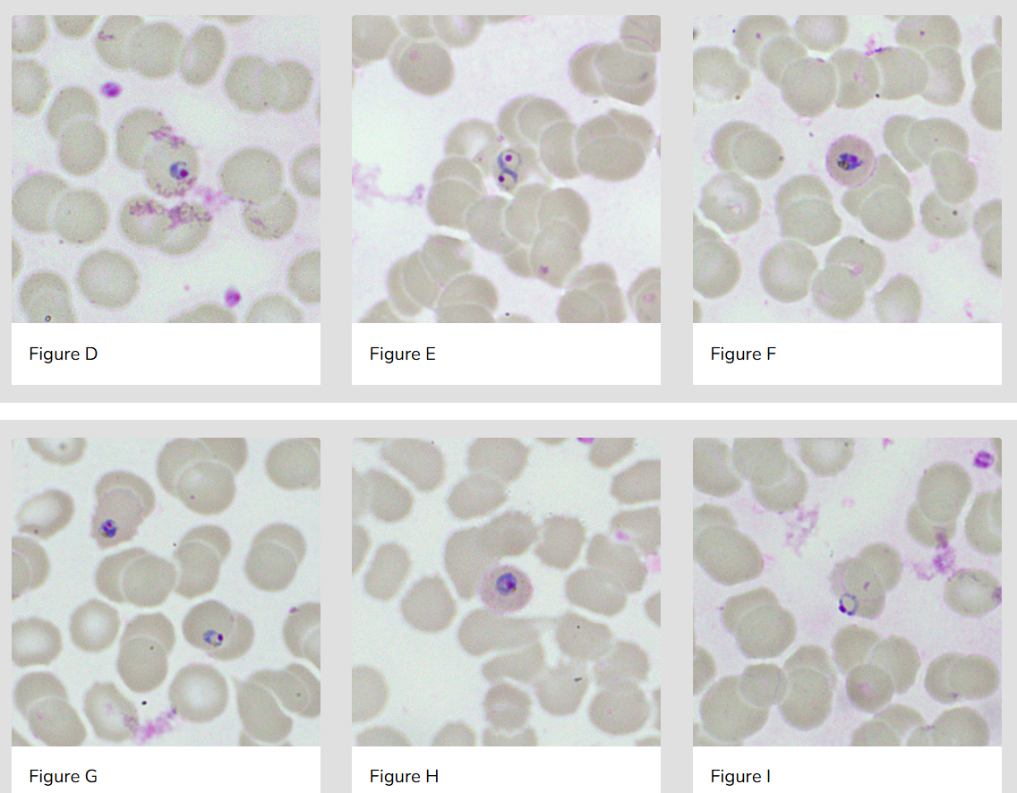
<p>Case #461</p><p><span>A 12-year-old boy, with symptoms of fever and chills after returning from a one month trip to an undisclosed West African country, was taken to a clinic for evaluation. It was determined that he had not taken any prophylactic medication for malaria prior to the trip. Blood smears were ordered, stained with Giemsa, and examined at 1000x magnification with oil at a pathology laboratory. Images were captured from a stained thin smear were sent to the DPDx Team for identification. Figures </span><strong>A – I </strong><span>show what was observed in some of the images submitted.</span></p>

Case #461

A 12-year-old boy, with symptoms of fever and chills after returning from a one month trip to an undisclosed West African country, was taken to a clinic for evaluation. It was determined that he had not taken any prophylactic medication for malaria prior to the trip. Blood smears were ordered, stained with Giemsa, and examined at 1000x magnification with oil at a pathology laboratory. Images were captured from a stained thin smear were sent to the DPDx Team for identification. Figures A – I show what was observed in some of the images submitted.

Plasmodium ovale
13
New cards
<p>Case #460</p><p><span>A 39 year-old man from Nigeria returned from visiting his country of birth with fever. A medical evaluation confirmed the fever and also detected low hemoglobin. A Binax-NOW rapid test for malaria showed a strong T1 band and a weak T2 band (Figure </span><strong>A</strong><span>). A blood smear was ordered and stained with Giemsa. Examination of a stained thin smear showed what is shown in Figures </span><strong>B</strong><span>–</span><strong>I</strong><span> and the parasite density was calculated to be 0.5%.</span></p>

Case #460

A 39 year-old man from Nigeria returned from visiting his country of birth with fever. A medical evaluation confirmed the fever and also detected low hemoglobin. A Binax-NOW rapid test for malaria showed a strong T1 band and a weak T2 band (Figure A). A blood smear was ordered and stained with Giemsa. Examination of a stained thin smear showed what is shown in Figures B–I and the parasite density was calculated to be 0.5%.

Plasmodium falciparum



