Bio 1a 2

0.0(0)
Studied by 0 people
call kaiCall Kai
learnLearn
examPractice Test
spaced repetitionSpaced Repetition
heart puzzleMatch
flashcardsFlashcards
GameKnowt Play
Card Sorting

1/28

encourage image

There's no tags or description

Looks like no tags are added yet.

Last updated 3:24 AM on 10/4/26
Name
Mastery
Learn
Test
Matching
Spaced
Call with Kai
Chat

No analytics yet

Send a link to your students to track their progress

29 Terms

1
New cards

Principles of chromosomes replication

Chromosomes are copied by a semi-conservative mechanism where the parental strands are copied to form a hybrid duplex, rather than used to make a fully new duplex with conservation of the parental duplex.

Explanation:
Semi-conservative means each new DNA molecule has one old parental strand and one newly made strand. The original DNA strands separate, and each one is used as a template to make a matching new strand.

Replication proceeds bidirectionally on circular bacterial and linear eukaryotic chromosomes.

Explanation:
Replication starts at an origin and moves in two directions at the same time. This creates two replication forks moving away from the origin. Bacteria usually have circular chromosomes, while eukaryotes have linear chromosomes.

Lagging strands polymerize away from the replication fork to form short “Okazaki” fragments that are stitched together by the action of a repair DNA polymerase and DNA ligase.

Explanation:
DNA polymerase can only build DNA in the 5′ to 3′ direction. Because of this, the lagging strand cannot be made continuously. It is made in short pieces called Okazaki fragments. These fragments are later connected together. DNA polymerase replaces the RNA primer with DNA, and DNA ligase seals the remaining gaps.

A special RNA “primase” makes a short oligonucleotide on the leading and lagging template strand to initiate replication by a separate chromosome replication DNA polymerase.

Explanation:
DNA polymerase cannot start making DNA by itself. Primase first makes a short RNA primer. That primer gives DNA polymerase a place to start adding DNA nucleotides. The leading strand usually needs one primer, while the lagging strand needs many primers because every Okazaki fragment needs its own starting point.

2
New cards

The meson stahl experiment

  • Bacteria were first grown in ¹⁵N (heavy nitrogen) → DNA became heavy.

  • Bacteria were transferred to ¹⁴N (light nitrogen) → new DNA strands used light nitrogen.

  • After the 1st replication, DNA showed one intermediate-density band.

  • After the 2nd replication, DNA showed two bands: one light and one intermediate.

  • DNA was separated by centrifugation based on density.

  • These results were later used to determine how DNA replicates.


It showed that DNA replicates semi-conservatively.

That means each new DNA molecule contains:

  • 1 original parental strand

  • 1 newly made strand

  • this means the band has this

Why the experiment showed this:

  • After the 1st replication, all DNA had intermediate density → each molecule had one heavy old strand + one light new strand.

  • After the 2nd replication, there were intermediate and light bands → some DNA was still hybrid, while some was completely light.


<ul><li><p>Bacteria were first grown in <strong>¹⁵N (heavy nitrogen)</strong> → DNA became heavy.</p></li><li><p>Bacteria were transferred to <strong>¹⁴N (light nitrogen)</strong> → new DNA strands used light nitrogen.</p></li><li><p>After the <strong>1st replication</strong>, DNA showed <strong>one intermediate-density band</strong>.</p></li><li><p>After the <strong>2nd replication</strong>, DNA showed <strong>two bands</strong>: one <strong>light</strong> and one <strong>intermediate</strong>.</p></li><li><p>DNA was separated by <strong>centrifugation based on density</strong>.</p></li><li><p>These results were later used to determine how DNA replicates.</p></li></ul><p></p><p>It showed that <strong>DNA replicates semi-conservatively</strong>.</p><p>That means each new DNA molecule contains:</p><ul><li><p><strong>1 original parental strand</strong></p></li><li><p><strong>1 newly made strand</strong></p></li><li><p><strong>this means the band has this </strong></p></li></ul><p>Why the experiment showed this:</p><ul><li><p>After the <strong>1st replication</strong>, all DNA had <strong>intermediate density</strong> → each molecule had one heavy old strand + one light new strand.</p></li><li><p>After the <strong>2nd replication</strong>, there were <strong>intermediate and light bands</strong> → some DNA was still hybrid, while some was completely light.</p></li></ul><p></p>
3
New cards

Bacterial chromosome replication vs eukaryotic chromosome replication

Bacterial chromosome replication

  • DNA is circular

  • Usually has one origin of replication

  • Replication moves in both directions from the origin

  • Creates one replication bubble

  • The bubble has two replication forks

  • Ends with two circular daughter DNA molecules

Eukaryotic chromosome replication

  • DNA is linear

  • Has multiple origins of replication

  • Each origin forms its own replication bubble

  • Each bubble has two replication forks

  • Bubbles grow and eventually merge together

  • Ends with two linear daughter DNA molecules

Easy distinction:
Bacteria = circular + one origin
Eukaryotes = linear + many origins

<p>Bacterial chromosome replication</p><ul><li><p>DNA is <strong>circular</strong></p></li><li><p>Usually has <strong>one origin of replication</strong></p></li><li><p>Replication moves <strong>in both directions</strong> from the origin</p></li><li><p>Creates <strong>one replication bubble</strong></p></li><li><p>The bubble has <strong>two replication forks</strong></p></li><li><p>Ends with <strong>two circular daughter DNA molecules</strong></p></li></ul><p>Eukaryotic chromosome replication</p><ul><li><p>DNA is <strong>linear</strong></p></li><li><p>Has <strong>multiple origins of replication</strong></p></li><li><p>Each origin forms its own <strong>replication bubble</strong></p></li><li><p>Each bubble has <strong>two replication forks</strong></p></li><li><p>Bubbles grow and eventually <strong>merge together</strong></p></li><li><p>Ends with <strong>two linear daughter DNA molecules</strong></p></li></ul><p><strong>Easy distinction:</strong><br><strong>Bacteria = circular + one origin</strong><br><strong>Eukaryotes = linear + many origins</strong></p>
4
New cards

Leading vs Lagging strand

Leading strand

  • Synthesized continuously

  • DNA is built 5′ → 3′

  • Follows the replication fork as it opens

Lagging strand

  • Synthesized discontinuously

  • Made as short DNA pieces called Okazaki fragments

  • Each Okazaki fragment is made 5′ → 3′

  • The fragments are later joined together by DNA ligase

  • This produces one continuous daughter DNA strand

Why is the lagging strand made in pieces?

  • DNA polymerase can only add nucleotides 5′ → 3′

  • Because the two template strands run in opposite directions, one strand can be made continuously while the other must be made in fragments

Okazaki experiment

  • Okazaki used velocity sedimentation to separate newly synthesized DNA by size

  • Small DNA fragments sedimented more slowly

  • Large DNA strands sedimented faster

  • The presence of small newly made fragments supported the idea that the lagging strand is synthesized in pieces

Overview of the replication fork

Primase

  • Makes a short RNA primer

  • Gives DNA polymerase a 3′ OH starting point

  • Needed because DNA polymerase cannot start DNA from nothing

DNA polymerase III

  • Main enzyme that builds the new DNA strand

  • Adds DNA nucleotides to the 3′ end

  • Synthesizes DNA 5′ → 3′

  • Makes the leading strand continuously and the lagging strand as Okazaki fragments

DNA polymerase I

  • Removes the RNA primers

  • Replaces those RNA nucleotides with DNA

DNA ligase

  • Seals the remaining gaps between DNA pieces

  • Joins Okazaki fragments together into one continuous strand


<p><strong>Leading strand</strong></p><ul><li><p>Synthesized <strong>continuously</strong></p></li><li><p>DNA is built <strong>5′ → 3′</strong></p></li><li><p>Follows the replication fork as it opens</p></li></ul><p><strong>Lagging strand</strong></p><ul><li><p>Synthesized <strong>discontinuously</strong></p></li><li><p>Made as short DNA pieces called <strong>Okazaki fragments</strong></p></li><li><p>Each Okazaki fragment is made <strong>5′ → 3′</strong></p></li><li><p>The fragments are later <strong>joined together by DNA ligase</strong></p></li><li><p>This produces one continuous daughter DNA strand</p></li></ul><p><strong>Why is the lagging strand made in pieces?</strong></p><ul><li><p>DNA polymerase can only add nucleotides <strong>5′ → 3′</strong></p></li><li><p>Because the two template strands run in opposite directions, one strand can be made continuously while the other must be made in fragments</p></li></ul><p><strong>Okazaki experiment</strong></p><ul><li><p>Okazaki used <strong>velocity sedimentation</strong> to separate newly synthesized DNA by size</p></li><li><p><strong>Small DNA fragments sedimented more slowly</strong></p></li><li><p><strong>Large DNA strands sedimented faster</strong></p></li><li><p>The presence of small newly made fragments supported the idea that the lagging strand is synthesized in pieces</p></li></ul><p>Overview of the replication fork</p><p><strong>Primase</strong></p><ul><li><p>Makes a short <strong>RNA primer</strong></p></li><li><p>Gives DNA polymerase a <strong>3′ OH starting point</strong></p></li><li><p>Needed because DNA polymerase cannot start DNA from nothing</p></li></ul><p><strong>DNA polymerase III</strong></p><ul><li><p>Main enzyme that <strong>builds the new DNA strand</strong></p></li><li><p>Adds DNA nucleotides to the <strong>3′ end</strong></p></li><li><p>Synthesizes DNA <strong>5′ → 3′</strong></p></li><li><p>Makes the leading strand continuously and the lagging strand as <strong>Okazaki fragments</strong></p></li></ul><p><strong>DNA polymerase I</strong></p><ul><li><p>Removes the <strong>RNA primers</strong></p></li><li><p>Replaces those RNA nucleotides with <strong>DNA</strong></p></li></ul><p><strong>DNA ligase</strong></p><ul><li><p>Seals the remaining gaps between DNA pieces</p></li><li><p>Joins <strong>Okazaki fragments</strong> together into one continuous strand</p></li></ul><p></p>
5
New cards

Nucleotide addition / polymerization

  • the precursor molecules for DNA and RNA are nucleoside triphosphates.

    • DNA uses dNTPs

    • RNA uses NTPs

  • Nucleic acids are polymerized by forming phosphodiester bonds in the sugar-phosphate backbone.

  • During polymerization, the 3′ OH of the growing strand attacks the phosphate of the incoming nucleotide.

  • The incoming nucleotide loses pyrophosphate (PPi), which helps drive the reaction forward.

  • New nucleotides are added only to the 3′ end, so nucleic acids are synthesized 5′ → 3′.

DNA vs RNA starting point

  • DNA polymerase cannot start de novo

    • it needs a pre-existing 3′ OH

    • usually provided by a primer

    • A primer is a short piece of nucleic acid that gives DNA polymerase a place to start.

  • RNA polymerase can start de novo

    • it does not need a primer


<ul><li><p>the precursor molecules for <strong>DNA and RNA are nucleoside triphosphates</strong>.</p><ul><li><p>DNA uses <strong>dNTPs</strong></p></li><li><p>RNA uses <strong>NTPs</strong></p></li></ul></li><li><p>Nucleic acids are polymerized by forming <strong>phosphodiester bonds</strong> in the sugar-phosphate backbone.</p></li><li><p>During polymerization, the <strong>3′ OH of the growing strand attacks the phosphate of the incoming nucleotide</strong>.</p></li><li><p>The incoming nucleotide loses <strong>pyrophosphate (PPi)</strong>, which helps drive the reaction forward.</p></li><li><p>New nucleotides are added only to the <strong>3′ end</strong>, so nucleic acids are synthesized <strong>5′ → 3′</strong>.</p></li></ul><p>DNA vs RNA starting point</p><ul><li><p><strong>DNA polymerase cannot start de novo</strong></p><ul><li><p>it needs a <strong>pre-existing 3′ OH</strong></p></li><li><p>usually provided by a <strong>primer</strong></p></li><li><p><span>A </span><strong>primer</strong><span> is a short piece of nucleic acid that gives DNA polymerase a place to start.</span></p></li></ul></li><li><p><strong>RNA polymerase can start de novo</strong></p><ul><li><p>it does <strong>not</strong> need a primer</p></li></ul></li></ul><p></p>
6
New cards

DNA helices and single strand binding protein

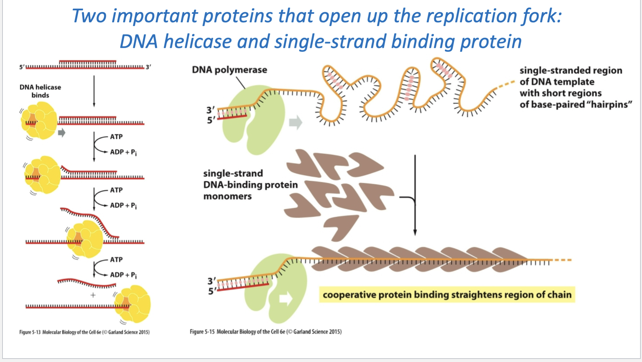
1. DNA helicase

  • Unwinds the DNA double helix

  • Separates the two parental DNA strands by breaking the hydrogen bonds between bases

  • Uses ATP for energy

  • As helicase moves forward, it opens more DNA and creates the replication fork

Think: Helicase = unzipper

2. Single-strand binding proteins (SSBs)

  • Bind to the DNA strands after helicase separates them

  • Keep the separated strands from pairing back together

  • Prevent the single strands from folding into structures like hairpins

  • Keep the DNA stretched out so DNA polymerase can copy it

Think: SSB = holds the unzipped DNA open

But helices cause torsional stress to build up and causes the fork to form supercoils so its like knots think about when you pull a water hose it coils up

Helicase

  • unwinds the double helix

  • separates the two DNA strands

  • as it keeps opening DNA, the still-double-stranded DNA ahead of it gets overwound

  • this creates supercoils / torsional stress

If that stress kept building up, the DNA would get too tightly twisted and replication would have trouble continuing.

That’s where topoisomerase comes in:

  • makes a temporary nick/cut in the DNA

  • lets the DNA rotate and relax

  • removes the supercoiling

  • then reseals the DNA

So for your flashcard:

Helicase unwinds DNA → causes supercoiling ahead of the fork → topoisomerase relieves the twisting stress by temporarily cutting and resealing DNA.
<p>1. DNA helicase</p><ul><li><p><strong>Unwinds the DNA double helix</strong></p></li><li><p>Separates the two parental DNA strands by breaking the <strong>hydrogen bonds between bases</strong></p></li><li><p>Uses <strong>ATP for energy</strong></p></li><li><p>As helicase moves forward, it opens more DNA and creates the <strong>replication fork</strong></p></li></ul><p>Think: <strong>Helicase = unzipper</strong></p><p>2. Single-strand binding proteins (SSBs)</p><ul><li><p>Bind to the DNA strands <strong>after helicase separates them</strong></p></li><li><p>Keep the separated strands from <strong>pairing back together</strong></p></li><li><p>Prevent the single strands from folding into structures like <strong>hairpins</strong></p></li><li><p>Keep the DNA stretched out so <strong>DNA polymerase can copy it</strong></p></li></ul><p>Think: <strong>SSB = holds the unzipped DNA open</strong></p><p>But helices cause torsional stress to build up and causes the fork to form supercoils so its like knots think about when you pull a water hose it coils up</p><p><strong>Helicase</strong></p><ul><li><p>unwinds the double helix</p></li><li><p>separates the two DNA strands</p></li><li><p>as it keeps opening DNA, the still-double-stranded DNA ahead of it gets <strong>overwound</strong></p></li><li><p>this creates <strong>supercoils / torsional stress</strong></p></li></ul><p>If that stress kept building up, the DNA would get too tightly twisted and replication would have trouble continuing.</p><p>That’s where <strong>topoisomerase</strong> comes in:</p><ul><li><p>makes a temporary <strong>nick/cut</strong> in the DNA</p></li><li><p>lets the DNA rotate and relax</p></li><li><p>removes the supercoiling</p></li><li><p>then reseals the DNA</p></li></ul><p>So for your flashcard:</p><p><strong>Helicase unwinds DNA → causes supercoiling ahead of the fork → topoisomerase relieves the twisting stress by temporarily cutting and resealing DNA.</strong></p>
7
New cards

DNA Repair + Telomeres

DNA repair / sequence fidelity

  • Cells use multiple repair mechanisms to keep DNA replication highly accurate

  • DNA polymerases can detect and correct some mismatched bases

  • Other repair enzymes scan DNA for mismatches and distortions

  • If DNA repair fails, mutations can accumulate

  • Repair defects are associated with diseases, including some cancers

Telomeres

  • Telomeres = repetitive DNA sequences at the ends of linear eukaryotic chromosomes

  • Their main job is to protect chromosome ends

  • They prevent chromosome ends from being mistaken for damaged/broken DNA

Main distinction:
DNA repair = protects the DNA sequence
Telomeres = protect the ends of chromosomes

8
New cards

Ways to correct tanslation

DNA polymerase proofreading its own work

  • DNA polymerase adds a nucleotide.

  • If the new base is mismatched, DNA polymerase detects that the 3′ end is wrong.

  • The polymerase shifts the DNA to its editing site.

  • Its 3′ → 5′ exonuclease activity removes the incorrect nucleotide.

  • Then DNA polymerase goes back to the polymerizing site and continues adding the correct nucleotides.

So the key idea is:

DNA polymerase builds DNA 5′ → 3′, but proofreads backward 3′ → 5′ to remove mistakes.

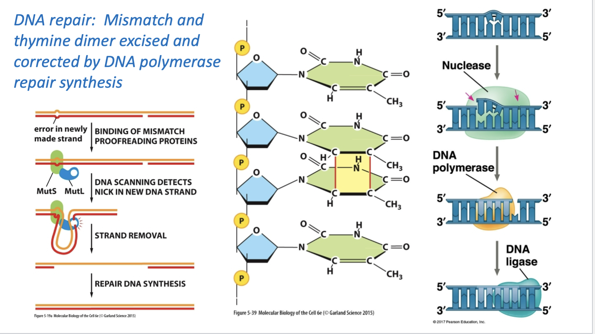
1. Mismatch repair

This fixes a base-pairing mistake that escaped DNA polymerase proofreading.

  • Repair proteins recognize the mismatch

  • The cell identifies the newly made strand

  • A section containing the mistake is removed

  • DNA polymerase fills the gap with the correct nucleotides

  • DNA ligase seals the backbone

MutS and MutL in mismatch repair

  • MutS recognizes and binds the mismatched base pair

  • MutL binds with MutS and helps coordinate the repair process

  • The repair system identifies the newly synthesized strand

  • The incorrect section is removed

  • DNA polymerase fills in the correct DNA

  • DNA ligase seals the strand

Easy way to remember:

MutS = spots the mismatch
MutL = helps organize the repair

So this is basically:

detect mismatch → remove wrong section → DNA polymerase replaces it → ligase seals it

2. Thymine dimer repair

The middle/right side is showing damage called a thymine dimer, where two neighboring thymines become abnormally linked.

  • Nuclease cuts out the damaged DNA section

  • DNA polymerase fills in the missing DNA

  • DNA ligase seals the final gap

The big difference from the previous slide is:

Proofread = DNA polymerase proofreads while DNA is being replicated
Repair = repair systems fix mistakes or damage after they are detected
<p><strong>DNA polymerase proofreading its own work</strong></p><ul><li><p>DNA polymerase adds a nucleotide.</p></li><li><p>If the new base is <strong>mismatched</strong>, DNA polymerase detects that the 3′ end is wrong.</p></li><li><p>The polymerase shifts the DNA to its <strong>editing site</strong>.</p></li><li><p>Its <strong>3′ → 5′ exonuclease activity</strong> removes the incorrect nucleotide.</p></li><li><p>Then DNA polymerase goes back to the polymerizing site and continues adding the correct nucleotides.</p></li></ul><p>So the key idea is:</p><p><strong>DNA polymerase builds DNA 5′ → 3′, but proofreads backward 3′ → 5′ to remove mistakes.</strong></p><p>1. Mismatch repair</p><p>This fixes a base-pairing mistake that escaped DNA polymerase proofreading.</p><ul><li><p>Repair proteins recognize the <strong>mismatch</strong></p></li><li><p>The cell identifies the <strong>newly made strand</strong></p></li><li><p>A section containing the mistake is removed</p></li><li><p><strong>DNA polymerase</strong> fills the gap with the correct nucleotides</p></li><li><p><strong>DNA ligase</strong> seals the backbone</p></li></ul><p>MutS and MutL in mismatch repair</p><ul><li><p><strong>MutS</strong> recognizes and binds the <strong>mismatched base pair</strong></p></li><li><p><strong>MutL</strong> binds with MutS and helps coordinate the repair process</p></li><li><p>The repair system identifies the <strong>newly synthesized strand</strong></p></li><li><p>The incorrect section is removed</p></li><li><p><strong>DNA polymerase</strong> fills in the correct DNA</p></li><li><p><strong>DNA ligase</strong> seals the strand</p></li></ul><p>Easy way to remember:</p><p><strong>MutS = spots the mismatch</strong><br><strong>MutL = helps organize the repair</strong></p><p>So this is basically:</p><p><strong>detect mismatch → remove wrong section → DNA polymerase replaces it → ligase seals it</strong></p><p>2. Thymine dimer repair</p><p>The middle/right side is showing damage called a <strong>thymine dimer</strong>, where two neighboring thymines become abnormally linked.</p><ul><li><p><strong>Nuclease</strong> cuts out the damaged DNA section</p></li><li><p><strong>DNA polymerase</strong> fills in the missing DNA</p></li><li><p><strong>DNA ligase</strong> seals the final gap</p></li></ul><p>The big difference from the previous slide is:</p><p><strong>Proofread = DNA polymerase proofreads while DNA is being replicated</strong><br><strong>Repair = repair systems fix mistakes or damage after they are detected</strong></p>
9
New cards

What happens to the last RNA primer?

  • On the lagging strand, DNA is made in Okazaki fragments

  • Each fragment starts with an RNA primer

  • Normally, the RNA primer is removed and replaced with DNA

The problem is at the very end of a linear chromosome:

  • The last RNA primer gets removed

  • But there is no 3′ OH upstream for DNA polymerase to extend from

  • So DNA polymerase cannot fill that final gap

  • This would make the chromosome shorter after each round of replication

How telomerase fixes it

  • Telomerase binds to the 3′ end

  • It uses its own RNA template

    • Telomerase contains a short RNA oligonucleotide (short chain of nucleotides) sequence

    • That RNA acts as a template

    • Telomerase uses it to add many repetitive, noncoding DNA sequences to the chromosome end

    • Those repeats form the telomere

    So the key correction is: the RNA itself does not become the long sequence. It is the template used to build the long repetitive DNA sequence.

  • It adds repetitive DNA sequences to extend the chromosome end

  • That gives the cell extra template so the lagging strand can be completed


10
New cards

gene expression

the process of using DNA information to make RNA and usually a protein

<p><span>the process of using DNA information to make RNA and usually a protein</span></p>
11
New cards

Genetic complementation test

This slide is showing how a genetic complementation test tells you whether two mutations are in the same gene or different genes.

A — Mutations are in different genes → complementation

Here:

  • m1 is a mutation in Gene 1

  • m2 is a mutation in Gene 2

When you put the two mutant copies together:

  • one chromosome has a working Gene 1

  • the other chromosome has a working Gene 2

So together, the cell can still make:

  • Protein 1

  • Protein 2

✅ Normal function is restored → the mutations complement each other → they are in different genes.

B — Mutations are in the same gene → no complementation

Here:

  • m1 is in Gene 1

  • m2 is also in Gene 1

So neither chromosome has a functional copy of Gene 1.

Result:

  • ❌ No Protein 1

  • ✅ Protein 2 is still made

Therefore the mutant phenotype remains.

❌ They do not complement → the mutations are in the same gene.

The shortcut to memorize is:

Complementation = different genes
No complementation = same gene

<p><span>This slide is showing how a <strong>genetic complementation test</strong> tells you whether two mutations are in the <strong>same gene or different genes</strong>.</span></p><p><span><strong>A — Mutations are in different genes → complementation</strong></span></p><p><span>Here:</span></p><ul><li><p><span><strong>m1</strong> is a mutation in <strong>Gene 1</strong></span></p></li><li><p><span><strong>m2</strong> is a mutation in <strong>Gene 2</strong></span></p></li></ul><p><span>When you put the two mutant copies together:</span></p><ul><li><p><span>one chromosome has a working <strong>Gene 1</strong></span></p></li><li><p><span>the other chromosome has a working <strong>Gene 2</strong></span></p></li></ul><p><span>So together, the cell can still make:</span></p><ul><li><p><span><strong>Protein 1</strong></span></p></li><li><p><span><strong>Protein 2</strong></span></p></li></ul><p><span data-name="check_mark_button" data-type="emoji">✅</span><span> Normal function is restored → the mutations <strong>complement each other</strong> → they are in <strong>different genes</strong>.</span></p><p><span><strong>B — Mutations are in the same gene → no complementation</strong></span></p><p><span>Here:</span></p><ul><li><p><span><strong>m1</strong> is in Gene 1</span></p></li><li><p><span><strong>m2</strong> is also in Gene 1</span></p></li></ul><p><span>So neither chromosome has a functional copy of Gene 1.</span></p><p><span>Result:</span></p><ul><li><p><span data-name="cross_mark" data-type="emoji">❌</span><span> No Protein 1</span></p></li><li><p><span data-name="check_mark_button" data-type="emoji">✅</span><span> Protein 2 is still made</span></p></li></ul><p><span>Therefore the mutant phenotype remains.</span></p><p><span data-name="cross_mark" data-type="emoji">❌</span><span> They <strong>do not complement</strong> → the mutations are in the <strong>same gene</strong>.</span></p><p><span>The shortcut to memorize is:</span></p><p><span><strong>Complementation = different genes</strong><br><strong>No complementation = same gene</strong></span></p>
12
New cards

Transcription in prokaryotes and eukaryotes

  1. Gene expression can be controlled at transcription

    • Transcription = making RNA from DNA.

    • If a cell makes more mRNA from a gene, it can usually make more of that gene’s protein.

    • So controlling transcription is a major way cells control gene expression.

  2. Only one DNA strand is used as the template for a given transcript

    • DNA has two strands.

    • For a particular gene, RNA polymerase normally reads one strand as the template strand.

    • Different genes can use different DNA strands.

  3. Prokaryotic transcription starts at a promoter

    • RNA polymerase + regulatory proteins bind to a promoter.

    • The DNA locally unwinds.

    • RNA polymerase reads the DNA template 3′ → 5′.

    • It builds RNA 5′ → 3′.

A really important rule to memorize:

DNA template read: 3′ → 5′
RNA made: 5′ → 3′

  1. Elongation and termination

    • RNA polymerase moves along the DNA.

    • DNA opens briefly in front of RNA polymerase and then re-anneals/re-zips behind it.

    • The newly made RNA separates from the DNA as it gets longer.

    • When RNA polymerase reaches a termination signal, transcription ends and the RNA is released.

  2. Eukaryotic transcription is more complicated
    Eukaryotes have more regulatory proteins and their initial RNA must usually be processed before becoming mature mRNA.

The big 3 processing steps are:

  • 5′ cap added

  • Introns removed by splicing

  • 3′ poly-A tail added

So a simple way to remember the difference is:

Prokaryotes: DNA → mRNA, relatively direct
Eukaryotes: DNA → pre-mRNA → processing → mature mRNA

And then that mature mRNA can be used for translation into protein.

13
New cards

Gene expression is often controlled at the level of transcription

Gene expression can be controlled at transcription

  • Transcription = making RNA from DNA.

  • If a cell makes more mRNA from a gene, it can usually make more of that gene’s protein.

  • So controlling transcription is a major way cells control gene expression.

Gene A is transcribed a lot, so it makes many RNA molecules, which then get translated into many A proteins.

Gene B is transcribed much less, so it makes less RNA, which leads to less B protein.

So the key idea is:

More transcription → more mRNA → more protein
Less transcription → less mRNA → less protein


<p><strong>Gene expression can be controlled at transcription</strong></p><ul><li><p>Transcription = making <strong>RNA from DNA</strong>.</p></li><li><p>If a cell makes more mRNA from a gene, it can usually make more of that gene’s protein.</p></li><li><p>So controlling transcription is a major way cells control <strong>gene expression</strong>.</p></li></ul><p>Gene A is transcribed a lot, so it makes <strong>many RNA molecules</strong>, which then get translated into <strong>many A proteins</strong>.</p><p>Gene B is transcribed much less, so it makes <strong>less RNA</strong>, which leads to <strong>less B protein</strong>.</p><p>So the key idea is:</p><p><strong>More transcription → more mRNA → more protein</strong><br><strong>Less transcription → less mRNA → less protein</strong></p><p></p>
14
New cards

Transcripts may use either DNA strand as a template, but usually not both for the same gene

Only one DNA strand is used as the template for a given transcript

  • DNA has two strands.

  • For a particular gene, RNA polymerase normally reads one strand as the template strand.

  • Different genes can use different DNA strands.

  • we can see in the photo that there is nearly no overlap


<p><strong>Only one DNA strand is used as the template for a given transcript</strong></p><ul><li><p>DNA has two strands.</p></li><li><p>For a particular gene, RNA polymerase normally reads <strong>one strand as the template strand</strong>.</p></li><li><p>Different genes can use different DNA strands.</p></li><li><p>we can see in the photo that there is nearly no overlap</p></li></ul><p></p>
15
New cards

sequence of events in the trasncirption of prokaryotic mRNA

  • Initiation

    • RNA polymerase binds to the promoter.

    • The promoter tells RNA polymerase where transcription should begin.

    • The DNA unwinds near the start point.

    • One strand becomes the template strand.

    • The other strand is the nontemplate strand.

    • RNA synthesis begins.

  • Elongation

    • RNA polymerase moves along the template DNA strand.

    • It reads the template DNA 3′ → 5′.

    • It builds the RNA 5′ → 3′.

    • The DNA opens in front of RNA polymerase and then rewinds behind it.

    • The growing RNA transcript comes out of the polymerase.

  • Termination

    • RNA polymerase reaches a termination signal.

    • The completed RNA transcript is released.

    • RNA polymerase comes off the DNA.


<ul><li><p><strong>Initiation</strong></p><ul><li><p>RNA polymerase binds to the <strong>promoter</strong>.</p></li><li><p>The promoter tells RNA polymerase where transcription should begin.</p></li><li><p>The DNA unwinds near the start point.</p></li><li><p>One strand becomes the <strong>template strand</strong>.</p></li><li><p>The other strand is the <strong>nontemplate strand</strong>.</p></li><li><p>RNA synthesis begins.</p></li></ul></li><li><p><strong>Elongation</strong></p><ul><li><p>RNA polymerase moves along the template DNA strand.</p></li><li><p>It reads the template DNA <strong>3′ → 5′</strong>.</p></li><li><p>It builds the RNA <strong>5′ → 3′</strong>.</p></li><li><p>The DNA opens in front of RNA polymerase and then <strong>rewinds behind it</strong>.</p></li><li><p>The growing RNA transcript comes out of the polymerase.</p></li></ul></li><li><p><strong>Termination</strong></p><ul><li><p>RNA polymerase reaches a termination signal.</p></li><li><p>The completed RNA transcript is released.</p></li><li><p>RNA polymerase comes off the DNA.</p></li></ul></li></ul><p></p>
16
New cards

eukaryotic transcription is more complex because the RNA has to be processed after transcription

Eukaryotic transcription is more complicated
Eukaryotes have more regulatory proteins and their initial RNA must usually be processed before becoming mature mRNA.

The big 3 processing steps are:

  • 5′ cap added

  • Introns removed by splicing

  • 3′ poly-A tail added

So a simple way to remember the difference is:

Prokaryotes: DNA → mRNA, relatively direct
Eukaryotes: DNA → pre-mRNA → processing → mature mRNA

And then that mature mRNA can be used for translation into protein.

  • remember mcn strcture with exons and introns


<p><strong>Eukaryotic transcription is more complicated</strong><br>Eukaryotes have more regulatory proteins and their initial RNA must usually be processed before becoming mature mRNA.</p><p>The big 3 processing steps are:</p><ul><li><p><strong>5′ cap added</strong></p></li><li><p><strong>Introns removed by splicing</strong></p></li><li><p><strong>3′ poly-A tail added</strong></p></li></ul><p>So a simple way to remember the difference is:</p><p><strong>Prokaryotes:</strong> DNA → mRNA, relatively direct<br><strong>Eukaryotes:</strong> DNA → pre-mRNA → processing → mature mRNA</p><p>And then that mature mRNA can be used for <strong>translation into protein</strong>.</p><ul><li><p>remember mcn strcture with exons and introns </p></li></ul><p></p>
17
New cards

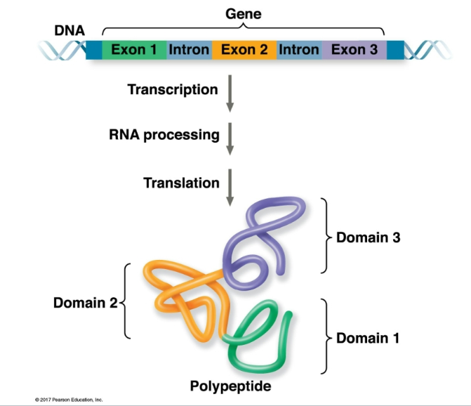
Eukaryotic coding sequences-exons- often correspond to independent domains of the protein

  • Exon 1 contributes to Domain 1

  • Exon 2 contributes to Domain 2

  • Exon 3 contributes to Domain 3

A protein domain is a region of a protein that can have its own structure or function.

So the flow is:

DNA with exons + introns → transcription → RNA processing → exons joined → translation → protein with different domains

In eukaryotes, introns are removed and exons are spliced together; exons often encode separate functional domains of a protein.

<ul><li><p>Exon 1 contributes to <strong>Domain 1</strong></p></li><li><p>Exon 2 contributes to <strong>Domain 2</strong></p></li><li><p>Exon 3 contributes to <strong>Domain 3</strong></p></li></ul><p>A <strong>protein domain</strong> is a region of a protein that can have its own structure or function.</p><p>So the flow is:</p><p><strong>DNA with exons + introns → transcription → RNA processing → exons joined → translation → protein with different domains</strong></p><p><strong>In eukaryotes, introns are removed and exons are spliced together; exons often encode separate functional domains of a protein.</strong></p><p></p>
18
New cards
<p>remember how to read and interpret this dont memorize </p>

remember how to read and interpret this dont memorize

You can see that this single mRNA has several separate regions, each with:

  • its own ribosome-binding site

  • its own AUG start codon

  • its own coding region

That means the same mRNA can be translated into:

  • protein α

  • protein β

  • protein γ

  • each mRNA can translate different start signals with the start signal in between


<p>You can see that this single mRNA has several separate regions, each with:</p><ul><li><p>its own <strong>ribosome-binding site</strong></p></li><li><p>its own <strong>AUG start codon</strong></p></li><li><p>its own coding region</p></li></ul><p>That means the same mRNA can be translated into:</p><ul><li><p><strong>protein α</strong></p></li><li><p><strong>protein β</strong></p></li><li><p><strong>protein γ</strong></p></li><li><p><strong>each mRNA can translate different start signals with the start signal in between</strong></p></li></ul><p></p>
19
New cards

redundancy of the genetic code

Redundancy means that more than one codon can code for the same amino acid.

For example, leucine can be coded by several different codons:

  • UUA

  • UUG

  • CUU

  • CUC

  • CUA

  • CUG

So even if one nucleotide changes, the amino acid sometimes does not change.

That matters because it can make the genetic code more tolerant of mutations. Some mutations are silent mutations, meaning the DNA or mRNA codon changes, but the same amino acid is still added to the protein.

20
New cards

Single nucleotide mutations in the 3rd codon position are usualyl silenced

Still gly this is because of redundancy

<p>Still gly this is because of redundancy</p>
21
New cards

missense mutation

mutations in the 1st or 2nd nucleotide position of a codon are more likely to change the amino acid, because those positions are usually more important for determining which amino acid is coded.

<p>mutations in the <strong>1st or 2nd nucleotide position of a codon</strong> are more likely to change the amino acid, because those positions are usually more important for determining which amino acid is coded.</p>
22
New cards

stop chain mutation

Remmeber stop chains are UAA UAG and UGA


<p>Remmeber stop chains are UAA UAG and UGA</p><p></p>
23
New cards

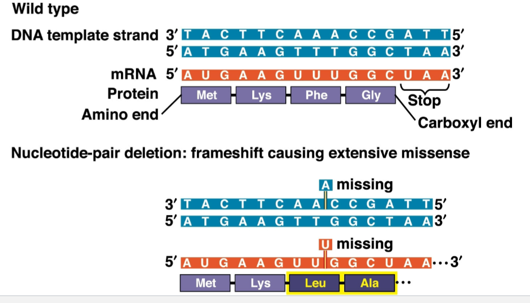
Single nucleotide deletion (or insertion) shifts the reading frame downstream of the insertion

very substatial because think about a story if you take a part out of a story or add a part the story would not make sense

more substantial depending on how early it arrives
<p>very substatial because think about a story if you take a part out of a story or add a part the story would not make sense</p><p>more substantial depending on how early it arrives</p>
24
New cards

ribosome during translation

The ribosome has 2 subunits:

  • Small ribosomal subunit: binds the mRNA

  • Large ribosomal subunit: helps form the protein by making peptide bonds

Inside the ribosome are 3 important tRNA-binding sites:

  • A site = Aminoacyl site/ Acceptor site

    • The new tRNA enters here

    • It carries the next amino acid

  • P site = Peptidyl site

    • Holds the tRNA carrying the growing polypeptide chain

    • This is where the protein chain is being built

  • E site = Exit site

    • The empty tRNA moves here before leaving the ribosome

The easiest way to memorize the order is:

A → P → E

Arrive → Protein → Exit

So during elongation:

  1. A charged tRNA enters the A site

  2. Its amino acid gets added to the growing chain

  3. The tRNA shifts to the P site

  4. The old empty tRNA shifts to the E site and leaves


25
New cards

translating the code to the placement of amino acids in a polypeptide

1. Transfer RNA – tRNA – folds into a tertiary structure recognized by enzymes that

attach specific amino acids to the 3’OH terminus. A folded loop of the tRNA presents

the triplet information to base pair to a codon.

2. Amino acids are attached to the 3’ terminus of a tRNA in a two-step process

requiring ATP to form a high energy phosphoanhydride bond between the α phosphate

of ATP and the aa COO- which is then transferred to the 3’OH of the tRNA with the

release of AMP and PPi.

3. The full ribosome has two large multisubunit RNA-protein complexes which contain

three sites engaged in polypeptide assembly: A (the acceptor site), P (the peptide-

tRNA site and E (the exit site free tRNA). mRNA is positioned along the surface of the

smaller ribosome subcomplex. A growing polypeptide chain threads through a

channel in the large subcomplex.

4. Each step in the cycle of polypeptide growth is organized by GTPases involved in

initiation, elongation and chain termination. Peptide bonds are formed by a catalytic

site in the large subunit with RNA and protein involved in catalysis.

26
New cards

tRNA

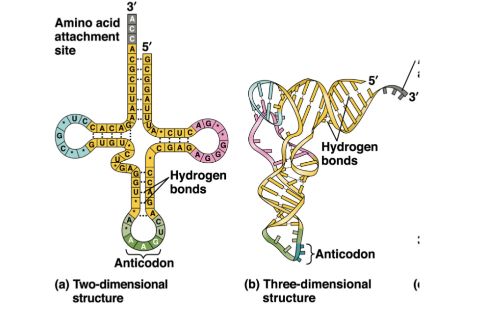
tRNA, or transfer RNA, is basically the molecule that brings the correct amino acid to the ribosome during translation.

Think of it as an adapter between the mRNA code and amino acids.

It has two especially important parts:

  • The 3′ end at the top is the amino acid attachment site. A specific amino acid gets attached there.

  • The anticodon at the bottom is a 3-base sequence that pairs with a complementary codon on the mRNA.

For example, if the mRNA codon is:

5′-AUG-3′

the tRNA has a complementary anticodon that pairs with it, and that tRNA carries methionine.

The left side of the figure is the simplified 2D cloverleaf shape, while the right side shows the actual folded 3D shape of tRNA.
<p><strong>tRNA, or transfer RNA</strong>, is basically the molecule that <strong>brings the correct amino acid to the ribosome during translation</strong>.</p><p>Think of it as an <strong>adapter</strong> between the mRNA code and amino acids.</p><p>It has two especially important parts:</p><ul><li><p>The <strong>3′ end</strong> at the top is the <strong>amino acid attachment site</strong>. A specific amino acid gets attached there.</p></li><li><p>The <strong>anticodon</strong> at the bottom is a 3-base sequence that pairs with a complementary <strong>codon on the mRNA</strong>.</p></li></ul><p>For example, if the mRNA codon is:</p><p><strong>5′-AUG-3′</strong></p><p>the tRNA has a complementary anticodon that pairs with it, and that tRNA carries <strong>methionine</strong>.</p><p>The left side of the figure is the simplified <strong>2D cloverleaf shape</strong>, while the right side shows the actual folded <strong>3D shape</strong> of tRNA.</p>
27
New cards

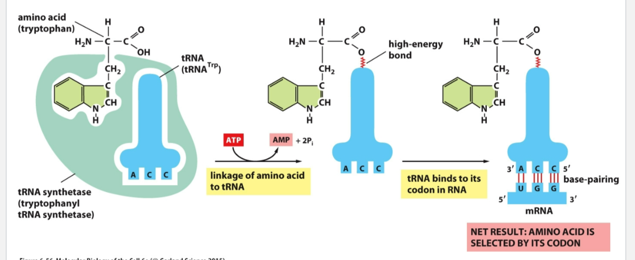
how a tRNA gets “charged” with the correct amino acid

  • On the left, the enzyme aminoacyl-tRNA synthetase recognizes:

    • the correct amino acid

    • the correct tRNA

In this example, the amino acid is tryptophan and the tRNA is tRNAᵀʳᵖ.

  • The enzyme uses ATP to help attach tryptophan to the 3′ end of the tRNA.

  • ATP is converted to AMP + PPi.

  • This forms a high-energy covalent bond between the amino acid and the tRNA.

Now the tRNA is called a charged tRNA or aminoacyl-tRNA.

Then on the right:

  • The anticodon of the tRNA base-pairs with the matching mRNA codon

  • Here, the mRNA codon is UGG

  • UGG codes for tryptophan


<ul><li><p>On the left, the enzyme <strong>aminoacyl-tRNA synthetase</strong> recognizes:</p><ul><li><p>the correct <strong>amino acid</strong></p></li><li><p>the correct <strong>tRNA</strong></p></li></ul></li></ul><p>In this example, the amino acid is <strong>tryptophan</strong> and the tRNA is <strong>tRNAᵀʳᵖ</strong>.</p><ul><li><p>The enzyme uses <strong>ATP</strong> to help attach tryptophan to the <strong>3′ end of the tRNA</strong>.</p></li><li><p>ATP is converted to <strong>AMP + PPi</strong>.</p></li><li><p>This forms a <strong>high-energy covalent bond</strong> between the amino acid and the tRNA.</p></li></ul><p>Now the tRNA is called a <strong>charged tRNA</strong> or <strong>aminoacyl-tRNA</strong>.</p><p>Then on the right:</p><ul><li><p>The anticodon of the tRNA base-pairs with the matching <strong>mRNA codon</strong></p></li><li><p>Here, the mRNA codon is <strong>UGG</strong></p></li><li><p>UGG codes for <strong>tryptophan</strong></p></li></ul><p></p>
28
New cards

how a peptide bond forms during translation

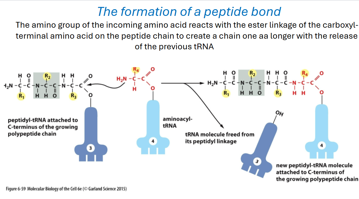
It connects directly to the ribosome A and P sites

On the left, the growing polypeptide chain is attached to the tRNA in the P site. A new amino acid arrives attached to another tRNA in the A site.

Then the key reaction happens:

  • the amino group of the new amino acid attacks the bond holding the growing peptide to the old tRNA

  • a new peptide bond forms

  • the entire growing polypeptide chain gets transferred onto the tRNA in the A site

  • the old tRNA is released from the peptide

So before:

P-site tRNA = holds growing chain
A-site tRNA = holds new amino acid

After the peptide bond forms:

A-site tRNA = now holds the longer growing chain
P-site tRNA = empty

Then the ribosome moves forward and the empty tRNA can eventually leave through the E site.

The protein grows from the N-terminus toward the C-terminus, so each new amino acid is added to the C-terminal end.
<p>It connects directly to the ribosome <strong>A and P sites</strong></p><p>On the left, the growing polypeptide chain is attached to the tRNA in the <strong>P site</strong>. A new amino acid arrives attached to another tRNA in the <strong>A site</strong>.</p><p>Then the key reaction happens:</p><ul><li><p>the <strong>amino group</strong> of the new amino acid attacks the bond holding the growing peptide to the old tRNA</p></li><li><p>a new <strong>peptide bond</strong> forms</p></li><li><p>the entire growing polypeptide chain gets transferred onto the tRNA in the A site</p></li><li><p>the old tRNA is released from the peptide</p></li></ul><p>So before:</p><p><strong>P-site tRNA = holds growing chain</strong><br><strong>A-site tRNA = holds new amino acid</strong></p><p>After the peptide bond forms:</p><p><strong>A-site tRNA = now holds the longer growing chain</strong><br><strong>P-site tRNA = empty</strong></p><p>Then the ribosome moves forward and the empty tRNA can eventually leave through the <strong>E site</strong>.</p><p>The protein grows from the <strong>N-terminus toward the C-terminus</strong>, so each new amino acid is added to the <strong>C-terminal end</strong>.</p>
29
New cards

Prokaryotes transcription and translation

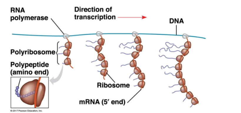
In prokaryotes, transcription and translation can happen at the same time.

Here’s what is happening:

  • RNA polymerase is moving along the DNA and making mRNA.

  • As soon as part of the mRNA comes out, ribosomes can bind to it immediately.

  • Those ribosomes start translating the mRNA into protein before transcription is even finished.

That is what “coupled” means here.

The long mRNA has many ribosomes attached to it at once. That group is called a:

polyribosome / polysome

So one mRNA can be translated by many ribosomes at the same time, producing many copies of protein quickly.

The reason prokaryotes can do this is that they do not have a nucleus, so there is no nuclear membrane separating DNA transcription from ribosome translation.

So the main takeaway is:

Prokaryotes: transcription and translation are coupled

  • RNA polymerase makes mRNA

  • ribosomes begin translating that mRNA immediately

  • multiple ribosomes can translate the same mRNA at once

This is different from eukaryotes, where transcription happens in the nucleus and translation happens in the cytoplasm, so the two processes are separated.
<p><strong>In prokaryotes, transcription and translation can happen at the same time.</strong></p><p>Here’s what is happening:</p><ul><li><p><strong>RNA polymerase</strong> is moving along the DNA and making mRNA.</p></li><li><p>As soon as part of the mRNA comes out, <strong>ribosomes can bind to it immediately</strong>.</p></li><li><p>Those ribosomes start translating the mRNA into protein <strong>before transcription is even finished</strong>.</p></li></ul><p>That is what “coupled” means here.</p><p>The long mRNA has many ribosomes attached to it at once. That group is called a:</p><p><strong>polyribosome / polysome</strong></p><p>So one mRNA can be translated by many ribosomes at the same time, producing many copies of protein quickly.</p><p>The reason prokaryotes can do this is that they <strong>do not have a nucleus</strong>, so there is no nuclear membrane separating DNA transcription from ribosome translation.</p><p>So the main takeaway is:</p><p><strong>Prokaryotes: transcription and translation are coupled</strong></p><ul><li><p>RNA polymerase makes mRNA</p></li><li><p>ribosomes begin translating that mRNA immediately</p></li><li><p>multiple ribosomes can translate the same mRNA at once</p></li></ul><p>This is different from eukaryotes, where transcription happens in the <strong>nucleus</strong> and translation happens in the <strong>cytoplasm</strong>, so the two processes are separated.</p>