D103 MT Review
 Note
Note Call Kai
 Learn
Learn Practice Test
 Spaced Repetition
 Match
Match Flashcards
 Knowt Play
1/83

There's no tags or description
Looks like no tags are added yet.
Name | Mastery | Learn | Test | Matching | Spaced | Call with Kai | Chat |
|---|
No analytics yet
Send a link to your students to track their progress
84 Terms
How does dysfunction of organization affect the cell?
Leads to disease
Ex) centrosome, golgi membrane, ER network, mitochondria
Model Systems
Simple (easy, cheap, quick)
Must have conserved mechanism
Availability of genetics (ie genome sequenced)
Light microscopy limitation?
Resolving power is ½ wavelength of source light, need shorter light to see smaller molecules (ex electron microscopy)
General phospholipid structure
Glycerol backbone with diff head group and tails
Lipid distribution in a membrane
Asymmetrical, can get curvature bc of smaller head sizes
Mobility of phospholipids
Addition of heat leads to a more fluid membrane
Regulation of membrane fluidity
Length of chains → shorter chains get less interaction, more fluid membrane
Saturation degree → highly saturated = more kinks, more fluid
Cholesterol levels → stiffens membrane and increases thickness, less fluid
Transmembrane proteins
Must have hydrophobic domains that are inserted into the membrane
Hydropathy plot
Top area refers to hydrophobicity, number of peaks surpassing 20-25 residues coincide with a trans membrane domain
Membrane transport proteins
All have transmembrane domains
Allow water soluble membranes to pass through
Undergo a conformational change
Transporters: Uniporter
Passive transport down a gradient
Transporters: Symporter
Moves two molecules at once, movement of one particle down its gradient provides energy to move another particle against its gradient in the same direction
Transporters: Antiporter
Moves two molecules at once in diff directions, energy gained from movement of one particle down its gradient
Pumps
Use ATP to move a molecule up its gradient
Ion channels
Passive transport of an ION down its gradient, opens due to binding of some ligand
Vmax
Each protein is working at max rate
Trans-epithelial transport of glucose
Na+/Glu symporter takes advantage of the low Na+ concentration in the lumen to move glucose
This low Na+ concentration is maintained by a Na+/K+ pump on the blood vessel membrane
Glucose then moves to blood by a simple uniporter
Role of mitochondria
Takes up food, breaks it down into energy (ATP) through ox phos
Oxidative phosphorylation
Food from cytosol → moves into inner membrane → acetyl-CoA → TCA makes electron carriers → ETC (on inner membrane) → generates proton gradient → ATP synthase (inner mito membrane) uses gradient to make ATP
UCP (uncoupling protein)
Uncouples proton gradient from ATP synthase
Forms an alternative pathway for protons back into the mito matrix
Releases energy as heat
Key features of mitochondria
Double membrane
Abundant (proportional to energy req), distributed throughout the cell
Dynamic as they regularly fuse and divide as they move around the cell
Inner mitochondrial membrane
Many cristae to increase surface area
High protein content
Has a phospholipid with 4 tails → EXTREMELY hydrophobic an robust (impermeable)
Outer mitochondrial membrane
Has porins, permeable to small molecules
mtDNA
Mitochondria have their own DNA that is transcribed/translated on demand but in house machinery
Most mito proteins are still encoded by nuclear DNA
Dense packing/no introns
No proof-reading/repair so mutation rate is really high
Passed down from mother (mtDNA is not evenly distributed into daughter cells so can have diff levels of mutational severity)
Immunofluorescent staining
Primary antibody: inject whatever protein into a host that will make antibodies for it
must have diff animals if staining multiple things
Secondary antibody: inject primary into ANOTHER animal, it will recognize the animal antibody, has a fluorescent light
Cytoskeletal elements
Microfilament actin (F-actin)
Intermediate filament
Microtubules (tubulin)
V dynamic due to monomer construction and regulatory proteins that control de/polymerization
Fluorescent tagging
Tag genes with GFP or fluorescent marker
Then transcription/translation results in protein that glows
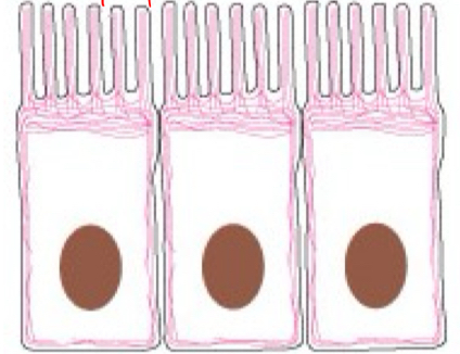
F-actin
Cell shape and migration
Made of G-actin subunits
Assembly depends on ATP hydrolysis (reqs energy)
Has + and - end
Nucleation: monomers slowly form stable complexes
Cytosolic concentration of G-actin determines growth/shrinkage
Above Cc = growth
Below Cc = shrinkage
Cc is different for +/- end, so growth rates are different
Addition is faster at + end
Treadmilling
Cc- > Ccyto > Cc+
+ end continuously grows (to reach Ccyto) while - end continuously shrinks (to reach Ccyto)
Cell maintains the same filament content
Very dynamic behavior lends to flexibility → cell can change to make quick responses to diff signals
Regulated by accessory proteins
Sequestering proteins and G-actin activators: regulate concentration of available G-actin (neg/pos)
capping proteins: protest from depolymerization
Nucleators: nuclear assembly or linear and branched actin
Actin coordinated cell migration
Protrusions form at leading edge due to polymerization
Atttach to substratum
Cell body moves using myosin generated force
Aka polymerization, adhesion, and translocation
Small GTPases
Rho (stress fiber and contraction), Rac (protrusion formation), and Cdc42 (morphology for metastasis)
All regulate cell migration through actin → do so by regulating downstream effectors
They themselves are regulated by GEF and GAP
GEF activates the protein by phosphorylation to GTP bound state
GAP deactivates protein by dephosphorylation to GDP state
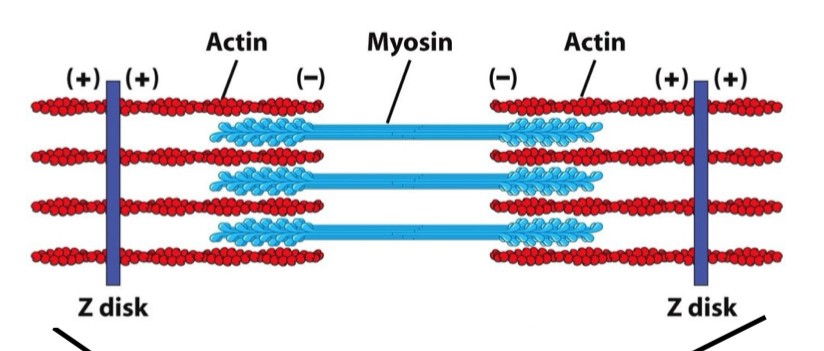
Myosin
Actin-based motor proteins
Two binding sites: ATP (for energy) and actin
Basic unit = sarcomere
Actin + end is capped, - end is held by myosin
Ca++ binds to troponin, allowing tropomyosin to lift away and reveal myosin binding site for ATP
Myosin then “walks” to + end to contract
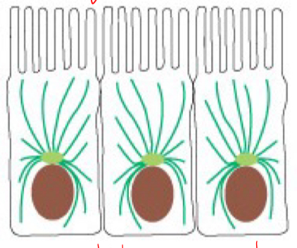
Tubulin
organize organelles and mediate intracellular transport
composed of heterodimers
alpha is GTP bound
beta is GDP bound but can be exchanged with soluble GTP
elongation occurs at + end
GTP cap refers to GTP accumulation at the + end
rate of hydrolysis from GTP to GDP is slower than addition of dimer subunits
Tubulin dynamics
catastrophe refers to accidental loss of GTP cap, leads to rapid shrinkage
rescue refers to regain of GTP cap and rapid growth
cap loss/gain controlled by local GTP-bound tubulin concentration
Structural consequences of GTP hydrolysis
loss of GTP cap
curling of microtubule filaments
MTOC
microtubule organizing center
where mts grow from, provide a nucleation site
during interphase, centrosome is the main MTOC
nucleating sites are gamma-tubulin rings, grow from plus end while minus end is capped
Cargo transport with microtubules
kinesin carries cargo to the plus end (K+ like potassium)
dynein moves cargo to the minus end
has two head domains: one for ATP and one for the mt
movement requires ATP hydrolysis
organelle membranes contain motor receptors to determine which motors can bind and deposit their cargo where
MT during mitosis
significant increase in MT nucleation during mitosis
more dynamic bc they have to search for chromosomes (do so by continuously reaching out (growing/shrinking)
FRAP (fluorescence recovery after photobleaching)
expression GFP fusion protein, photo bleach, determine how fast GFP signal recovers
premise is that the more dynamic neighboring cells are, the faster they move to fill up that hole and the faster the signal recovers
Taxol
binds to mts and inhibits mt depolymerization → inhibits dynamics and thus halts the process of mitosis
kills of hyper prolific cancer cells
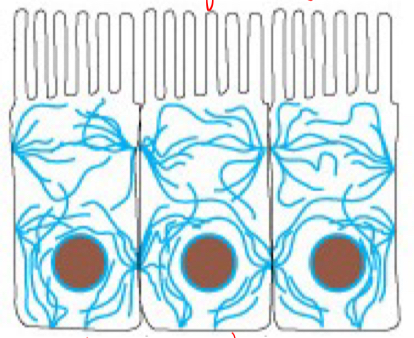
Intermediate filament
gives cell mechanical strength
ex keratin in epithelia or nuclear lamins
polymer formation is spontaneous, however depolymerization requires energy
Lamins
provide structural support for cell
contain nuclear localization agent NLS
Nuclear envelope during mitosis
must breakdown
requires energy to dissemble lamina
regulated by kinase induced phosphorylation
Keratin
a type of intermediate filament in the epithelial cells that connect neighboring tissue
Desmosome
a cadherin-keratin structure that links IF with neighboring cells and ECM
cadherin and keratin also require an anchor protein to mediate their connection
cell to cell
Cadherin
Ca++ dependent adhesion
have Ca++ binding sites, when bound they straighten out the structure and mediate cell to cell connection
cells that express the same cadherin turn to interact with each other
Cadherin and cancer
dysregulation frequently associated with cancer
loss of cadherin leads to increased migration and invasion of cells
epithelial cells undergo EMT and metastasize
Hemidesmosome
cell junction that anchors IF to the basal lamina
cell to ECM
Adherens junction
cadherin links an actin bundle to another bundle in a neighboring cell
cell to cell
Focal adhesion
uses integrins (alpha and beta subunit) to link the cytoskeleton (actin and IF) to the ECM
binding mediated by adaptor proteins
cell to ECM
Integrins
a transmembrane protein used to link cytoskeleton to ECM
binding of the ligand causes conformational change
outside-in activation: ECM ligand binds to activate integrin and thus intracellular events
inside-out activation: actin associated protein binding activates integrins and then extracellular events
inactive form is compact, active form is stretched out
Actin polymerization
induces integrin activation and focal adhesion
ex of inside-out activation
ECM
collection of extracellular molecules that provide structural/biochemical support for surrounding cells
contains insoluble fibers such as collagen/elastin/etc
Collagen
extremely important ECM component
vitamin c is a cofactor for collagen processing
Principles for protein transport
has to have a sorting signal
signal has to get recognized by a specific receptor
receptor should bring protein to translocation machinery
transported protein gets released
energy input for transport
processing required for function
Sorting signal
must be necessary and sufficient
may be removed upon arrival
signal sequences or patch (which is made of distant aa residues that make a patch when processed)
Protein transport into nucleus
nuclear pores constitutively open, but require NLS
importin proteins bind to the cargo when they recognize NLS
move into nucleus where Ran GTPase binds to importin, leaving cargo
GTPase activated by GEF in nucleus
Ran GTPase and importin move out through nuclear pore to cytosol, where GAP converts it to Ran GDPase and releases importin
Import into mitochondrial matrix
sorting signal is amphipathic helix
proteins imported in unfolded state, require chaperones
transport machinery TOM (outer) and TIM (inner)
driving force for import is ATP and a proton gradient
ER
stores Ca++ for muscle contraction
receives, modifies, and transports proteins
synthesizes lipids
smooth: vesicle formation
rough: protein import, secretory pathway, folding processed here
Protein import to ER
SRP or signal recognition particle is recognized by SRP receptor
translocated by translocons
GTP hydrolysis powers translocation
Translocation of soluble molecule
ER signal sequence is read then cleaved by signal peptidase after translocation
goes straight through
Translocation of transmembrane molecule
ER signal sequences is read then once the stop-transfer sequence is read, the protein stops moving through the membrane and the signal sequence is cleaved
Topology of ER transmembrane protein
C-terminal in cytosol
N-terminal in cytosol
ER proteins get glycosylated when they enter the ER
N-linked: initiated in ER, also in golgi
O-linked: only in the golgi
both just add a sugar tree to the protein
ER proteins are associated with chaperones
prevent premature folding
v strong bond to protein, requires energy to break
chaperones also recognize incorrect glycosylation
Folding issues and UPR
glucosidases remove sugar tree
if folded incorrectly, exit the ER
if not, another protein adds the tree back
misfolded proteins leads to ER stress
UPR tries to solve the issue by halting protein prod, influx, and increasing chaperones
when the cell gives up, it will undergo ERAD to degrade misfolded proteins and induce apoptosis
ERAD
when misfolded protein gets pulled out of ER and then degraded in proteosome
Secretory pathway
mediated by vesicles
clathrin: trans-golgi network, endocytosis
COPI: golgi to golgi, golgi to ER
COPII: ER to golgi
divided into early and late stages
Proteins involved with vesicle budding/trafficking
assemble GTPase → coat assembly from donor compartment
Rab GTPase → vesicle docking
SNARE → mediate fusion with target compartment
Vesicle formation
small GTPase recruited to donor membrane
inner and outer coat proteins recruited
coat proteins selectively bind and recruit cargo
vesicle buds
Vesicle fusion
vesicle gets uncoated to expose v-SNARE proteins
Rab initiates docking by finding an effector on the target
v-SNARE binds to t-SNARE
vesicle fuses to target compartment
SNARE proteins get hydrolyzed and then recycled
Botox
inhibits muscle contraction by blocking synaptic vesicle fusion
bind and cleave SNARE proteins
Early secretory pathway
ER to cis-golgi (COPII)
Cis-golgi to ER and golgi to golgi (COPI)
Late secretory pathway
proteins are modified in golgi
Protein trafficking between ER and golgi
forward is mediated by COPII
backward is mediated by COPI
for retrieving recycled proteins
maintaining membrane balance
missorted ER proteins
KDEL
protein retrieval signal
KDEL receptors are super pH sensitive, in the golgi it will have high affinity for KDEL and help move it back to the ER
Constitutive secretion
default, always on
for proteins in PM and ECM
Regulatory secretion
stored, wait for signal stimulation
Lysosome
major function is to eat/digest things with its many enzymes
most efficient at low pH maintained by proton pump
clathrin vesicles transport cargo to lysosome
Vesicle transport to lysosome
M6P sorting signal
pH sensitive, dissociates in the more acidic lysosome environment
clathrin vesicle buds, congregate to form early endosome which congregate with each other to form late endosome
fusion
Protein modification
only permanent one is lipid/GPI anchor
Ubiquitination
any protein containing a lysine residue can be modified
ubiquitin also has a lysine, can bind to more of itself for polyubiquitination
the more you add the greater the mw
three steps mediated by E1-3
DUB also removes ubiquitin
Polyubiquitination
targets proteins for degradation through proteasome
modify in cytosol
Phosphorylation
modification of Ser, Thr, Tyr
adds negative charge