NU 236 Care of the Adult II: Surgical Respiratory
 Call Kai
 Learn
Learn Practice Test
 Spaced Repetition
 Match
Match Flashcards
 Knowt Play
1/77

There's no tags or description
Looks like no tags are added yet.
Name | Mastery | Learn | Test | Matching | Spaced | Call with Kai | Chat |
|---|
No analytics yet
Send a link to your students to track their progress
78 Terms

Lung cancer
second most common type of cancer and #1 cancer killer worldwide, with a 5-year survival rate of 28.6%
Risks: Lung Cancer
Cigarette smoking (85-95%)
Prolonged exposure to secondhand smoke, asbestos, radiation, air pollution, chemicals, or coal mining
Primary Prevention: Lung Cancer
NO smoking
Avoid radon, asbestos, and secondary smoke
Second-Hand Smoke
environmental tobacco smoke; increases risk of lung cancer in non-smokers, SIDs, heart disease, and asthma in children
Adolescents and Tobacco
often use flavored vapes; can help quit with tobacco control programs such as mass media campaigns, higher prices, smoke-free laws, policies, evidence-based school programs, and bans
Three Steps to Quitting Smoking
Want IT (set a QUIT date and stick to it)
Plan IT (make a list of triggers and find something to distract you)
Do IT (reward yoruself when you do not smoke)
Stages of Change
1. Precontemplation
2. Contemplation
3. Preparation
4. Action
5. Maintenance
When teaching smokers, always meet them ________ using the stages of change.
where they are at!
Smoking Cessation Options
Counseling/support groups
Nicotine patches, gum, inhalers, zyban, Chantix
Secondary Prevention: Lung Cancer
Often asymptomatic early
Screenings ONLY for smokers
Screenings for Lung Cancer
recommended is annual CT screening of current/former smokers aged 50-80 with 20 pack/yr history, and applies to those who have quit within the past 15 years
Clinical Manifestations: Lung Cancer
Persistent cough
Hemoptysis
Chest pain
Recurring pneumonia/bronchitis
Anorexia
Weight loss
Fatigue
Dyspnea
Hoarseness
Hemoptysis
bloody sputum
Diagnosis: Lung Cancer
Sputum for cytology
Biopsy via bronchoscopy
Transthoracic needle biopsy
Bronchoscopy
the visual examination of the bronchi using a bronchoscope, often to obtain a biopsy

Pre-Procedure: Bronchoscopy
Informed consent and IV access
NPO unless emergent
Post-Procedure: Bronchoscopy
NPO until swallow reflex returns d/t risk of aspiration from numbness
No driving 24hr
Complications: Bronchoscopy
Frank bleeding (NOT blood-streaked sputum, expected)
Vitals, O2 sat, and lung sounds
Possible pneumothorax

Transthoracic needle biopsy
technique of obtaining a biopsy specimen of lung tissue by which a needle is passed through the chest, often guided by CT imaging to create a culture for analysis
When else are transthoracic needles used?
pleural effusion to remove fluid

Pleural Effusion
abnormal accumulation of fluid in the pleural space, decreasing the ability of the lungs to expand
Causes: Pleural Effusion
HEART FAILURE
Bacterial pneumonia
Malignancies
Cirrhosis with ascites
Pancreatitis
TB
Signs and Symptoms: Pleural Effusion
SHORTNESS OF BREATH
Chest pain (sharp or stabbing) and increased with deep inspiration
May have other local/systemic symptoms and may look like TB, bacterial pnuemonia, or CHF
Assessment: Pleural Effusion
Decreased or absent breath sounds
Dullness upon percussion
Decreased fremitus/resonance
Abnormal CXR/CT
Labs
Thoracentesis
Pleural biopsy
Labs: Pleural Effusion
Cytology for malignancy (may not always show cancer)
WBC/RBC
Blood and pleural fluid cultures
Presence of proteins, amylase, glucose

Thoracentesis
surgical puncture to remove fluid from the pleural space with immediate relief; done from the back where the pleural gutter is deepest
Positioning: Thoracentesis
sitting on the side of the bed and leaning over the table with head down
Complications: Thoracentesis
Pneumothorax (d/t lung puncture)
Bleeding
Pneumothorax
atmospheric air enters the pleural lining, causing lung collapse

Max amount of fluid removed in a thoracentesis is..
1L at a time
Nursing Interventions: Thoracentesis
Informed consent and explanation
Pre-procedure x-ray
Proper positioning and remain still
Vitals, especially O2 sat, and skin color throughout procedure
Apply dressing post-op, monitor vitals, and encourage deep breathing
Mediastinoscopy
visual examination of the mediastinal structures, including the heart, trachea, esophagus, bronchus, thymus, and lymph nodes
Thoroscopy
visual examination of the thoracic cavity
Pack Years
# of packs/day X # of years smoked
TNM Staging for Lung Cancer
T = primary tumor
N = regional lymph nodes
M = distant metastasis
Used to determine extent of disease and prognosis, and the patterns of spread depend on type of lung cancer and location (Stage I and II are surgical)
Types of Lung Cancer
Non-small cell lung cancer (NSCLC)
Small cell lung cancer (oat cell) (VERY aggressive)

Non-Small Cell Cancers
adenocarcinoma, squamous cell, large cell; first treated with usual respiratory interventions and possible surgery, radiation, and chemotherapy
Surgeries: NSCLC
Thoracotomy
Wedge resection
Lobectomy
Pneumoectomy
Post-Op: Lung Surgery
Assess cardiac/respiratory status
O2 admin
Incentive spirometry q1-2 hrs
Pain management
NO positioning on operative side
Turn, cough, deep breath
HOB > 30-45
Active range of motion to shoulder
Chest tube drainage system
Chest Tube
catheter inserted through the thorax into the chest cavity for removing air or fluid; used after chest or heart surgery or pneumothorax.
Hemothorax
a collection of blood in the pleural cavity

3 Chambers of Chest Tubes
Collection (measurement)
Water seal (one-way valve)
Suction (wall)
Chest Tube Assessment
Air leak
Bubbling
Crepitus
Dependant loops
Proper seal
Drainage measurement every shift
Below chest level
Is bubbling right after insertion expected? What about after three days?
YES, as fluid is being removed; NO, this means that there is a leak
Crepitus
air in the subcutaneous tissue
What should be kept at the bedside for a patient with a chest tube?
Bottle of unopened sterile water
Clamps
Sterile 4X4
Sterile vaseline gauze
Tape
Sterile basin
Always make sure chest tube systems are ___________ and ________.
connected, taped
How often do you get a CXR with a chest tube?
Before placement and after placement
1x/day
Chest Tube Dislodgement
Cover with a dry sterile dressing taped on 3 sides
If there is an air leak tape the dressing on 3 sides only to allow air to escape and prevent the formation of a tension pneumothorax
Notify HCP and prepare for reinsertion
Chest Tube Disconnection
DO NOT CLAMP
Immediately place end of chest tube in container of sterile saline or water until a new drainage system can be connected
Why do we place the chest tube in sterile water?
acts as a temporary one-way water seal that lets air and fluid escape from the pleural space while blocking outside air from rushing back into the chest
Concerns: Lung Cancer
Dyspnea
Pain
Anxiety
Coping
Altered nutrition
Activity intolerance
Stigma/judgment
D/C Teaching: Lung Cancer
Symptom management
When to call MD (pain, fever)
Safe use of O2 and narcotics at home
Smoking cessation
Facilitate expression of fears/death and referrals to hospice/home care
Smoking Cessation Teaching
done at EVERY encounter, provide resources
Laryngeal Cancer
cancer of the throat (vocal chords, larynx, or other areas of the throat and often occurs between ages 50-65 years and is 9x more common in men than women
Risks: Laryngeal Cancer
SMOKING
Alcohol abuse
Exposure to pollutants/radiation
Voice strain
Clinical Manifestations: Laryngeal Cancer
Persistent hoarseness or sore throat
Painless neck mass
"Lump in throat"
Burning of throat
Dysphagia
Change in voice quality
Dyspnea
Hemoptysis
Weakness
Foul breath
Diagnostics: Laryngeal Cancer
Laryngoscopy
Biopsy
Chest x-ray to rule out lung mets
Treatment: Laryngeal Cancer
Radiation and chemotherapy
Surgery (partial laryngectomy and total laryngectomy)
Partial laryngectomy
surgical removal of part of the larynx for small tumors; patient can STILL speak
Total laryngectomy
surgical removal of the entire larynx for large tumors; patient will NOT be able to speak and will have a stoma

Radical Neck Dissection
surgical removal of lymph nodes in the neck
Pre-Op: Total Laryngectomy
Establish a method of communication and encourage expression of feelings about changes of body image/voice loss
Rehab/trach care and suctioning
Post-Op: Total Laryngectomy
Position on side with HOB < 30-45
Humidification to trach site/stoma
Maintain airway and suction PRN
Monitor pulse ox, lung sounds, sputum, etc.
Administer TPN/G-Tube feeds
Patient education
Speech therapy
Communication Options: Post Laryngectomy
Lip motions
White board
Communication board
Esophageal speech
Mechanical devices (electrolarynx/implantable)
Visit from a laryngectomee
Safety: Laryngectomy
Teach self-care for trach/suctioning technique
Caution swimming, showering, aerosol sprays
How to use humidity at home
ROM with arms/shoulders/neck
Fluid intake 3000mL/day
Medic alert bracelet
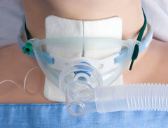
Tracheostomy
surgical creation of an opening into the trachea through the neck that can be temporary or permanent; required when client is on vent for > 7 days and uses local/general anesthesia in 2-10 minutes
Types of Tracheostomies
Cuffed
Uncuffed
Metal
Plastic
Single or double lumen
Stoma
surgically created during surgery for total laryngectomy and can take 8-10 hours and is done under general anesthesia; required to remove ALL cancerous tissue and is always permanent
Metal laryngectomy tube or plastic button at site
initially keeps stoma open
What should you keep at the bedside for a trach?
Suture removal set
Suction
Additional trach (current size/smaller)
Obterator
Indications: Tracheostomy
Acute airway obstruction when oral/nasal intubation is not feasible
Severe OSA
Prolonged intubation and need for mechanical ventilation
Airway reconstruction post-laryngeal trauma/cancer surgery
____________ is always preferred to suctioning.
coughing

Suctioning
indicated for noisy secretions, dyspnea, falling O2 sat, crackles, restlessness, tachypnea, client request and can be done nasopharyngeal, oral, trach, or stoma
Suctioning Technique
Apply suction no longer than 5-10 sec.
Wait 1-2 mins between suction passes
Risks: Suctioning
Hypoxia
Trauma
infection
Vagal stimulation
Bronchospasm
Nursing Concerns: Stoma
Alteration in airway clearance
Impaired communication
Pain
Anxiety
Care for stoma
Key Points: Surgical Respiratory
Positioning is IMPORTANT post-op
Immediate post-op/LT care of stoma/trach
Primary concern is airway, but do not lose sight of pain, emotional support, teaching, and communication