A&P Lab 8 - Histology & Muscles
 Call Kai
 Learn
 Practice Test
 Spaced Repetition
 Match
 Flashcards
 Knowt Play
1/84

There's no tags or description
Looks like no tags are added yet.
85 Terms
Which muscle tissue types have striations?
Cardiac & Skeletal muscle

What creates striations in skeletal and cardiac muscle?
Sections of thick and thin filaments arranged into sarcomeres
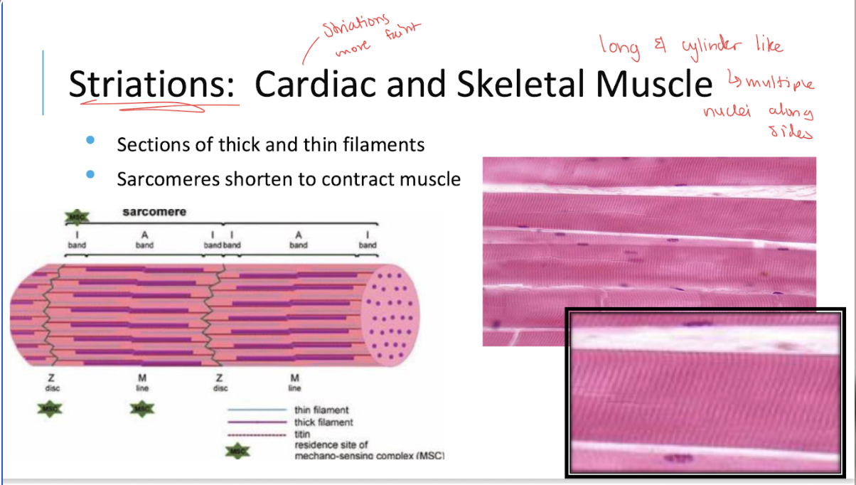
What happens to sarcomeres during muscle contraction?
They shorten
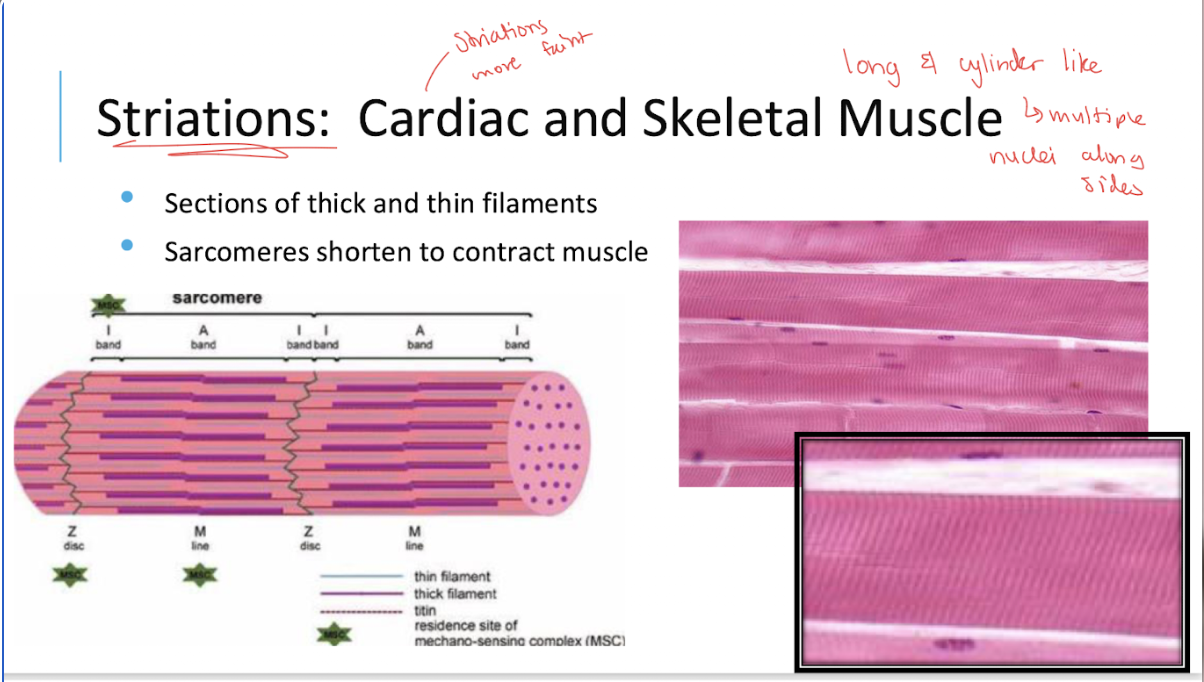
What are the characteristics of skeletal muscle?
Voluntary contractions
long straight fibers
multiple nuclei
nuclei arranged near the cell walls
all or nothing contractions

What does it mean that skeletal muscle contractions are all-or-none?
An individual muscle fiber either contracts fully when stimulated above threshold or does not contract.

What is notable about the nerve supply of skeletal muscle fibers?
Each skeletal muscle fiber has its own nerve supply
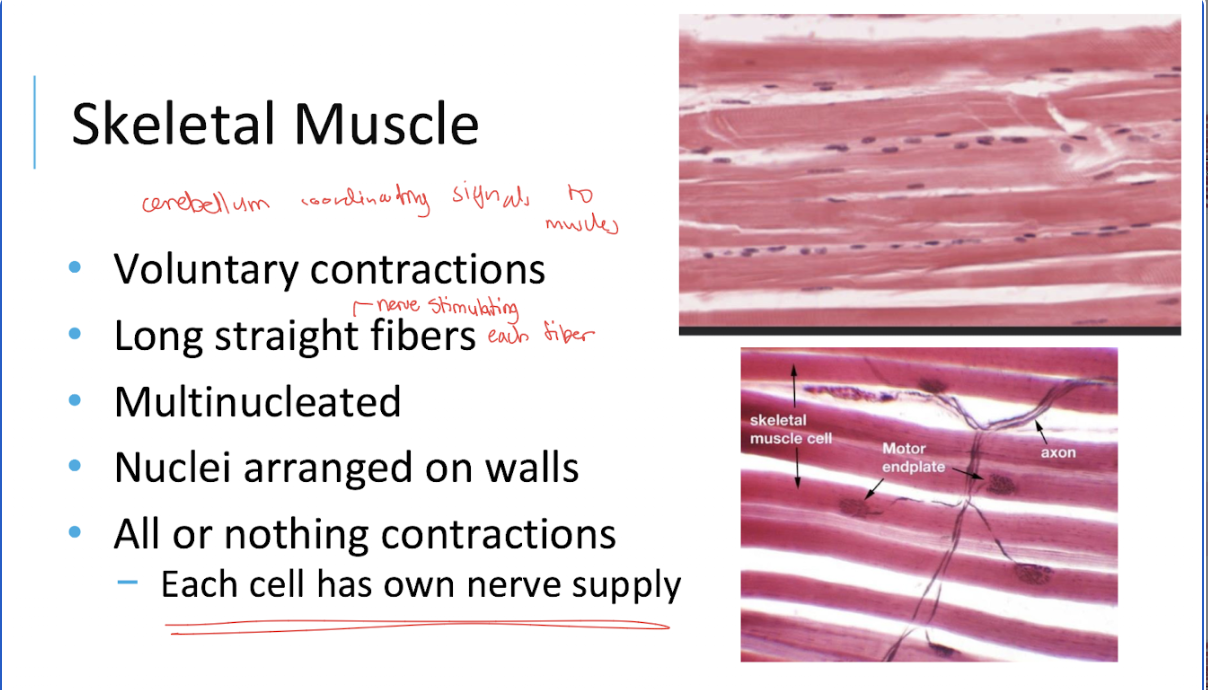
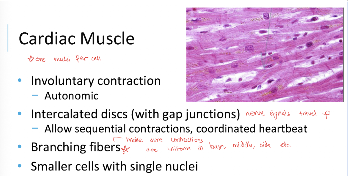
What are the characteristics of cardiac muscle tissue?
Involuntary contractions
branching fibers
relatively small cells with usually one nucleus
intercalated discs.
What is the function of intercalated discs in cardiac muscle?
They contain gap junctions that allow coordinated electrical activity and sequential contractions of the heart.
make sure contractions are uniform @ base, middle, side of heart etc.
What controls cardiac muscle contraction?
It is involuntary and regulated by the heart’s intrinsic conduction system and autonomic influences

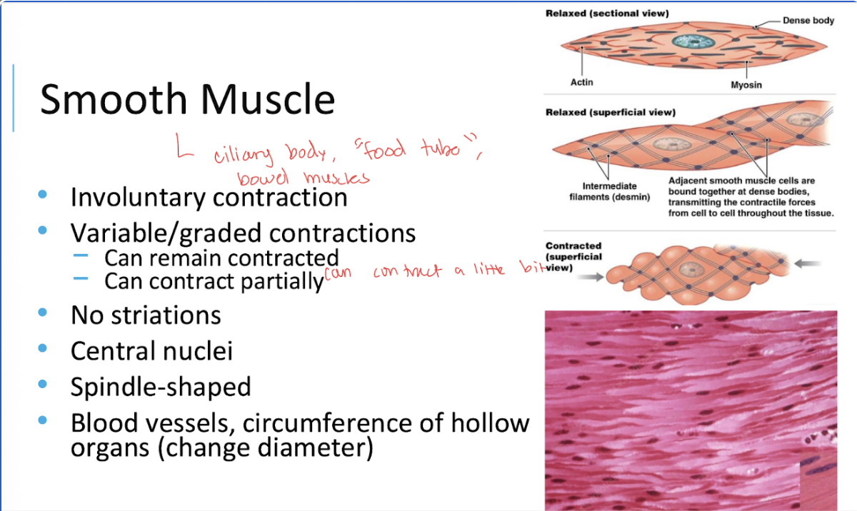
What are the characteristics of smooth muscle tissue?
Involuntary contractions
no striations
central nuclei
spindle-shaped cells
variable or graded contractions
What does it mean that smooth muscle contractions are graded?
Smooth muscle can contract partially or maintain a sustained contraction.
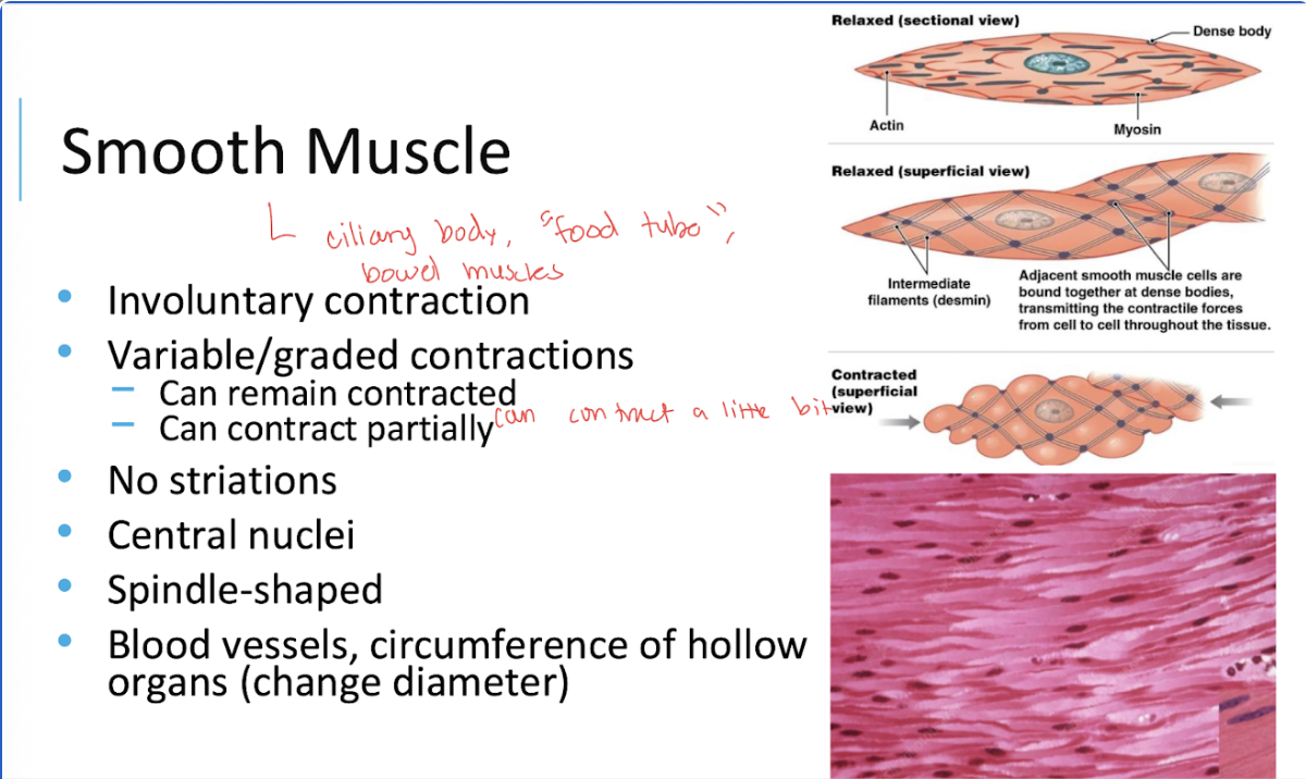
Where is smooth muscle found, and what does it do?
In blood vessel walls and around hollow organs, where it changes their diameter

How can skeletal, cardiac, and smooth muscle be distinguished under a microscope?
Skeletal: striated, long straight fibers, multiple peripheral nuclei.
Cardiac: striated, branching fibers, intercalated discs.
Smooth: no striations, spindle-shaped cells, central nuclei.
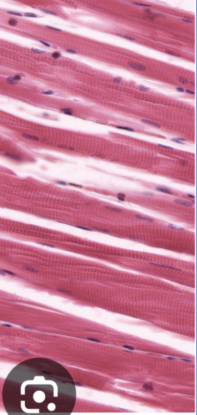
This is…
…skeletal muscle

This is…
…smooth muscle

This is…
…cardiac muscle

This is…
…smooth muscle
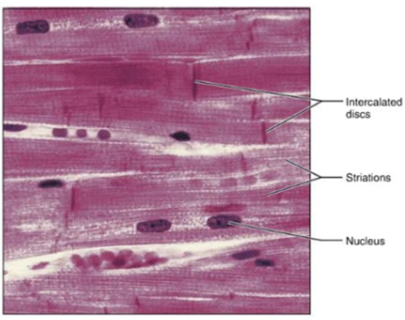
This is…
…cardiac muscle
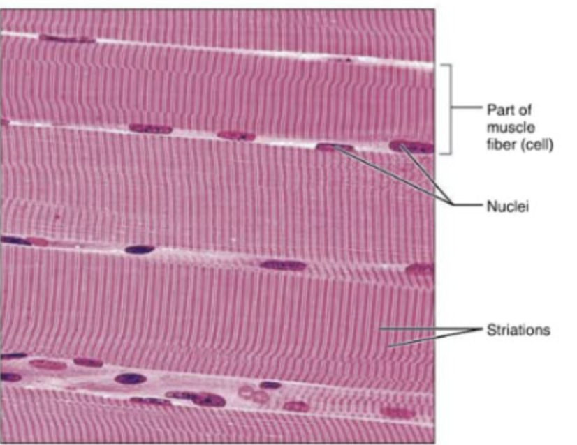
This is…
…skeletal muscle
What is a muscle’s origin?
The attachment generally considered more fixed during a particular action.
What is a muscle’s insertion?
The attachment generally considered to move toward the origin during a concentric contraction.
What are the three dimensions of the thoracic cavity that change during breathing?
Vertical, anteroposterior (front-to-back), and transverse (side-to-side).
What is the primary muscle of inspiration?
The diaphragm


The diaphragm….
….can be passive (autonomic) or conscious (skeletal)
contracts = flattens = increases pleural space (inspiration)
relax = expiration
external intercostals assist

What is number 1?
Rectus abdominis (most superficial)
straight fibers
flexion of torso (sit-ups)

What is number 2?
Transversus abdominis (deepest)
parallel horizontal lines
core muscles, breathing, girdle that holds everything in place

What is number 3?
Internal oblique
opposite to external oblique
the two opposite to each other work together (ie. left external oblique works with right internal oblique)

What is number 4?
External oblique
hands in pocket
fibers point down
The right external oblique and the left internal oblique work together as synergists to
rotate your trunk and spine to the left

the left external oblique and the right internal oblique work together to
rotate your trunk to the right

What is the pelvic diaphragm?
A group of muscles forming the muscular floor of the pelvis.
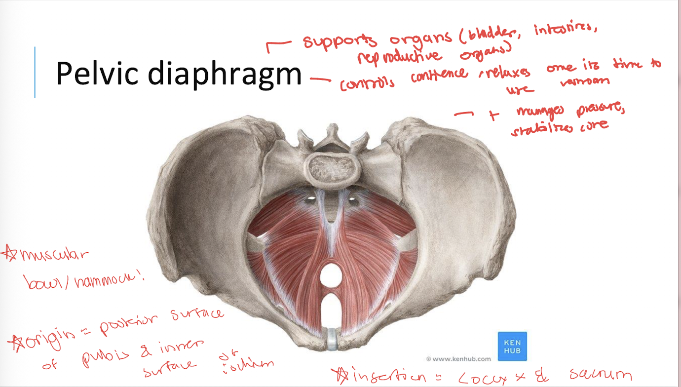
What are the main functions of the pelvic diaphragm?
It supports pelvic organs and contributes to pelvic floor function (see image)


What is number 1?
Gluteus Maximus

What is number 2?
Gluteus Medius

What is number 3?
Gluteus Minimus

What is number 1?
Pelvic diaphragm

What is number 1?
Diaphragm
inferior view

What is number 2?
Sternum

What is number 3?
Xiphoid process
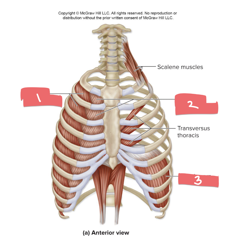
What is number 1?
External intercostals

What is number 2?
Internal intercostals

What is number 3?
Diaphragm

What is the first one?
External intercostals

What is the second one?
Internal intercostals
What is the mnemonic for the external and internal intercostals?
“E for I and I for E”
external intercostals for inspiration
internal intercostals for forced expiration.

What is number 1?
External oblique
point down
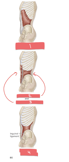
What is number 2?
Internal Oblique
point up

What is number 3?
rectus abdominis

What is number 4?
Transverus abdominis

What is number 1?
Errector spinae
origin = sacrum, iliac crest, & lower spinous processes
insertion = ribs (true & false) & upper spinous processes
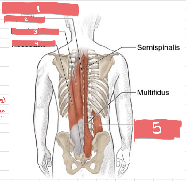
What is number 2?
Spinalis muscle
extends spine (mostly upper)

What is number 3?
Longissimus
extends & side bends spine AND head

What is number 4?
Iliocostalis
extends & side-bends spine

What is number 5?
Quadratus luborum
lateral flexion, pelvic hiking
spine extension, rib stabiization
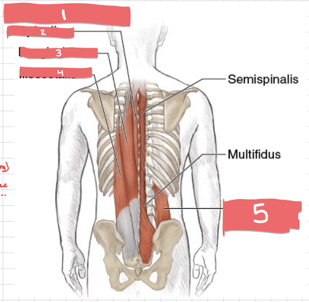
Origin of Quadratus luborum? Insertion?
Origin = iliac crest
insertion = 12th rib (floating) & lumbar vertebrae

What is number 1?
Transversus abdominis

What is number 2?
rectus abdominus

What is number 3?
external oblique

What is number 4?
internal oblique

This is…
…cardiac muscle

This is…
…skeletal muscle
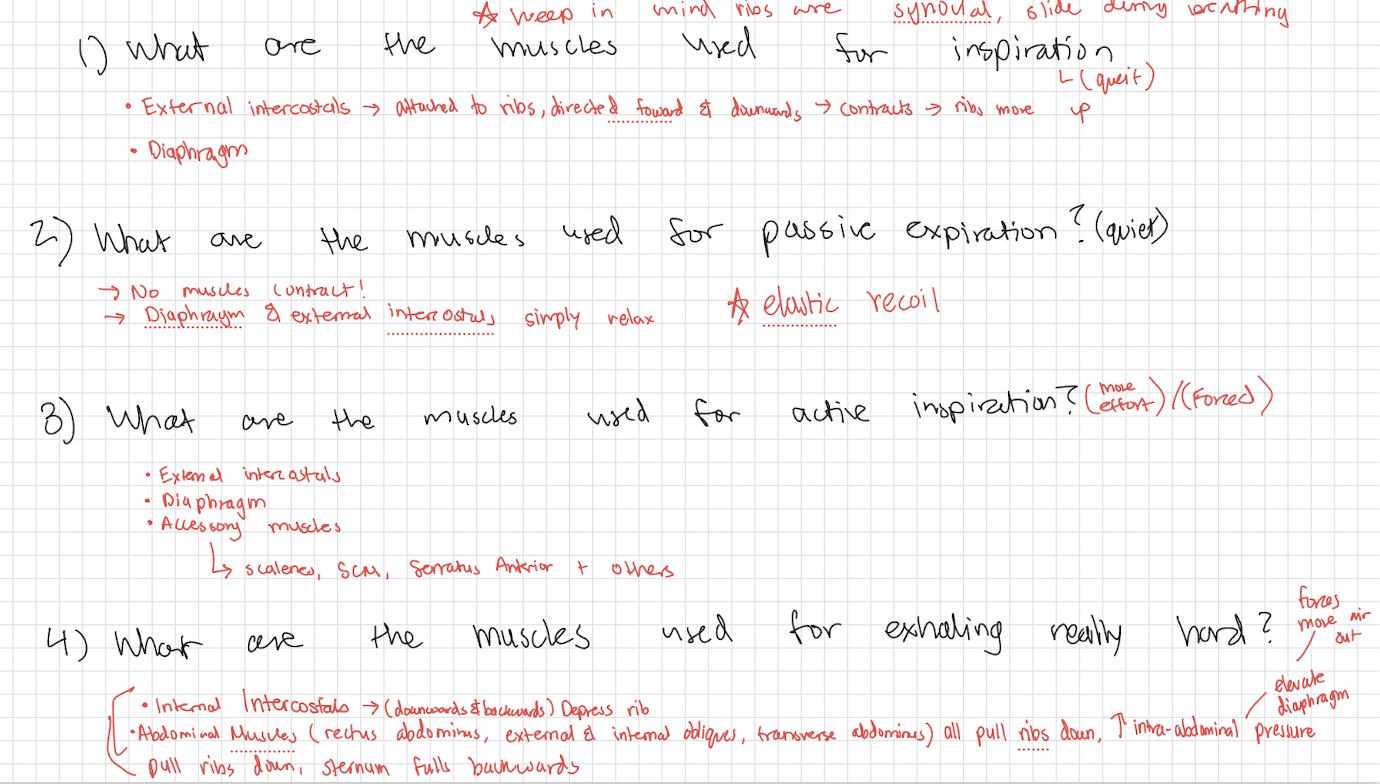
What muscles are used for inspiration (quiet)?
The diaphragm
external intercostals.
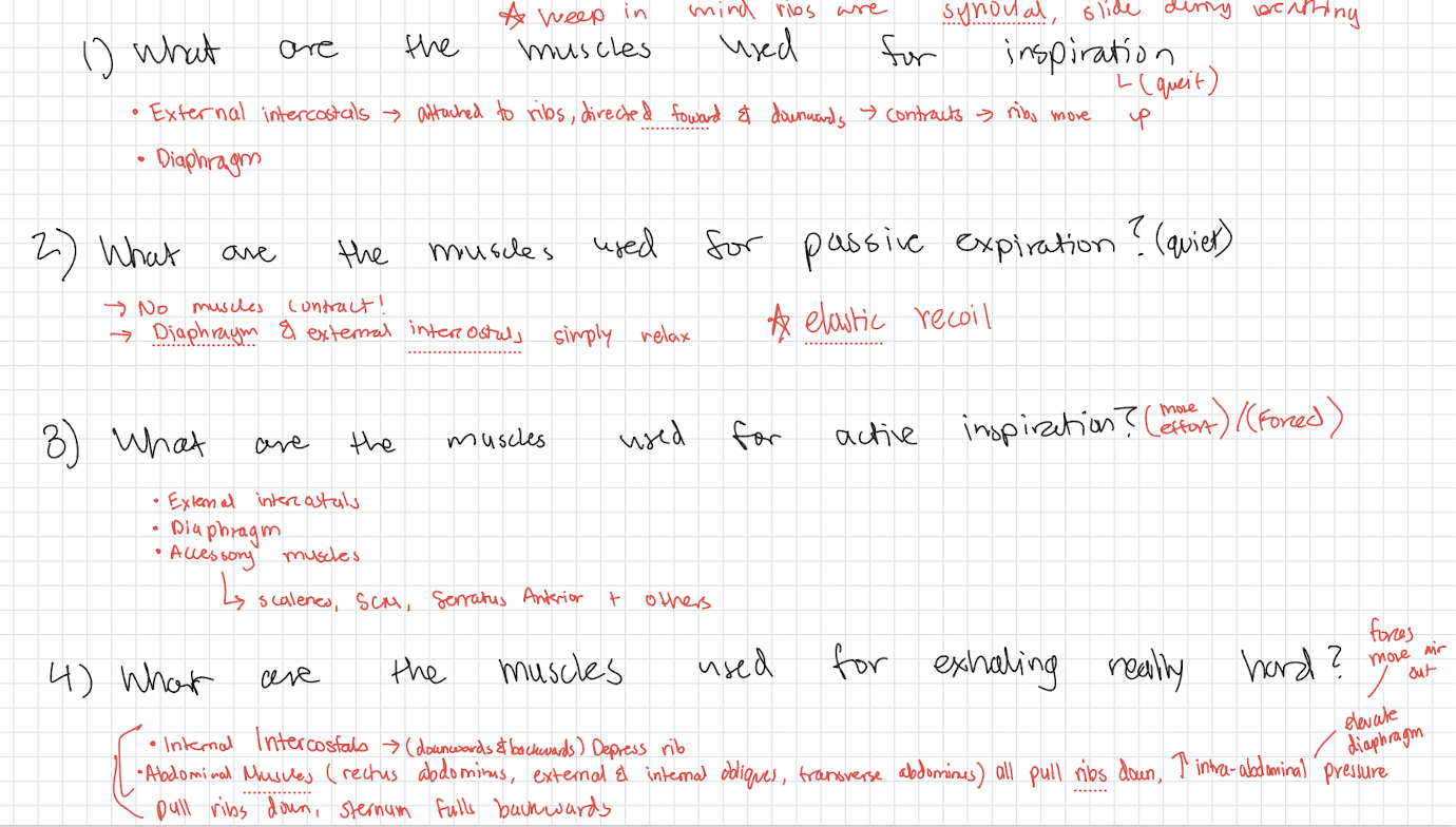
What happens when the diaphragm contracts during inspiration?
It flattens, increasing the vertical dimension of the thoracic cavity
lungs expand and air enters.
What do the external intercostals do during inspiration?
They contract and pull up sternum & elevates ribs
increasing the front-to-back diameter of the thoracic cavity

What muscles are actively used for passive expiration? (quiet)
None
the diaphragm and external intercostals relax, and elastic recoil helps move air out of the lungs.

What happens to the thoracic cavity during passive expiration?
The diaphragm returns toward its dome shape
the ribs move down
the sternum moves backward
thoracic volume decreases.

What muscles are used for active inspiration (forced)?
The diaphragm & external intercostals
→ working more forcefully to increase thoracic cavity volume and draw air in.
accessory muscles as well

What muscles are used for active expiration (forced)?
internal intercostals and abdominal muscles
→ rectus abdominis, external oblique, internal oblique, and transversus abdominis.

What do the internal intercostals do during forced expiration?
They pull down sternum & depresses ribs, decreasing thoracic cavity dimensions

How do the abdominal muscles assist forced expiration?
They contract, increase intra-abdominal pressure, pull the ribs down, and push the diaphragm upward to force more air out.

What articulates at an intervertebral cartilaginous joint?
The bodies of adjacent vertebrae, with an intervertebral disc between them.
intervertebral disc = connects adjacent vertebral bodies, helps absorb forces, and permits limited movement between vertebrae
What type of joint is found between adjacent vertebral bodies?
A cartilaginous symphysis.
What articulates at an intervertebral facet joint?
The superior articular facet of one vertebra and the inferior articular facet of an adjacent vertebra
planar synovial joint → Gliding movements that contribute to spinal movement; the amount and direction depend on the region of the vertebral column
What articulates at the first sternocostal joint?
The first rib’s costal cartilage and the manubrium of the sternum.
cartilaginous → synchondrosis
What articulates at the second through seventh sternocostal joints?
The costal cartilages of ribs 2–7 with the sternum.
Synovial joints, planar
What bones articulate at the sacroiliac joint?
The sacrum and ilium
Synovial
What type of joint is the pubic symphysis, and what tissue is present?
A cartilaginous symphysis containing fibrocartilage.
What movements occur in the sagittal plane?
Flexion and extension.
Flexion (bending) and extension (straightening) move body parts forward (anterior) or backward (posterior). When you view the body from the side (a sagittal view), these actions trace a path that stays completely within the left-right dividing plane

What movements occur in the coronal (frontal) plane?
Lateral flexion and elevation/depression movements, depending on the structure.
• Vertical Motion in a Frontal Alignment: Elevation (raising a structure like the scapula/shoulder blade or mandible) and depression (lowering it) occur strictly in a superior-inferior direction.
• Coronal Framing: Although elevation and depression move up and down, the physical structures involved—the shoulder girdle, scapulae, and lower jaw—lie within the front-to-back (coronal/frontal) slice of the body rather than swinging forward/backward in the sagittal plane or twisting in the transverse plane

What movement occurs in the transverse plane?
Rotation to the left or right.
Think of your body like a
skewer pushed straight down through the top of your head to the floor.
When you spin or twist around that skewer, your body parts sweep out a flat, horizontal circle. That flat circle is the transverse plane.

What is anterior pelvic tilt?
Rotation of the pelvis forward.

What is posterior pelvic tilt?
Rotation of the pelvis backward.

What is lateral flexion of the vertebral column?
Bending the trunk or spine to the left or right.

What are rib elevation and rib depression?
Rib elevation raises the ribs; rib depression lowers them.
What type of joint are the sternocostal joints?
Rib 1: Synchondrosis (primary cartilaginous joint) - rigid anchor with virtually no movement.
Ribs 2–7: Synovial plane / planar joints - allows smooth gliding for breathing expansion.