Chest and Lung Exam Part 1
 Call Kai
 Learn
Learn Practice Test
 Spaced Repetition
 Match
Match Flashcards
 Knowt Play
1/50

There's no tags or description
Looks like no tags are added yet.
Name | Mastery | Learn | Test | Matching | Spaced | Call with Kai | Chat |
|---|
No analytics yet
Send a link to your students to track their progress
51 Terms
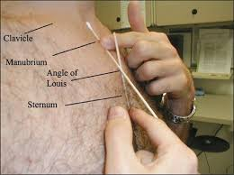
Vertical Axis Landmarks: Sternal Angle (Angle of Louis) Location
5 cm inferior to sternal notch
horizontal ridge joining manubrium to body of sternum
Visceral vs. Parietal Pleurae vs. Pleural Space
visceral:
lacks sensory nerves
serous membrane
covers outer surfaces of each lung
parietal:
innervated by intercostal+phrenic nerves
lines pleural cavity along rib cage+upper surface of diaphragm
pleural space:
contains serous pleural fluid
space between visceral+parietal pleurae
Muscles of Respiration
principal:
diaphragm
internal+external intercostals
accessory:
sternocleidomastoid
scalenes:
-anterior
-middle
-posterior
other:
abdonimals:
-rectus abdominis
-external oblique
-internal oblique
-transversus abdominis
ROS
dyspnea (shortness of breath)
-orthopnea→supine
-paroxysmal nocturnal dyspnea→sitting+HS
cough
hemoptysis
chest pain
daytime sleepiness
snoring
disordered sleep
-snoring
-choking sensation
-morning headache
apnea (stops breathing for 10+ seconds)
Orthopnea vs. Paroxysmal Nocturnal Dyspnea
orthopnea: shortness of breath when supine
paroxysmal nocturnal dyspnea: shortness of breath at night
relieved by sitting up
Dyspnea Health Hx
onset:
-acute
-chronic
triggers:
do certain activities/environmental exposures trigger sx/make sxs worse
severity:
-how severe
-are they affective ADL
associated sxs
medical hx
-respiratory dxs
-social hx→smoking
mxs
Cough Hx
response to
-irritant of trachea
-larynx
-bronchi
ex: allergens
dust
foreign body
extreme temp air
inflammation of respiratory tract
tumors
ask
duration
-acute→u3 wks
-subacute→3-8 wks
-chronic→8+ wksproductivity
triggers
associated sxs
medical hx
mxs
Hemoptysis Health Hx
blood tinged sputum
onset
amount
-small
-frank/largelocation
-mouth
-pharynx
-respiratory tract
-GI tract→darker color+hematemesis (“coffee ground emesis”)
associated/other sxs
medical hx
mxs→anticoagulants
Chest Pain Health Hx
aka: angina
location/quality
-sharp
-dull
-recent radiation exposureassociated sxs
mx history
mxs
key details
pain breathing in→pleuritic pain
lung tissue→no pain fibers→irritation/inflammation of pleural surface
chostocondritis→muscle pain from coughing
Associated Sxs
fever
sweats
chills
weakness
dizziness
lightheaded
syncope
recent weight change
exertional chest pain
palpitations
orthopena (dyspnea when supine)
paroxysmal nocturnal dyspnea (dyspnea at night+sitting up)
edema
reflux/burning sensation
Pertinent Past Medical Hx
childhood/adult illnesses:
asthma
COPD
tuberculosis
pneumonia
cancer
cystic fibrosis
pneumothorax
accidents/injuries: chest trauma
immunizations+vaccinations:
flu shot
pneumococcal vaccine
COVID vaccine
screening tests:
last chest x-ray
last ppd
surgeries
chest
lung
cardiac
hospitalizations: asthma→ask if ever intubated
Family Hx
cancer
tuberculosis
asthma
cystic fibrosis
emphysema
chronic bronchitis
pulmonary embolism
Pertinent Psycho-Social Hx
home situation:
flight of stairs
community-type living
occupation: exposure to physical/environmental hazards
travel: foreign countries
sleep:
daytime sleepiness
snoring
apnea
morning headache
waking up with sense of choking
exercise: exercise-induced dyspnea
tobacco:
smoking hx
second-hand smoke exposure
vaping
illicit mx use
General Survey
vitals
facial expression
color:
-cyanosis
-pallor
pursed-lip breathing
nasal flaring
use of accessory muscles
tripod position (leaning forward to breathe)
tachypnea (rapid breathing)
stridor/other breathing sounds
clubbing of digits
shape of chest
Physical Exam (General) List
general survey:
rate
rhythm
depth
effort of breathing
signs of distress
IPPA
inspect chest
palpate chest
percuss chest
auscultate chest
Normal Breathing
adults→14-20/min
infants→ 44/min
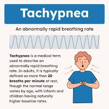
Tachypnea
rapid shallow breathing
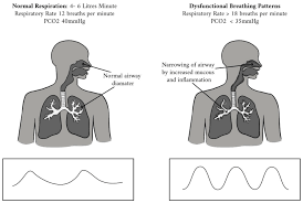
Hyperpnea/Hyperventilation
rapid deep breathing
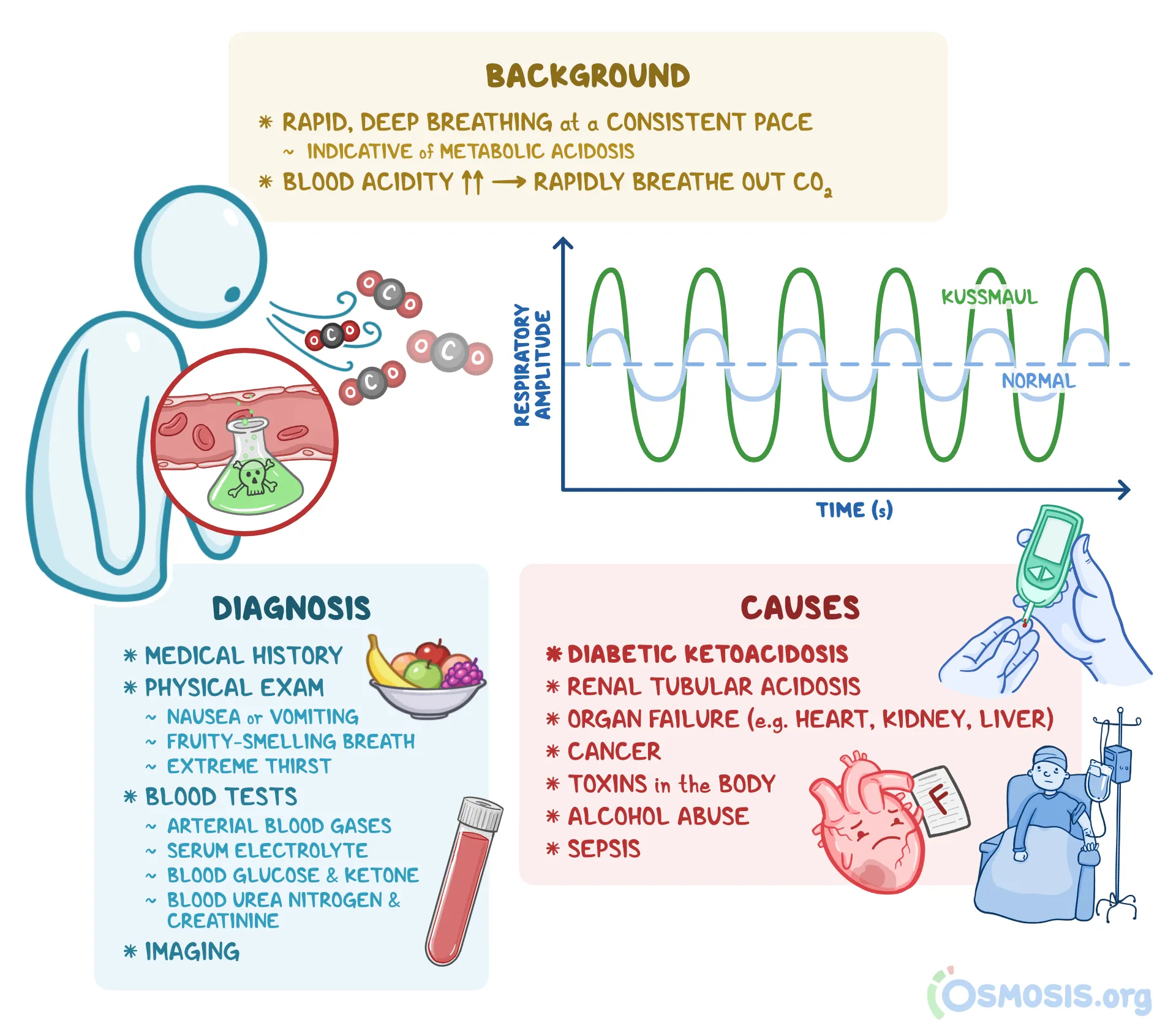
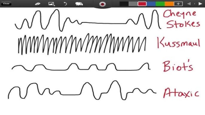
Kussmaul Breathing
systemic acidosis→compensatory overbreathing
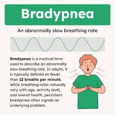
Bradypnea
slow breathing
±increase in tidal volume
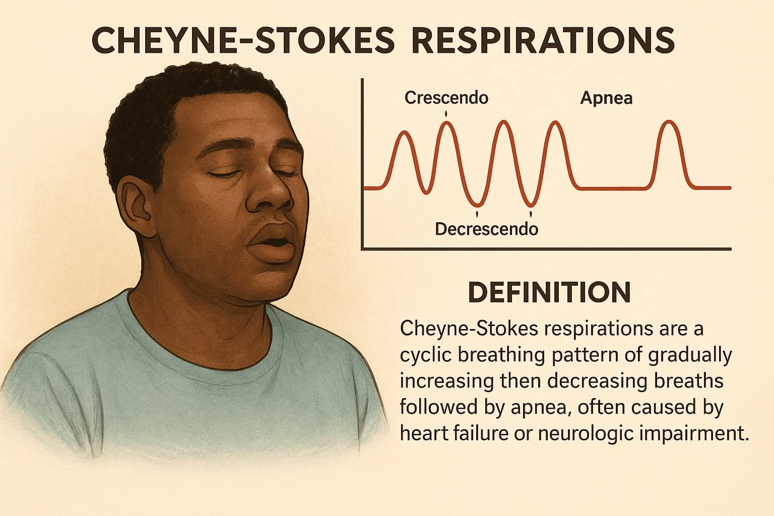
Cheyne-Stokes Breathing
hyperpnea alternating with apnea
Ataxic/Biot Breathing
unpredictable irregularity
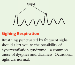
Sighing Respiration
hyperventilation syndrome
Obstructive Breathing
prolonged expiration secondary to airway narrowing
Normal Adult Thorax/Chest
lateral diameter greater than AP diameter
ratio: 0.7-0.9
aging: increased diameter
chest wall stiffer→decreased movement
weakened respiratory muscles
lessened elastic coil
skeletal changes
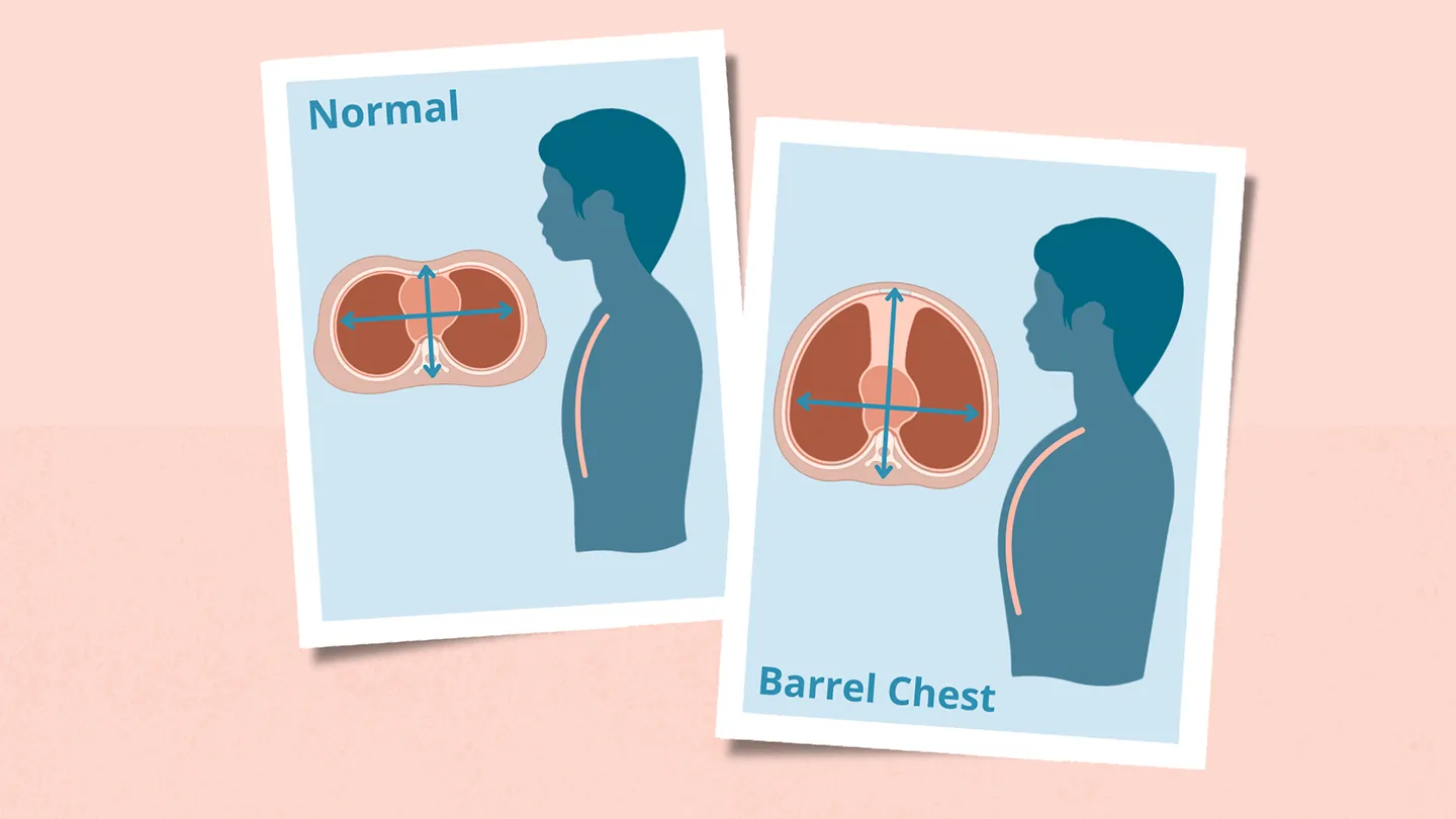
Barrel Chest
increased AP diameter
normal: infancy
aging
DX: COPD
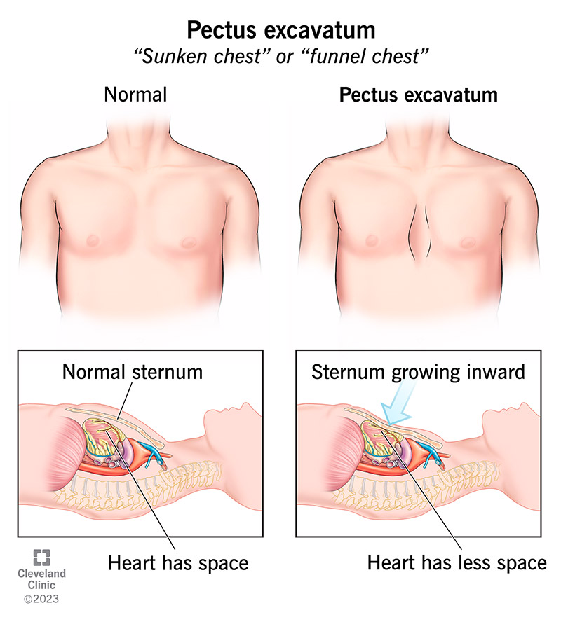
Pectus Excavatum/Funnel Chest
depression in lower part of sternum
compressed heart+great vessels→murmur
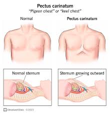
Pectus Carinatum/Pidgeon Chest
sternum sticks out→increased AP diameter
depressed costal cartilage adjacent to protruding sternum
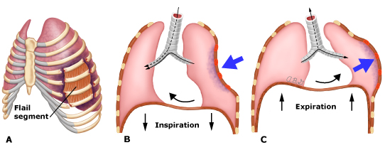
Traumatic Flail Chest
multiple rib fractures→paradoxical movements of thorax/chest
inspiration→depression of injured area
expiration→injured area bulges outward
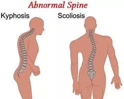
Thoracic Kyphoscoliosis
abnormal spinal curvature+vertebral rotation→deformed chest
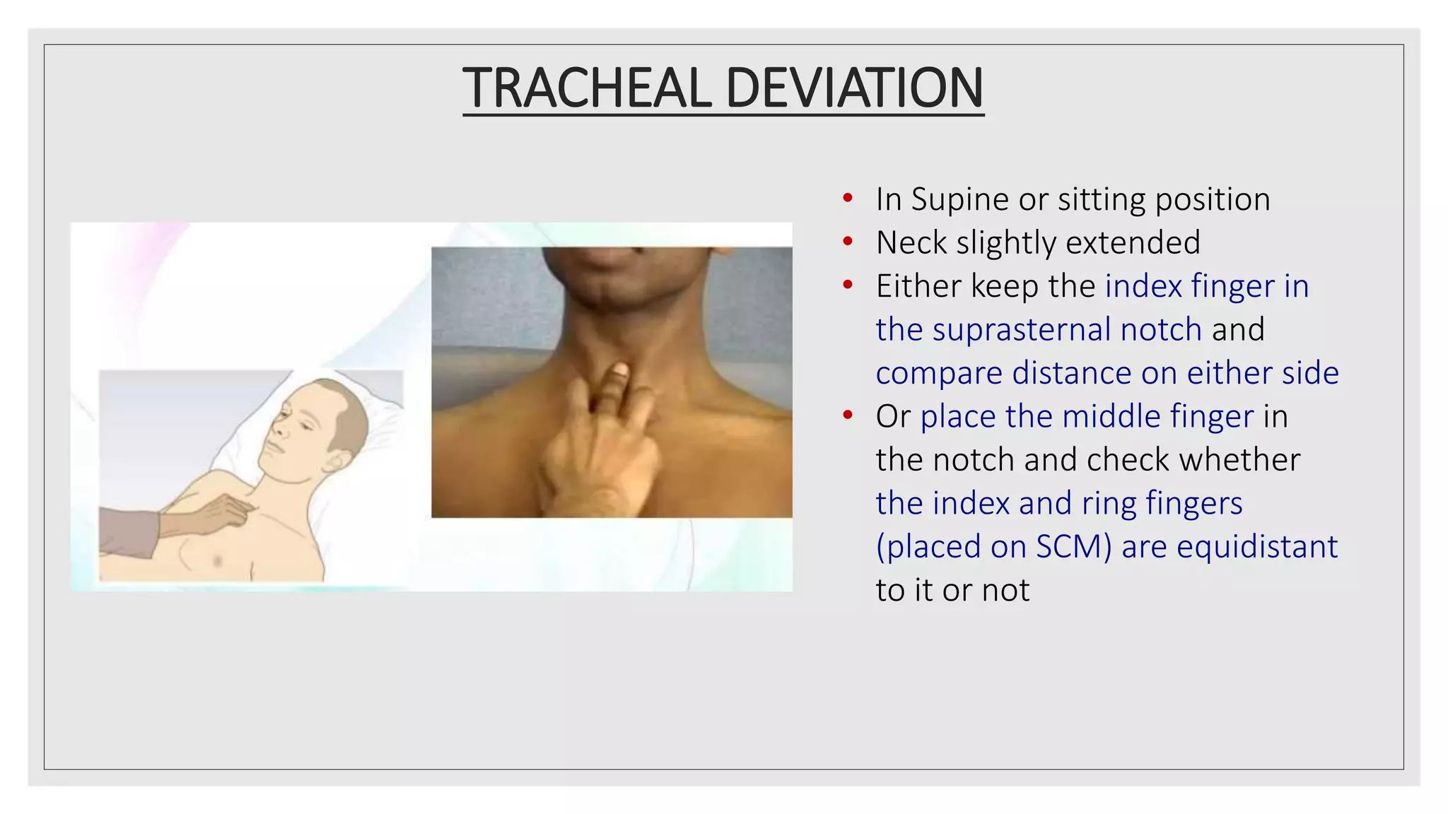
Palpation: Tracheal Deviation
grab trachea with thumb+index finger below cricoid cartilage
ask pt to take deep breath in
Physical Exam: Palpation (General)
anterior chest:
pt sitting/supine position
discomfort→arms slightly abducted
P: anterior chest
tenderness
abnormalities
posterior+lateral chest:
pt sitting
arms:
posterior: across chest
lateral: on hips
P: posterior+lateral chest
posterior+lateral chest
tenderness
abnormalities
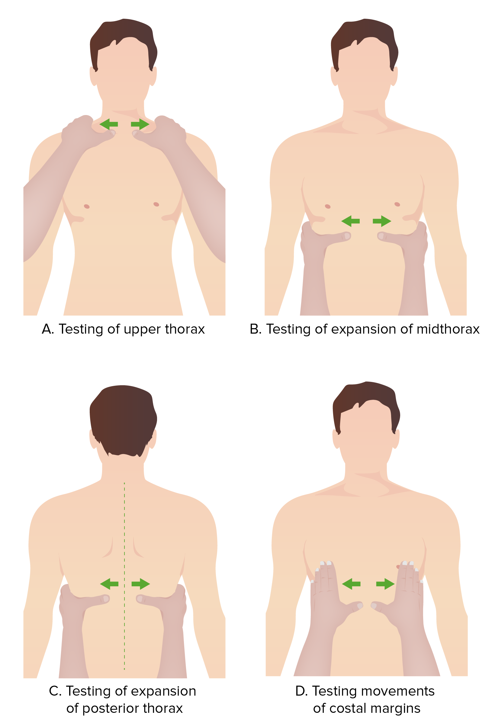
Palpation: Thoracic/Chest Expansion
aka: lung excursion
chest expansion
place thumb on each costal margin posteriorly
-at level of+parallel to 10th rib→form loose skin fold
-pt unable to sit→anterior chest→hands along each costal margin→form loose skin foldask pt to inhale deeply
observe:
-extent of respiratory movement
-symmetry of respiratory movement
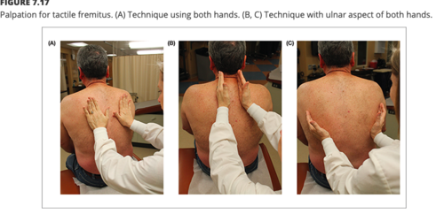
Palpation: Tactile Fremitus
process
use ulnar surface of hand
ask pt to put hands on hips
ask pt to say+repeat “99”
3 anterior lung field positions
2 mid-clavicular
1 lower anterior axillary line region
4 posterior lung field positions
3 mid-scapular line
1 lower posterior axillary line region
increased fremitus→enhances the transmission of vibration
consolidated tissue
inflammation around a lung abscess
decreased/absent fremitus→obstruction of transmission of vibration
DX:
thickened pleurae
pleural effusion
pneumothorax tumor
Physical Exam: Percussion Process
hyperextend middle finger on 1 hand
place distal IPJ firmly against pt’s skin
avoid surface contact→dampens out vibrations
end (not pad) of opposite middle finger→quick flick of wrist→strike dorsal aspect of first long finger
no long nails→can’t do percussion
Chest Anatomy+Anatomic Landmarks
superior
clavicle
neck tissue
anterior
ribs
sternum
lateral→ribs
inferior→diaphragm
Midsternal Line
runs vertically along center of sternum (breastbone)
divides chest in to L+R halves

Midclavicular Line
runs vertically from midpoint of clavicle (collarbone)
reference point for clinical assessments of chest+abdomen

Anterior Axillary Line
runs vertically from front/anterior axillary fold
-junction between chest+upper arm
helps ID specific landmarks on chest wall

Midaxillary Line
runs vertically from apex of axilla (armpit)
used to locate structures in the middle of the axillary region

Posterior Axillary Line
runs vertically from posterior axillary fold
helps ID specific landmarks on the back

Scapular Line
drops down from the inferior angle of the scapula (shoulder blade)
useful for
assessing back injuries
ID specific landmarks on the back
Vertebral Line
follows the path of the toracic spinous process (bony projections felt along the spine)
used for reference during spinal assessments+procedures
PE→Inspection (General)
I: posterior chest
anterior chest
lateral chest
pattern of breathing
note: shape
note: skin
lesions
rashes
scars
any visible signs of trauma
Inspection→Thorax
I: thorax
shape
-normal→lateral diameter greater than AP diameter
-0.7-0.9deformities
asymmetry
abnormal retractions of intercostal spaces
use of accessory muscles
tracheal position
Inspection→Skin
pallor
cyanosis
sinus tracts
bruising
rash
Fremitus
palpable vibrations transmitted through bronchopulmonary tree to chest wall
more prominent in interscapular areas than in lower lung fields
PE→Percussion General Locations
use “ladder” pattern
compare bilaterally
posterior (7 locations)
5 locations→apices to bases of lungs
2 locations→posterior axillary line regions (lateral)
anterior (6 locations)
4 locations→apices to base of lungs
2 locations→anterior axillary line regions (lateral)
PE→Percussion→Diaphragmatic Excursion
ID→descent of the diaphragm
measure
distance between dullness
-on full expirationfull inspiration
normal→3 to 5.5cm
PE→Auscultation (General)
assessing airflow through tracheobroncheal tree
listening to sounds generated by breathing
adventitious sounds
transmitted voice sounds
-bronchophony
-egophony
-whispered pectoriloquy
PE→Auscultation Process
ask pt to clear lungs/cough once/twice
ask pt to take deep breath in+out with open mouth each time the diaphragm of the stethoscope is placed against their skin
auscultate 7 locations on the posterior lung fields
-5→from apices to bases of lungs
-2→posterior axillary line regions (lateral)auscultate 6 locations on the anterior lung fields
-4→apices to bases of lungs
-2→anterior axillary line regions (lateral)
use “ladder” pattern→compare symmetric areas
listen to full inspiration+expiration