CHAPTER 24: SOFT TISSUE & DENTOALVEOLAR INJURIES
 Call Kai
 Learn
Learn Practice Test
 Spaced Repetition
 Match
Match Flashcards
 Knowt Play
1/41

Earn XP
Description and Tags
ppt
Name | Mastery | Learn | Test | Matching | Spaced | Call with Kai | Chat |
|---|
No analytics yet
Send a link to your students to track their progress
42 Terms
7-11 years of age
greatest incidence of soft tissue & dentoalveolar injuries
soft tissue injuries
abrasion, contusion, laceration
abrasion
causes:
by friction between an object and a soft tissue surface
iatrogenic example:
shank of a rotating bur accidentally touches the oral mucosa
key features:
surface wound
bleeding from damaged capillaries
tx:
copious saline irrigation
topical anesthetic for pain relief
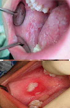
contusion
aka: bruise
indicates that some amount of tissue disruption has occurred within the tissues
results in subcutaneous or submucosal hemorrhage without a break in the soft tissue surface
causes:
by a blunt object
key features:
bruise
tissue disruption
no surface break
tx:
no treatment is required
in the early stages:
application of ice
pressure dressings
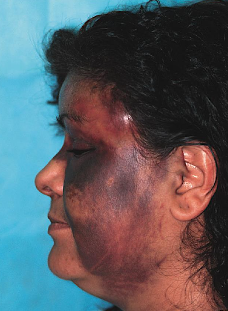
laceration
most frequent type of soft tissue injury
a tear in the epithelial and subepithelial tissues
commonly caused by a sharp object
knife & luxator
common sites:
lips, tongue, gingiva
alveolar mucosa
floor of the mouth
key features:
bruise
tissue disruption
no surface break
tx:
repositioning of tissues & suturing
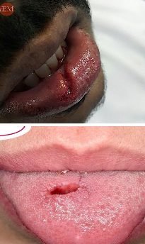
tx for soft tissue lacerations that need to be sutured
gingiva, alveolar mucosa, floor of the mouth
closed in 1 layer
mucosal layer
tongue or lip involving muscle
closed in 2 layers
muscle & mucosal layer
entire thickness of the lip
closed in 3 layers
oral, muscle, dermal layer
key principle:
all tissue layers should be properly positioned
number of layers depends on the location and depth of the laceration
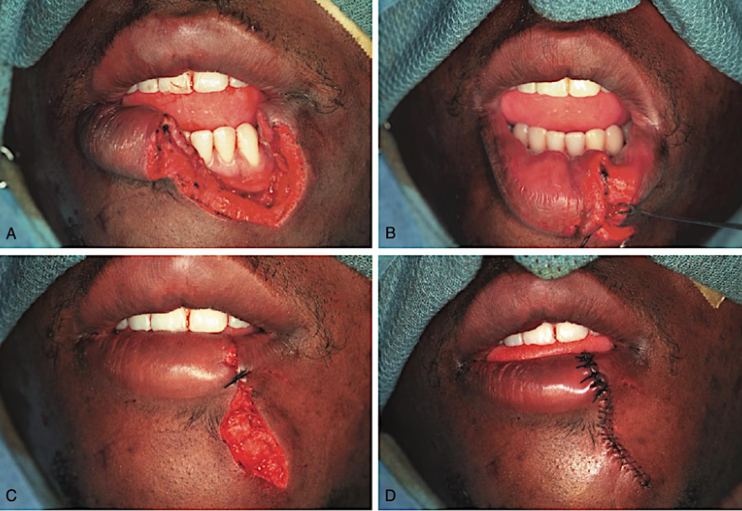
tx for soft tissue laceration with hard tissue injury
treat hard tissue first!
sutures may loosen or tear during manipulation
once lost, tissue closure is harder on a second attempt
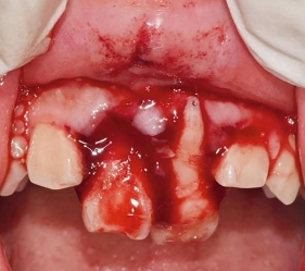
4 major steps in managing soft tissue injuries
cleansing
necessary to prevent any remaining debris
used by soap, brush, saline irrigation
debridement
removal of contused and devitalized tissue from a wound
removal of jagged pieces of surface tissue to enable linear closure
hemostasis
use of clamping and tying with ligatures
cauterization with an electrocoagulation unit
closure
use of sutures
not all wounds need to be sutured
clinical evaluation of dentoalveolar injury
1st: the extent of injury to the tooth
2nd: determine clinically if the tooth is stable, loosened or completely displaced from the socket
3rd: by digital manipulation, evaluate any suspected alveolar fracture
4th: radiographically, assess the stage of development of the root end of the tooth.
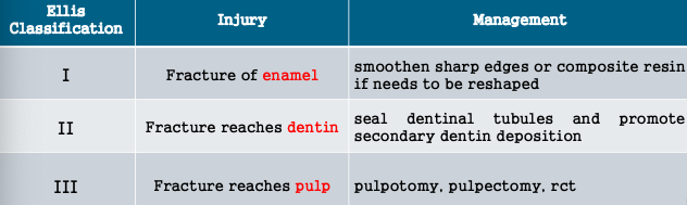
class I ellis classification
fracture of the enamel
class II ellis classification
fracture of enamel and dentin
class III ellis classification
fracture involving enamel, dentin, pulp
class IV ellis classification
fracture involving CEJ (nonvital tooth ; discolored)
class V ellis classification
avulsed tooth
class VI ellis classification
root fracture (may or may not involve the crown)
class VII ellis classification
luxation injuries (tooth displacement without fracture)
class VIII ellis classification
complete crown loss (fracture of the crown en masse)
class IX ellis classification
traumatic injuries to primary teeth
5 indications of pulp capping
the exposure is small
the patient is seen soon after injury
the patient had no root fractures
the tooth has not been displaced
no large or deep fillings exist that might indicate chronic inflammation within the pulp
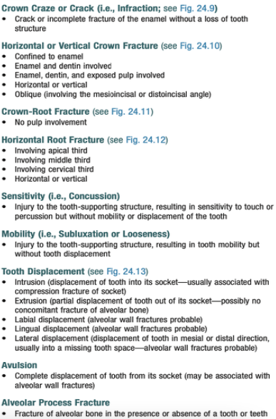
dentoalveolar injuries
intrusion
extrusion
avulsion
concussion
subluxation
crown craze
crown fracture
alveolar fractures
crown-root fracture
lateral displacement
horizontal root fracture
concussion
not loosened, not displaced
clinically tooth will be tender
damage to tooth without injury to supporting structures
tx: dis-occlude
subluxation
loosened but not displaced
clinically tooth will be tender
damage to tooth with injury to supporting structures
tx: splint for 2 weeks
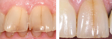
crown craze
aka: crack
normally limited to the enamel (ex: enamel infraction) and usually stop before reaching the dentinoenamel junction
tx:
periodic follow-up examinations
sealed with unfilled resin
crown fracture
treatment is determined by depth of tooth tissue involvement
see pic for management
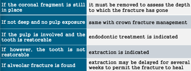
crown-root fracture
treatment depends on the location of the fracture and local anatomic variance
see pic for management
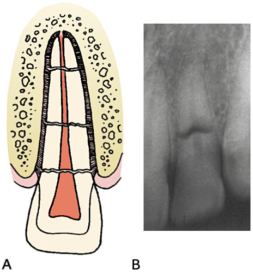
horizontal root fracture
treatment is determined by position of the fracture in relation to the gingival crevice
for cervical 3rd:
prognosis is worst due to exposure to oral environment
remove tooth fragment, endo treatment, post and core
for middle 3rd:
reposition and immobilization
for apical:
prognosis is best esp in the absence of masticatory forces
reposition and immobilization
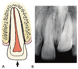
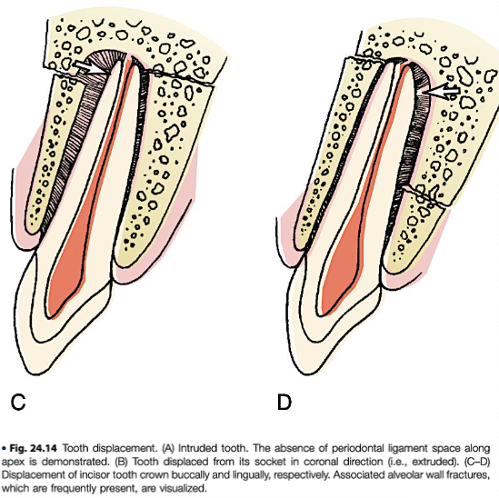
intrusion
socket sustains a compression fracture, causing the tooth to be displaced into a new position
for permanent teeth:
minimal intrusion → allow the tooth to re-erupt spontaneously
severe intrusion → reposition orthodontically or surgically
for primary teeth:
if the tooth touches the permanent tooth follicle → remove the deciduous tooth atraumatically
if it does not touch the permanent tooth follicle → observe and monitor the tooth
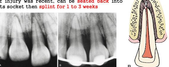
extrusion
displacement of the tooth out of its socket
if injury was recent & mild:
can be seated back into its socket
then splint for 1-3 weeks
lateral displacement
minimal
tooth is slightly displaced
no alveolar fracture
tx:
manual repositioning, splinting
severe
tooth is severely displaced with alveolar fracture
tx:
reposition tooth and bone
splinting, suturing
avulsion
most important factor → time
complete displacement of the tooth out of its socket
recommended storage media:
water
least desirable
hypotonic → causes cell lysis
saliva
keeps the tooth moist but is not ideal
incompatible osmolality and pH
contains bacteria
milk
best readily available alternative
compatible pH and osmolarity
helps maintain the vitality of PDL cells
hank’s balanced salt solution (HBSS)
most ideal storage medium
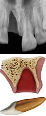
avulsed tooth for less than 20 mins
should be immediately rinsed in saline and replanted by the dentist
avulsed tooth for more than 20mins
place into hanks balanced salt solution for 30mins (this reduces incidence of ankylosis)
then in doxycycline (1mg / 20mL saline) for 5mins (this inhibits bacteria in the pulpal lumen)
replant and splint
5 factors considered before replanting avulsed teeth by Andreasen & Hjorting-Hansen
the avulsed tooth should have no advanced periodontal disease.
the alveolar socket should be reasonably intact to provide a seat for the avulsed tooth
no orthodontic contraindications (ex: significant crowding of teeth should exist)
extra-alveolar period should be considered
periods exceeding 2 hours → poor results
replanted within the first 30mins → excellent results
stage of root development should be evaluated
survival of the pulp is possible in teeth with incomplete root formation if replantation is accomplished within 2 hours after injury
alveolar fractures
may occur independently or concomitantly with tooth injuries
tx as with any fracture:
first, place the segment into its proper position
then, stabilize it until osseous healing occurs
2 factors necessary for healing:
reduction of fracture
restoration of the bony segments to their proper anatomic location
fixation of fracture
immobilization of the bony segments at the fracture site
in cases of tooth trauma
it is safe to assume that the pulp will undergo necrosis regardless of treatment.
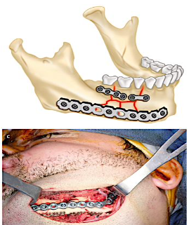
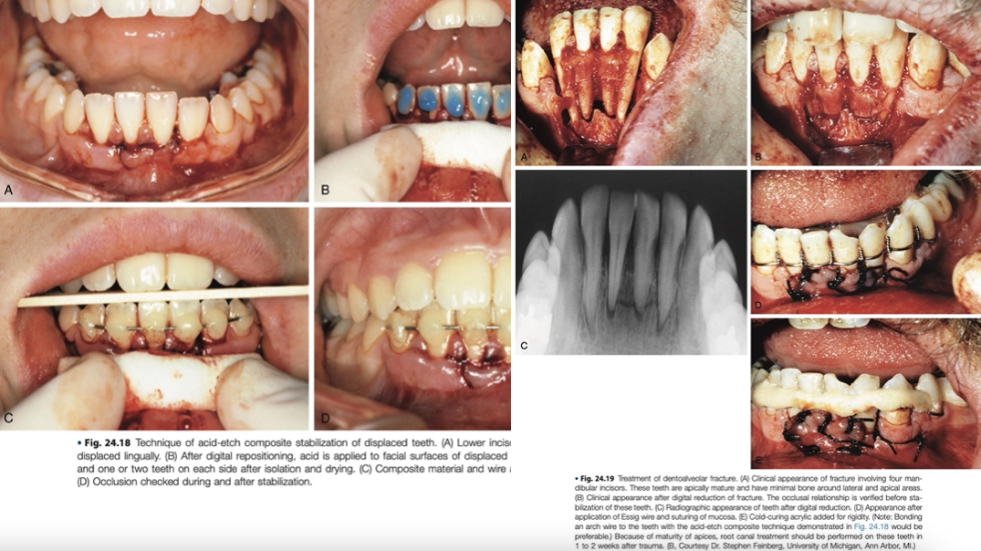
acceptable methods used to stabilize the segment in alveolar fractures
use of an acid-etched arch wire → also acceptable
ligate an arch bar to the teeth both mesial and distal to the segment and within the fractured alveolar segment → simplest
in cases of tooth trauma, when to do root canal?
if the apex of a mature tooth has moved more than 1mm in any direction, pulpal degeneration will occur
in cases of tooth trauma, when to do root canal if apex is close?
endodontic treatment be done after 2 weeks
helps minimize inflammatory root resorption by eliminating nonvital tissue from the pulp
in cases of tooth trauma, when to do root canal if apex is open?
endodontic treatment may be delayed for several weeks
observe, do follow-up exams bc chance of revascularization is high so no need for rct
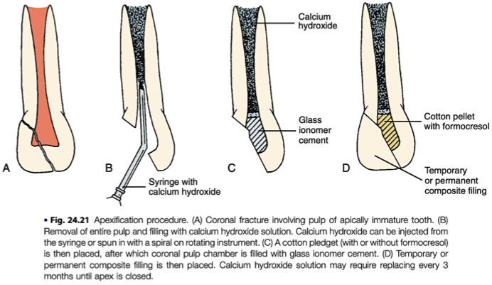
if not, do apexification
revascularization → return of blood flow to the pulp after an injury
apexification → for a non-vital immature permanent tooth with an open apex
years it take for the roots to fully form after the eruption of primary & permanent teeth
primary → 1-2 years
permanent → 2-3 years
stabilization periods for dentoalveolar injuries
mobile tooth → 7-10 days
tooth displacement → 2-3 weeks
root fracture → 2-4 months
replanted mature tooth → 7-10 days
replanted immature tooth → 3-4 weeks
classification of dentoalveolar injuries