Module 1: Introduction and Basic Concepts
 Call Kai
 Learn
 Practice Test
 Spaced Repetition
 Match
 Flashcards
 Knowt Play
1/62

There's no tags or description
Looks like no tags are added yet.
63 Terms
Abduction
Moving away from the midline
Adduction
Moving towards the midline
Peri-
Means "around”
Superior
On top of
Inferior
Below
Cranial (rostral)
Towards the head
Caudal
Towards the tail
Anterior (ventral)
Front of body or structure
Posterior (dorsal)
Back of body or structure
Medial
Towards the midline
Lateral
Away from the midline
Origin
Start of the structure, fixed structure of the muscle
Insertion
Target location of the structure, point of attachment that is moveable
Proximal
Closer to the origin
Distal
Further away from the origin
Superficial
Closer to the outer surface
Deep
Away from the outer surface
Central
Towards the centre
Peripheral
Toward the periphery
Intermediate
In between central and peripheral
Prone
Horizontal, face down
Supine
Horizontal, face up
Unilateral
One side
Bilateral
Both sides
Ipsilateral
Same side
Contralateral
Opposite side
Flexion
Bending at a joint
Extension
Straightening of a joint
Elevation
Superior movement
Depression
Inferior movement
Protrusion (protraction)
Anterior movement (puckering lips)
Retrusion (retraction)
Posterior movement (smiling)
Supination
Rotation so that dorsum lies dorsally or inferiorly
Pronation
Rotation so that the dorsum lies ventrally or superiorly
Epithelial tissue
Outer layer of mucous membranes; form a barrier to infectious agents, prevent loss of moisture
Connective tissue
Consist of cartilage, bone, blood, and fat
Cartilage
Strong and elastic, shock absorption, provides structure
Bone
Hardest connective tissue, forms the skeleton (average 206 bones), provides protection
Muscular
Provides movement through contraction
Cilia
Microscopic hair-like structures that move back and forth that help move foreign substances out of the structure. Helps move mucus out of the upper airway
Axial skeleton
Has 80 bones; supports the central portion of the body (skull, vertebral column, thoracic cage)

Appendicular skeleton
126 bones; consists of the upper and lower limbs

Girdles
Connect the appendicular to the axial skeleton; pectoral girdles and pelvic girdle

Neurocranium
Upper part of the skull that encloses your brain, includes 8 cranial bones (frontal, 2 parietal, 2 temporal, occipital, sphenoid and ethmoid)

Viscerocranium
Lower part of the skull which contains 14 facial bones (mandible, 2 maxilla bones, 2 palatine bones, 2 zygomatic bone, 2 nasal bones, vomer, 2 inferior nasal conchae, 2 lacrimal bones)

Mandible
The lower jawbone; it holds the lower teeth and is the main movable bone of the jaw
Maxillary bone (maxilla)
The upper jawbone; it holds the upper teeth and forms part of the nose, eye socket, and hard palate
Palatine bones
Two internal bones that form the back part of the hard palate (roof of the mouth)
Zygomatic bone
The cheekbone; forms part of the cheek, eye socket, and side of the face
Lacrimal bone
A small, thin bone on the inner side of the eye socket that helps form the wall of the orbit and contains the tear (lacrimal) duct
Foramen magnum
Exit for the spinal cord from the brain
Fontanel
A soft, flexible area between the skull bones of a newborn where the bones have not yet fully fused. It allows the skull to expand as the brain grows

Suture
Immovable joints between the bones of the skull that connect and hold the skull bones together.
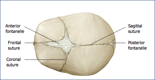
Anterior fontanelle
The largest soft spot (fontanelle) on a newborn’s skull; closes in infants by the time they reaches the age of 2 in most cases

Posterior fontanelle
A small soft spot at the back of a newborn’s skull; closes within the first 2 months after birth

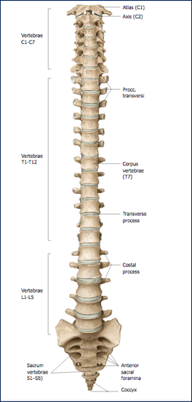
Vertebral column
33 bones (vertebrae); houses and protects the spinal nerve roots and spinal cord

Spinal cord
A long bundle of nerves that runs from the brain through the vertebral column and carries signals between the brain and the rest of the body (ends somewhere between T12 and L3)
Abdominal cavity
Houses visceral organs; separated from the thoracic space by the diaphragm muscle
Thoracic cage
The bony structure of the chest made up of the ribs, sternum, and thoracic vertebrae; it protects the heart and lungs
Intercostal spaces
The spaces between the ribs, containing muscles and nerves that help with breathing and movement of the chest wall
Sternum
The breastbone; a flat bone in the center of the chest that connects to the ribs
Anatomical orientation
You look at the top of the brain (Left is left, right is right)
Clinical orientation
You look at the brain from the person’s feet (Left is right, right is left)