Lecture 5: Proteins 2° and 3° structure
 Call Kai
 Learn
Learn Practice Test
 Spaced Repetition
 Match
Match Flashcards
 Knowt Play
1/17

Earn XP
Description and Tags
week 3:
Name | Mastery | Learn | Test | Matching | Spaced | Call with Kai | Chat |
|---|
No analytics yet
Send a link to your students to track their progress
18 Terms
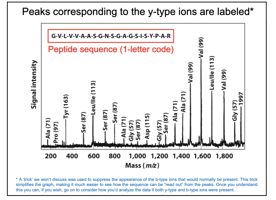
Sequencing using Mass Spec
1) purify to homogeneity
2) digest the protein into shorter peptides using protease
3) separate and sequence the resulting fragments using tandem mass spec (MS/MS)
B fragment vs Y fragment
B = N term
Y = C term
How to read peaks corresponding to y-type ions
we are seeing y-type ions
if peptide isn’t broken then will have largest mass (right side of graph)
if next signal is says Gly(57), that means a Glycine was lost
read amino acid sequence from right to left
peptide bond rotation
no freedom of rotation around peptide bond bc of resonance so can’t rotate a double bond
peptide bonds are ____
planar, there are 6 atoms in a single plane

which bonds can rotate in an amino acids
2 bonds adjacent to alpha C
phi Φ connects to C-N
psi ψ connects to C-C

ramachandran plot
many Φ and ψ angles are forbidden bc of steric clashes
dark blue areas show allowed angles

Why can Gly residues exist outside of normal regions in Ramachandran plot?
no big side chain to cause steric clash
Why are Pro residues confined to highly restricted areas?
bc the R group is covalently bonded to the backbone nitrogen
a-helix
C=O and N-H do H-bonding every 4 residues
no steric clashes
peptide bonds are planar
more H-bonds = more stable
helix is not hollow
right-handed
R groups come out
proline can’t form these
Glycine can’t form these bc has too much conformational flexibility so wants to be coiled
fibrous proteins summary
long strands or sheets
usually a single type of 2° structure and 3° is relatively simple
insoluble in water bc many hydrophobic amino acid residues
strength and flexibility
Why does collagen require a Gly at every third position?
Gly is at the very tight junctions between individual a-chains
1st x-ray crystal structure
myoglobin
made of a-helices
b-sheet
C=O forms H-bonds with N-H neighbor
consist of two or more strands
can be flat or twisted
no steric clashes + ideal angles
can be antiparallel or parallel
H-bonds in line when antiparallel
R groups come out
Hydrophobic Effect
biggest contribution to protein stability
fibrous proteins types
a-keratin
dimer of one long a-helix
collagen
trimer of left-handed helices
not made of a-helices
globular proteins
spherical shape
several types of 2° structure
ex: myoglobin
intrinsically disordered
no hydrophobic core
high density of charged amino acids
functional promiscuity