Global Brain Activity
 Call Kai
 Learn
 Practice Test
 Spaced Repetition
 Match
 Flashcards
 Knowt Play
1/27

There's no tags or description
Looks like no tags are added yet.
28 Terms
What are Brain Rhythms?
Rhythmic environment: temperature, day and night, tides…
Brain rhythms: sleeping and waking, breathing cycles, steps of walking, stages of night sleep
What is an Electroencephalogram?

How are Small Fields in Pyramidal Cells Generated?

What are the Basic Requirements for Signal Detection?
Basic requirements for signal detection:
1) A whole population of neurons must be active in synchrony to generate a large enough electrical field at the level of the scalp.
2) This population of neurons must be aligned in a parallel orientation so that they summate rather than cancel out.
What is Synchronous Activity?

EEG/ Brain Rhythms Correlate with Pathology and Behavioural States

Four Basic EEG Phenomena

How are Synchronous Rhythms Generated?

Generation of Synchronous Rhythms- One Neuron Oscillator

What are the Functions of Brain Rhythms?

What are the Behavioural Criteria that Classify Sleep?
Behavioural criteria:
Reduced motor activity
Decreased response to stimulation
Stereotypic postures
Relatively easy reversibility
How are Dolphins Adapted to Predators?

What are the Three Functional States of Sleep?

Summary Scheme of Sleep-wake States


What are the Functions of Sleep and Dreaming?
Conservation of metabolic energy
Cognition
Thermoregulation
Neural maturation and mental health
Question 1

Imaging Techniques- Structural Imaging
A. Structural imaging: Measures of the spatial configuration of types of tissue in the brain (static maps)
1.Computerised tomography, CT
2.Magnetic resonance imaging, MRI

Imaging Techniques- Functional Imaging
B. Functional imaging: Measures the moment-to-moment variable characteristics of the brain that may be associated with changes in cognitive processing (dynamic maps)
1.Positron emission tomography, PET
2.Functional magnetic resonance imaging, fMRI
Structural Imaging- CT Scans
CT scans
Based on the amount of X-ray absorption in different types of tissue.
Bone absorbs the most (the skull appears white), cerebrospinal fluid absorbs the least (the ventricles appear black) and the brain matter is intermediate (grey).
Used in clinical settings, e.g. to diagnose tumours or identify haemorrhaging or other gross brain anomalies.
Structural Imaging- MRI
MRI
2003 Nobel prize to Sir Peter Mansfield and Paul Lauterbur.
Completely safe, so people can be scanned many times.
Provides a much better spatial resolution.
Provides better discrimination between white and grey matter.
Can be adapted for detecting the changes in blood oxygenation associated with neural activity (fMRI).
What are the Sequence of Events in the Acquisition of an MRI Scan?
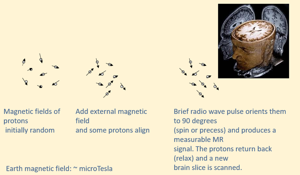
What is the Basic Physiology Underpinning Functional Imaging?

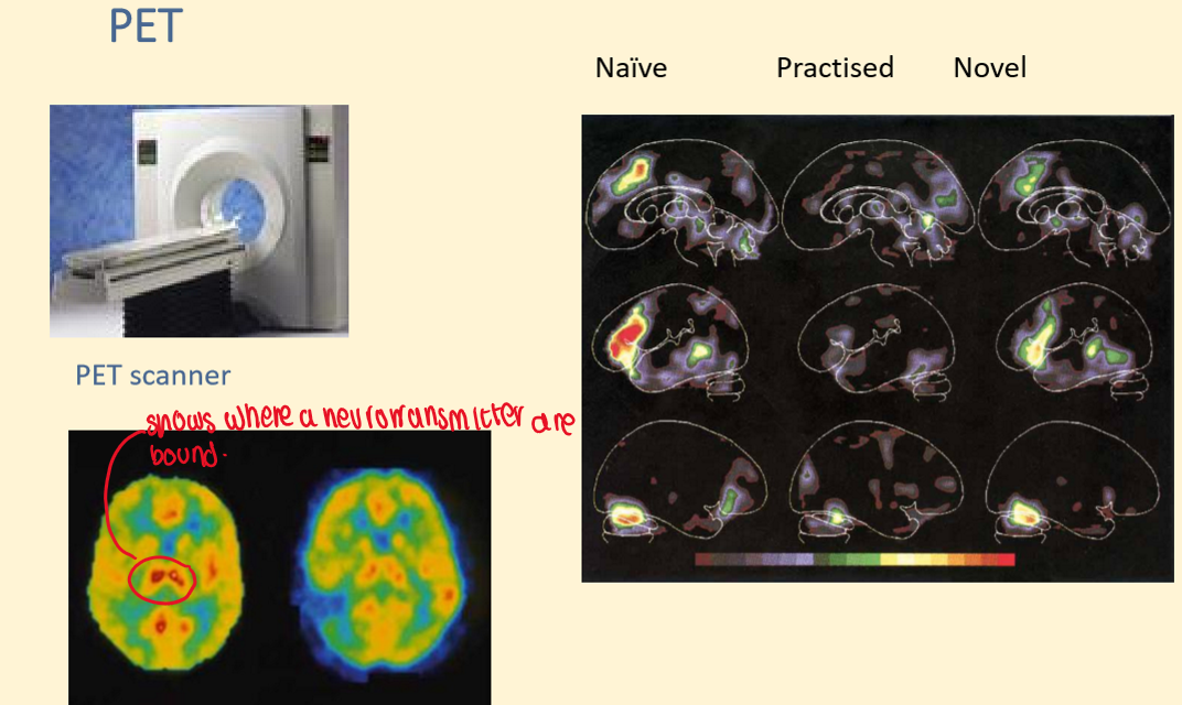
Example of PET Scans
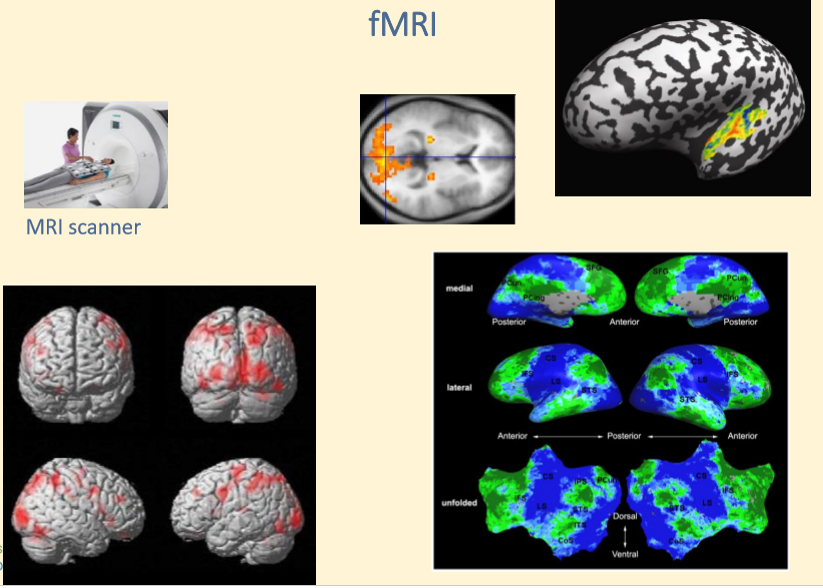
Example of fMRIs
Functional Imaging- PET
PET
Based on blood volume
Involves radioactivity (signal depends on radioactive tracer)
Participants scanned once or few times
Temporal resolution: 30”
Effective spatial resolution: 10mm
Sensitive to the whole brain
Can use pharmacological tracers
Functional Imaging- fMRI
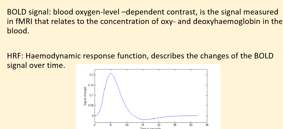
BOLD) fMRI
Based on blood oxygen concentration
No radioactivity (signal depends on deoxyhaemoglobin levels)
Participants scanned many times
Temporal resolution: 1-4”
Spatial resolution: 1mm
Some brain regions (e.g. near sinuses) are hard to image
How does fMRI Work?
Imaging Techniques Summary
