Chapter 6: Bone Tissue and the Skeletal System
 Call Kai
 Learn
 Practice Test
 Spaced Repetition
 Match
 Flashcards
 Knowt Play
1/60

Earn XP
Description and Tags
Define bone, cartilage, and the skeletal system; list and describe the functions of the skeletal system; classify bones according to their shapes; describe the function of each category of bones; identify the anatomical features of a bone; define and list examples of bone markings; describe the histology of bone tissue; compare and contrast compact and spongy bone; identify the structures that compose compact and spongy bone; describe how bones are nourished and innervated
61 Terms
Bone
also called osseous tissue, is a living, mineralized connective tissue that makes up most of the skeleton; its matrix contains collagen fibers, which help it resist pulling forces, and calcium phosphate crystals, which give it hardness
Cartilage
a firm, flexible connective tissue; its cells, called chondrocytes, maintain a water-rich matrix that helps cartilage withstand pressure
Skeletal System
the organ system made up of bones, cartilage, joints, and ligaments; it provides the body’s framework, protects internal organs, and works with muscles to produce movement
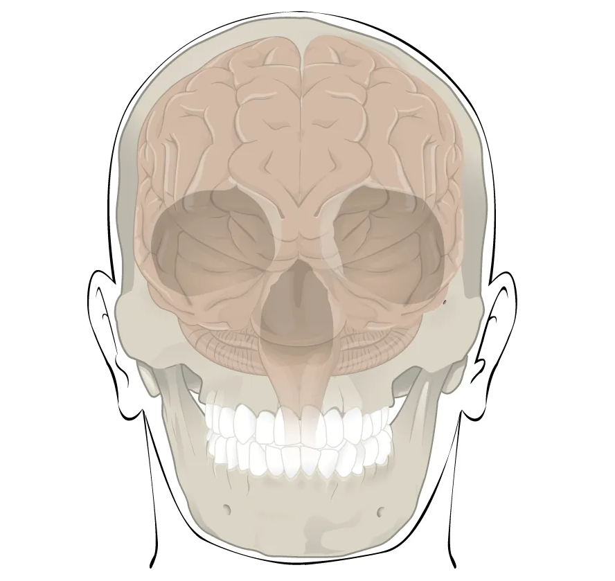
example of what?
bones protecting the brain
Orthopedist
also called an orthopedic surgeon, is a physician who diagnoses and treats injuries and disorders of the musculoskeletal system, including the bones, joints, muscles, tendons, and ligaments; use both surgical and nonsurgical treatments
Yellow Marrow
a type of bone marrow made mostly of adipocytes (fat cells); it’s found primarily in the medullary cavity of adult long bones, such as the femur, and its main function is to store fat as an energy reserve
Red Marrow
also called red bone marrow, is the soft tissue inside certain bones
Hematopoiesis
the process of forming red blood cells, white blood cells, and platelets; in adults, it occurs mainly in red bone marrow, where hematopoietic stem cells divide and give rise to all three

example of what?
head of femur showing red and yellow marrow
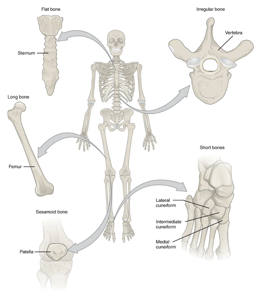
examples of what?
examples of classifications of bones
Long Bone
a bone that is longer than it is wide
it has a central shaft called the diaphysis and two ends called epiphyses
the shaft contains a medullary cavity that typically holds yellow marrow in adults
Function of the Long Bone
Leverage
Examples of Long Bones
femur, tibia, fibula, metatarsals, humerus, ulna, radius, metacarpals, phalanges
Short Bone
a roughly cube-shaped bone that is about as long as it is wide; it has a core of spongy bone surrounded by a thin layer of compact bone
Functions of the Short Bone
provide stability and support while allowing for some motion
Examples of the Short Bone
carpals and tarsals
Flat Bone
a thin, flattened, often slightly curved bone; it typically has two layers of compact bone with a layer of spongy bone between them
Functions of the Flat Bone
points of attachment for muscles; protectors of internal organs
Examples of the Flat Bone
sternum, ribs, scapulae, cranial bones
Irregular Bone
a bone with a complex shape that falls outside the long, short, flat, and sesamoid categories
Function of the Irregular Bone
protect internal organs
Examples of the Irregular Bone
vertebrae and facial bones
Sesamoid Bone
a bone embedded within a tendon, usually where the tendon crosses a joint; it helps protect the tendon from pressure and can improve the muscle’s leverage at the joint
Function of the Sesamoid Bone
protect tendons from compressive forces
Example of the Sesamoid Bone
Patellae
Diaphysis
the shaft, or long middle portion, of a long bone; it lies between the two ends, called epiphyses
Epiphysis
(plural epiphyses) is the expanded end of a long bone; each long bone has a proximal epiphysis closer to the trunk and a distal epiphysis farther from the trunk
Medullary Cavity
also called the marrow cavity, is the hollow space inside the diaphysis (shaft) of a long bone; it’s surrounded by compact bone and lined by a thin membrane called the endosteum
Compact Bone
also called cortical bone, is the dense, hard tissue that forms the outer layer of bones; it’s especially thick in the diaphysis (shaft) of long bones, where it surrounds the medullary cavity
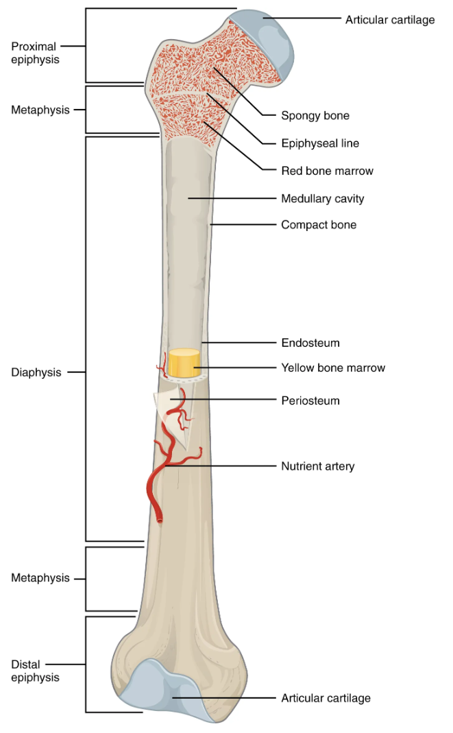
example of what?
anatomy of a long bone
Epiphyseal Plate
also called the growth plate, is a layer of hyaline cartilage in the metaphysis, between the shaft and end of a growing long bone
cartilage cells multiply on the side closer to the epiphysis (end), while cartilage is replaced by bone on the side closer to the diaphysis (shaft)
this process allows the bone to grow in length
Endosteum
a thin membrane that lines the internal surfaces of bone, including the medullary cavity, the trabeculae of spongy bone, and the canals within compact bone
Periosteum
a tough connective tissue membrane that covers the outer surface of a bone, except where articular cartilage covers the bone at a joint
Articular Cartilage
the smooth layer of cartilage that covers the ends of bones where they meet in a synovial joint, such as the knee; it reduces friction as the bones move and distributes pressure across the joint
Diploë
the layer of spongy bone between the inner and outer layers of compact bone in the flat bones of the skull; it consists of a network of thin bony struts called trabeculae, with marrow in the spaces between them
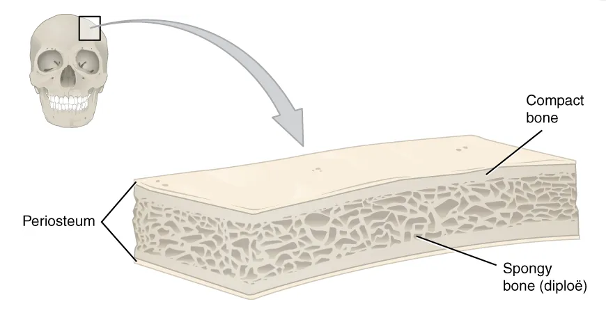
example of what?
anatomy of a flat bone
Articulation
also called a joint, is the place where two or more bones meet; some articulations allow a wide range of motion, like the shoulder joint, while others allow little or no movement, like the sutures between skull bones
Projection
a raised or protruding feature on the surface of a bone; projections can serve as attachment sites for muscles, tendons, or ligaments, or they can help form a joint
Hole
an opening in a bone
Articulator Bone Markings
features on a bone’s surface that meet another bone to form a joint; at synovial joints, these surfaces are usually covered by articular cartilage to reduce friction during movement
Example of Articular Bone Markings
knee joint
Head Bone Markings
a rounded, expanded articular surface at the end of a bone, often supported by a narrower region called the neck; it meets another bone to form a joint
Example of Head Bone Markings
head of femur
Facet Bone Markings
a smooth, nearly flat articular surface on a bone; it meets a matching surface on another bone to form a joint
Example of Facet Bone Markings
vertebrae
Condyle Bone Markings
a rounded, smooth projection, usually at the end of a bone, that forms part of a joint
Example of Condyle Bone Markings
occipital condyles
Projection Bone Markings
a raised feature that extends outward from the surface of a bone; some projections provide attachment sites for muscles, tendons, or ligaments, while others help form joints
Example of Projection Bone Markings
spinous process of the vertebrae
Protuberance Bone Markings
a bony prominence that bulges outward from the surface of a bone; it can serve as an attachment site for muscles or ligaments
Example of Protuberance Bone Markings
chin
Process Bone Markings
a projection that extends from a bone; it can serve as an attachment site for muscles or ligaments, or it can help form a joint
Example of Process Bone Markings
transverse process of vertebra
Spine Bone Markings
a sharp, slender, pointed projection from the surface of a bone; it can serve as an attachment site for muscles or ligaments
Example of Spine Bone Markings
ischial spine
Tubercle Bone Markings
a small, rounded projection on the surface of a bone; it often serves as an attachment site for a tendon or ligament
Example of Tubercle Bone Markings
tubercle of humerus
Tuberosity Bone Markings
a large, rounded, often rough projection on the surface of a bone; it typically serves as an attachment site for a tendon or ligament
Example of Tuberosity Bone Markings
deltoid tuberosity
Line Bone Markings
a low, narrow ridge on the surface of a bone; it often serves as an attachment site for muscles or other connective tissues