Med Term Chapter 12 - Eye
 Call Kai
 Learn
 Practice Test
 Spaced Repetition
 Match
 Flashcards
 Knowt Play
1/132

There's no tags or description
Looks like no tags are added yet.
133 Terms
eye
organ of vision
eyelids
folds of skin, soft tissue, and a thin layer of muscle located above and below the eye; protect the front of the eye and keep its surface moist by distributing tear film (mixture of tears and oil)
meibomian glands
(mī-BŌ-mē-an)
oil glands found in the upper and lower edges of the eyelids that help lubricate the eye (also called tarsal glands)

lacrimal apparatus
network of glands, ducts, canals, and sacs that produce and drain tears;
the lacrimal gland produces tears, which then flow through the lacrimal ducts to cover the surface of the eye. Tears drain into lacrimal canals, flow into the lacrimal sac (tear sac) and then into the nasolacrimal duct, which opens into the nasal cavity.
conjunctiva
mucous membrane lining the eyelids and covering the anterior portion of the sclera
sclera
outer protective layer of the eye; the portion seen on the anterior portion of the eyeball is referred to as the white of the eye
cornea
transparent anterior part of the sclera, which is anterior to the aqueous humor and lies over the iris. It allows the light rays to enter the eye.
uvea
layer of tissue beneath the sclera and cornea; contains the choroid, iris, and ciliary body
choroid
middle layer of the eye, which is interlaced with many blood vessels that supply nutrients to the eye
iris
pigmented muscular structure that regulates the amount of light entering the eye by controlling the size of the pupil
ciliary body
connects the choroid to the iris; produces aqueous humor and helps change the shape of the lens for focusing
pupil
opening in the center of the iris
lens
lies directly behind the pupil; its function is to focus and bend light
retina
innermost layer of the eye, which contains the vision receptors
macula
small portion of the retina at the back of the eye; responsible for detailed central vision
optic nerve
cranial nerve that carries visual impulses from the retina to the brain
aqueous humor
watery liquid found in the anterior cavity of the eye. It provides nourishment to nearby structures and maintains shape in the anterior part of the eye.
vitreous humor
jellylike substance found behind the lens in the posterior cavity of the eye that maintains its shape
aphakia
condition of without a lens (may be congenital, though often is the result of extraction of a cataract without the placement of an intraocular lens)
blepharitis
inflammation of the eyelid
blepharoptosis
drooping of the eyelid (also called ptosis)
conjunctivitis
inflammation of the conjunctiva (also called pink eye)
dacryocystitis
inflammation of the tear (lacrimal) sac
diplopia
double vision
endophthalmitis
(en-dof-thal-MĪ-tis)
inflammation within the eye
(Note: the o in the prefix endo- is dropped.)
iridoplegia
paralysis of the iris
iritis
inflammation of the iris
keratitis
inflammation of the cornea
keratomalacia
softening of the cornea (usually a bilateral condition associated with vitamin A deficiency)
oculomycosis
abnormal condition of the eye caused by a fungus
ophthalmalgia
pain in the eye
ophthalmopathy
(any) disease of the eye
ophthalmoplegia
paralysis of the eye (muscle)
phacomalacia
softening of the lens
retinoblastoma
tumor arising from a developing retinal cell (malignant, may be congenital; occurs mainly in children)
retinopathy
(any noninflammatory) disease of the retina (such as diabetic retinopathy)
scleritis
inflammation of the sclera
scleromalacia
softening of the sclera
uveitis
inflammation of the uvea
uveoscleritis
inflammation of the uvea and sclera
xerophthalmia
condition of dry eye (conjunctiva and cornea)
amblyopia
(am-blē-Ō-pē-a)
reduced vision in one eye caused by disuse or misuse associated with strabismus, unequal refractive errors, or otherwise impaired vision.
The brain suppresses images from the impaired eye to avoid double vision. (also called lazy eye)
anisometropia
(an-i-sō-ma-TRŌ-pē-a)
significant unequal refractive error between two eyes

astigmatism (Ast)
blurred vision caused by irregular curvature of the cornea or lens. Light refracts improperly, resulting in diffused, rather than points of light focusing on the retina. (Fig. 12.4C)

cataract
clouding of the lens of the eye
is derived from the Greek kato, meaning down, and raktos, meaning precipice. Together, the words were interpreted as waterfall. The individual with a cataract sees things as through a watery veil of mist or waterfall.
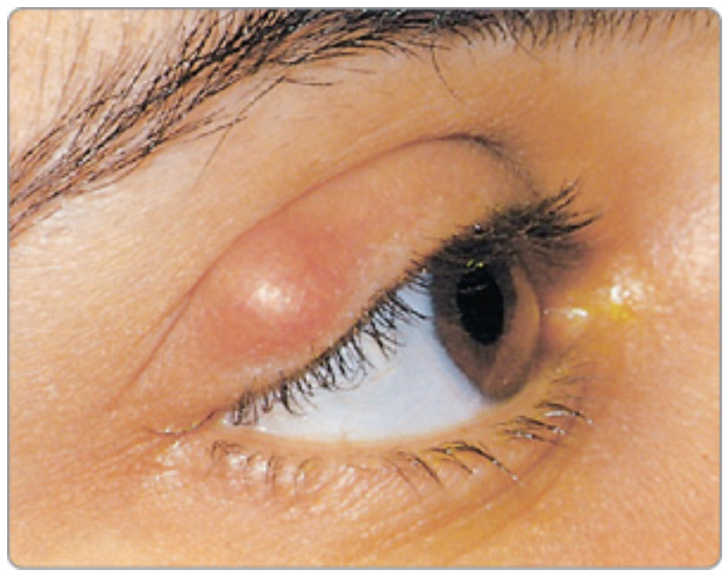
chalazion
(ka-LĀ-zē-on)
noninfected obstruction of an oil gland of the eyelid (also called meibomian cyst)

drusen
yellowish deposits located under the retina; commonly associated with aging and macular degeneration

glaucoma
eye disorder characterized by increase of intraocular pressure (IOP). If left untreated may progress to optic nerve damage and visual impairment or loss.
is composed of the Greek glaukos, meaning blue-gray or sea green, and oma, meaning a morbid condition. The term was given to any condition in which gray or green replaced the black in the pupil.

hyperopia
farsightedness

hyphema
hemorrhage within the anterior chamber of the eye; most often caused by blunt trauma (also called hyphemia)

keratoconus
(ker-a-tō-KŌ-nus)
progressive thinning of the cornea causing its normally rounded surface to bulge outward into a conelike shape and leading to vision impairment and, in some cases, vision loss; early symptoms, usually starting in the late teens and early twenties, include blurry vision and light sensitivity

macular degeneration
progressive deterioration of the portion of the retina called the macula, resulting in loss of central vision.
Age-related macular degeneration (AMD) is the leading cause of legal blindness in persons older than 65 years; onset occurs between the ages of 50 and 60.

myopia
nearsightedness
nyctalopia
(nik-ta-LŌ-pē-a)
poor vision at night or in faint light (also called night blindness)
nystagmus
(nis-TAG-mus)
involuntary, jerking movements of the eyes

pinguecula
(ping-GWEH-kū-la)
yellowish mass on the conjunctivea that may be related to long-term exposure to ultraviolet light, dry climates, and dust. A pinguecula that spreads onto the cornea becomes a pterygium.
presbyopia
(pres-bē-Ō-pē-a)
impaired vision as a result of aging

pterygium
(te-RIJ-ē-um)
thin tissue growing onto the cornea from the conjunctiva, usually caused by sun exposure

retinal detachment
separation of the retina from the choroid in the posterior portion of the eye resulting in a disruption of vision that may be permanent if treatment is delayed; onset may be gradual or sudden and is not painful

retinitis pigmentosa (RP)
group of hereditary diseases marked by the destruction of retinal cells that causes low vision and blindness; usually the first symptom is impairment of night vision, followed by loss of peripheral vision, and then loss of central vision

strabismus
(stra-BIZ-mus)
condition in which the eyes look in different directions; caused by dysfunction of the external eye muscles or an uncorrected refractive error (called cross-eyed when one eye turns in)

sty
infection of an oil gland of the eyelid (also spelled stye and also called hordeolum)
blepharoplasty
(BLEF-a-rō-plas-tē)
surgical repair of the eyelid

cryoretinopexy
(krī-ō-RE-tin-ō-pek-sē)
surgical fixation of the retina by using extreme cold (carbon dioxide)
dacryocystorhinostomy
(dak-rē-ō-sis-tō-rī-NOS-to-mē)
creation of an artificial opening between the tear (lacrimal) sac and the nose (to restore drainage into the nose when the nasolacrimal duct is obstructed or obliterated)
iridectomy
excision (of part) of the iris

iridotomy
incision into the iris

keratoplasty
(KER-a-tō-plas-tē)
surgical repair of the cornea (corneal transplant)
sclerotomy
incision into the sclera
enucleation
(ē-nū-klē-Ā-shun)
surgical removal of the eyeball (also, excision of a whole organ or mass without cutting into it)

LASIK (laser-assisted in situ keratomileusis)
(LĀ-sik) (ker-a-tō-mi-LOO-sis)
laser procedure that reshapes the corneal tissue beneath the surface of the cornea to correct astigmatism, hyperopia, and myopia.
It differs from photorefractive keratectomy (PRK) in that it reshapes corneal tissue beneath the surface rather than on the surface

phacoemulsification (PHACO)
method to remove cataracts in which an ultrasonic needle probe breaks up the lens, which is then aspirated

photorefractive keratectomy (PRK)
procedure for the treatment of astigmatism, hyperopia, and myopia in which a laser is used to reshape (flatten) the corneal surface by removing a portion of the cornea
posterior capsulotomy
laser procedure used to create an opening in the back center of the membrane holding an intraocular lens in place; performed to relieve vision impairment after cataract surgery (also called YAG laser capsulotomy)
retinal photocoagulation
intense beam of light from a laser condenses retinal tissue to seal leaking blood vessels, to destroy abnormal tissue or lesions, or to bond the retina to the back of the eye. Used to treat retinal tears, diabetic retinopathy, wet macular degeneration, glaucoma, and intraocular tumors.

scleral buckling
procedure to repair retinal detachment in which a piece of silicone is applied to the abnormal part of the sclera. This pushes in, or “buckles,” the sclera toward the middle of the eye, decreasing the traction on the retina. For larger detachments, an entire band may encircle the eyeball. Cryoretinopexy is often used to repair the hole in the retina prior to the buckling procedure.
trabeculectomy
surgical creation of an opening that allows aqueous humor to drain out of the eye to underneath the conjunctiva, where it is absorbed; used to treat glaucoma by reducing intraocular pressure. (Laser trabeculoplasty may also be used.)
vitrectomy
surgical removal of all or part of the vitreous humor (to provide access to interior structures of the eye; used to treat diabetic retinopathy, retinal detachment, or a hole in the macula)
fluorescein angiography (FA)
(flō-RES-ēn) (an-jē-OG-ra-fē)
radiographic imaging of blood vessels (of the eye with fluorescing dye)
keratometer
instrument used to measure (the curvature of) the cornea (used for fitting contact lenses)

ophthalmoscope
(of-THAL-mō-skōp)
instrument used for visual examination (of the interior) of the eye

ophthalmoscopy
(of-thal-MOS-ko-pē)
visual examination of the eye
optometry
measurement of vision (also measurement of the eye and visual processing system)
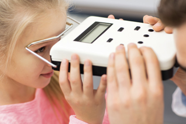
pupillometer
instrument used to measure (the diameter of) the pupil
pupilloscope
instrument used for visual examination of the pupil
retinoscopy
visual examination of the retina
tonometer
(tō-NOM-e-ter)
instrument used to measure pressure (within the eye, used to diagnose glaucoma)
tonometry
(tō-NOM-e-trē)
measurement of pressure (within the eye)

optical coherence tomography (OCT)
noninvasive test that uses light waves to take cross-sectional images of the retina; used to detect many eye conditions, including macular degeneration, glaucoma, retinopathy, and optic nerve damage.
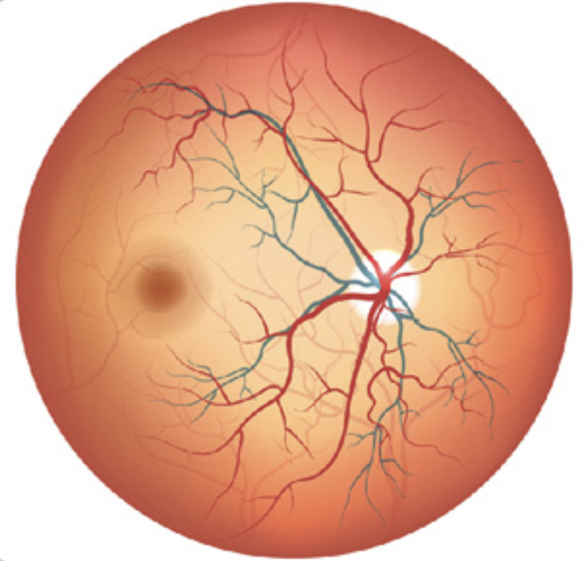
fundus examination
use of an ophthalmoscope to view the back of the eye, including the choroid, retina, macula, retinal blood vessels, and the optic disc (where the retina connects to the optic nerve); (also called fundoscopy)

slit lamp
horizontally mounted binocular microscope that uses a very narrow vertical beam of light (slit) to examine the eye in great detail. It is especially useful for viewing the conjunctiva, cornea, lens, and vitreous humor.

phoropter
instrument with many dials used to evaluate refractive errors and determine the lens curve and shape needed to correct vision. The patient reports which trial lens supports clearer vision in each eye, and then readings from the instrument are used to prescribe lenses.
anisocoria
(an-ī-sō-KŌR-ē-a)
condition of absence of equal pupil (size) (unequal size of pupils)
isocoria
(ī-sō-KŌR-ē-a)
condition of equal pupil (size)
leukocoria
condition of white pupil
photophobia
abnormal fear of (sensitivity to) light
pseudophakia
condition of false lens (placement of an intraocular lens during surgery to treat cataracts)
ophthalmologist
physician (surgeon) who studies and treats diseases of the eye
ophthalmology (Ophth)
study of the eye (branch of medicine that treats diseases of the eye)
binocular
pertaining to two or both eyes