Cardiovascular system
 Call Kai
 Learn
 Practice Test
 Spaced Repetition
 Match
 Flashcards
 Knowt Play
1/85

There's no tags or description
Looks like no tags are added yet.
86 Terms
heart
Acts like a pump and allows it to be both the lowest and highest pressure points, creating a pressure gradient within the heart and blood vessels. Allowing blood to flow out and into the heart
blood vessels/ vascular system
a set of tube or passageways for moving blood distributing nutrients and gases through the body
blood
A fluid connective tissue that contains dissolved substances ( things that are water soluble) and contains suspended substances (things that are insoluble and hydrophobic)

Explain the movement of blood around the heart


diastole
relaxation of the heart, the ventricles are filling up with blood, right and left AV valves are open, but the semilunar valves are closed


systole
heart contracting, specifically within the ventricular semilunar valves are open, but AV valves are closed ( done with blood in the atria)

when the atria are in systole and squeezing blood out of them …
ventricle is in diastole and receiving that blood
when the ventricle are in systole …
The atria are in diastole, receiving blood

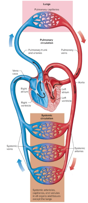
pulmonary circuit
carries oxygen-poor blood from the right ventricle to the lungs, where blood gets oxygenated and returns to the left atrium


systemic circuit
delivery oxygenated blood to all the systems
- oxygen-rich blood is carried from the left ventricle to all the organs of the body, and an exchange of O2 & CO2 occurs at every cell, and the oxygen-poor blood will return to the heart
Really long circuit and is under high pressure

endothelium
the inner layer of the heart that lines the inside of the chamber and blood vessels

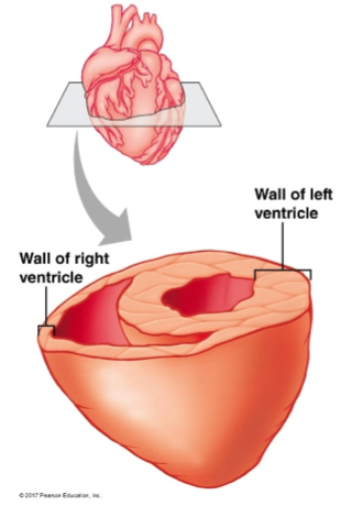
Myocardium
The thickest layer of the heart ( aka the muscular layer) generates the force needed to eject blood down stream into the blood vessels
More abundant in the left ventricle bc it leaves the heart to start the systemic circuit, which has more pressure
The left ventricle has the highest pressure due to its thick myocardial layer


epicardium
the outer dense connective tissue of the heart


coronary circulation
Blood circulation to the heart itself
The first branches of the aorta pass the aortic semilunar valve and perfuse the heart with oxygenated blood
You have to feed the heart before you feed any other parts of the body
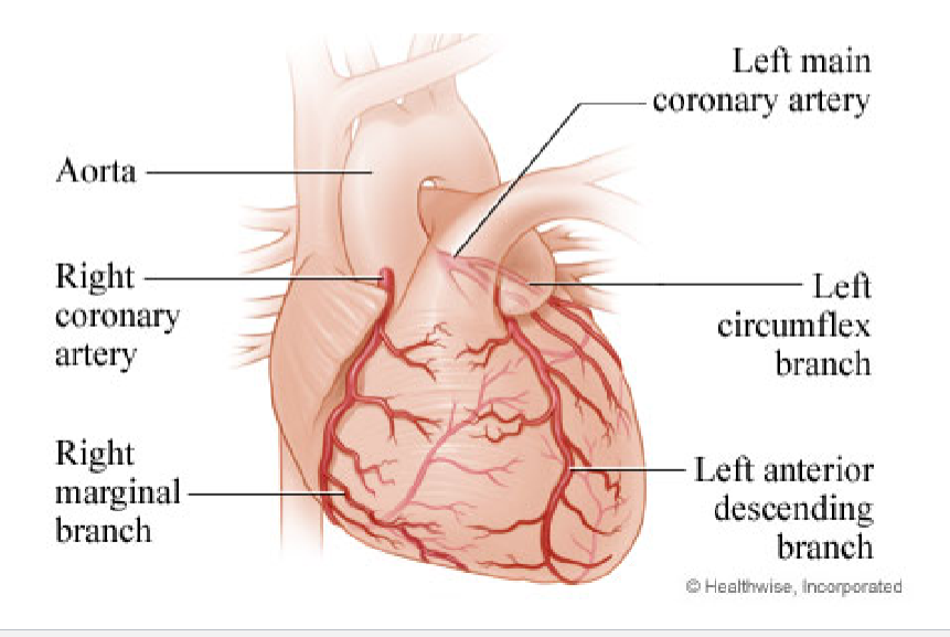
myocardial infraction ( aka a heart attack)
a blood clot in the coronary arteries that feed the heart with oxygenated blood
When cardiomyocytes die from this, they will not be replaced

the heart is autorhythmic meaning …
It can set its own rhythm and generate its own action potential, doesn’t need the NS to contract, but the autonomic NS can influence it to either increase or decrease its actions
contractile cells
Make their own AP and make up 90% of the cells in the heart
will have actin and myosin ( excitable cells 0
Contractible for the ventricle or chamber ( allowing the heart to act like a pump )
aka cardiomyocytes
autorhythmic cells
makes their own AP and is a percent of cells
Specialized and modified cells
They lack actin and myosin
Can initiate and conduct action potentials; tell contractile cells what to do, setting the pace and rhythm of the heart
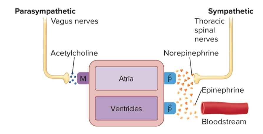
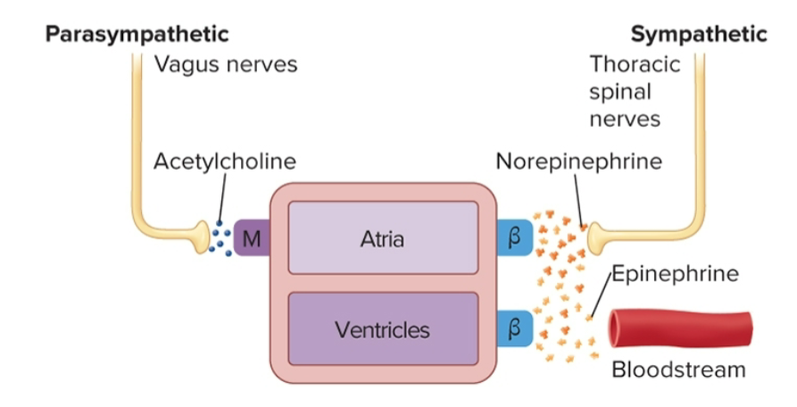
the autonomic NS can influence parasympathetic fibers …
through the vagus nerve will release ACh on muscarinic receptors loc only on autorhythmic cells
no parasympathetic NS on cardiomyocytes


The automatic NS can influence the sympathetic ganglion fibers …
by making them release norepinephrine, they innervate contraction cells of the ventricle & bind to autorhythmic cells
Epi produced by the adrenal medulla can bind to the same adrenergic receptor on the heart
Both epi and nori epi can bind to autorhythmic cells & contractile cells

myocardial cells
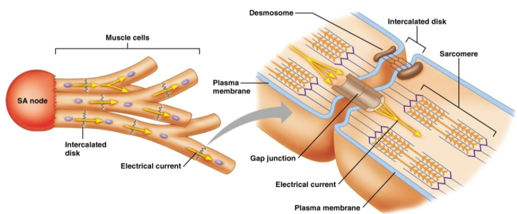
Cells that can contract and are branched
connected at intercolllated dics and connected by desomosomes


desmosomes
connect intercalated discs and help resist the mechanical stress put on the heart with each contraction
gap junctions
Used to connect adjacent cells, but they also provide a way for cells to communicate with one another tiny tunnels)
Allow the flow of AP from one cell to the next electrical synapse
allowing the heart to function in sync

Atria and ventricles have connective tissue between them, causing them to lack gap junctions ( CT tissue makes up for them). What does this do?
This allows the upper chamber and lower chamber to have a physical and functional separation
preventing the atria and ventricles from contracting like a big muscle but rather like a pump


The SA node spontaneously depolarizes at a rate of …
( 80-100AP /per min) The fasts of all the nodes and sets the pace of the heart, making it the pacemaker


If the SA node doesn’t work ….
The AV node will take over, but at a slower pace ( 30- 40 AP/min ) slower bc it usually uses AP from the SA node


Purkinjean fibers
The AV bundle splits and moves down the interventricular septum and innervates the left and right ventricles, specifically the myocardial layer


why is the HR not 80 -100 BPM at rest ?
At rest, the heart is in parasympathetic mode, making the parasympathetic NS more active, slowing down the nervous system to 70 BPM
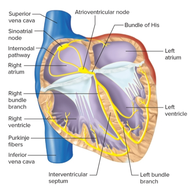

How AP spreads through the heart
When an SA node fires, it spreads from the right atrium through the internodal pathway and the interatrial pathway, allowing the atria to contract together through gap junctions
spreads like a wave through the atrial muscles, causing both of them to depolarize
When the AP reaches the AV node, it will cause the atria to contract at a unit
At the AV node, there will be a brief slowing of the conduction
From the AV node, it will go to the AV bundle, which carries it down through the left and right bundle branches to the Purkinje fibers, where it will travel up and depolarize the myocardium, causing the contraction of the ventricles
Ventricles contract from bottom to top

AV node transmission
From the AV to the ventricles, the only electrical connection between the atria and ventricles
Brief delay ( aka AV nodal delay = 0.1 sec) gives the atria time to fully contract and eject blood into the ventricles before they contract
ventricle excitation
The time the ventricles are depolarizing, and the wave of depolarization reaches the Purkinje fibers, causing the ventricle to contract from the bottom up

cardiac cells have, but their resting MP is …
-90 millivolts

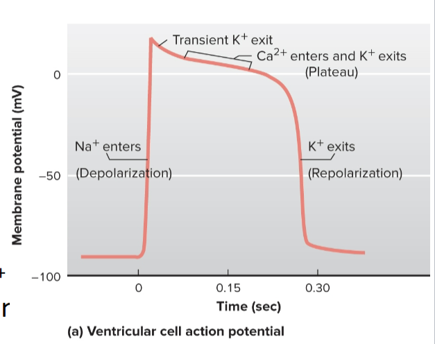
explain the Ap for the cardiac muscles
We open the VG Na+ channel on cardiomyocytes, and all the Na+ moves into the cell and causes rapid depolarization
At the peak of the AP (+30 mV), the VG Na+ channels close, and the VG K+ channels open, repolarizing the cell, but during this the L-type Ca2+ channel opens
The L-type channel lets in a positive charge (Ca) inside the cell, slowing down / pausing the repolarization of the cell, creating the plateau
The plateau creates a refractory period for the chambers, preventing the contraction of the heart to allow it to eject blood fully
When the plateau ends, the L-type Ca2+ channels close, stopping the movement of calcium into the cell, but during this process K+ channels are still open, so the cell repolarizes quickly, going back to -90mv

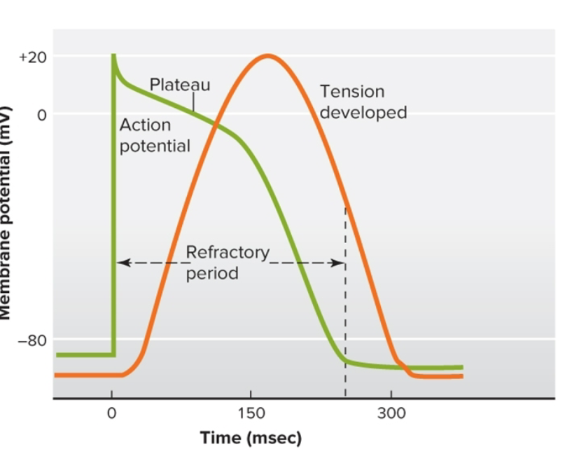
refractory period of cardiac cells
Lastly, as long as the muscle contraction prevents the summation of Ap
Contractile cells are contracting during the refractory period, but it’s not working due to the L- type channels bring in Ca
Without this period, the heart would be a constantly contracting muscle


autorhythmic cells are never at rest bc
There is no stable membrane potential


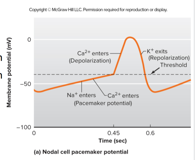
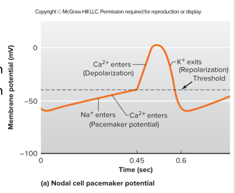
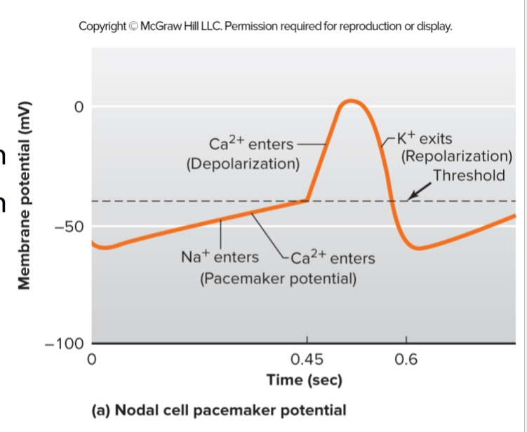
electrical activity of the autonomic cells (pacemaker cells)
When the Vg K+ channel closes at the end of repolarization, funny channels open
Funny channels allow both Na+ and K+ to cross the membrane, bringing more Na+ to repolarize the cell and K+ to slowly depolarize the cell, bringing it closer to threshold (getting us to about -50mv)
At -50 mV, the funny channels close, and sodium no longer moves into the cell, but another channel called a T-type Ca2+ channel opens for a short amount of time, continuing to depolarize, getting us to threshold, causing the opening of L-type Ca2+ channels, allowing Ca2+ to enter, finishing the depolarization of the cell

What do L type channel do in myocardial cells ?
these channel open at + 30 mv and while the K+ channels are repolarizing the cell
Their main job during the AP of myocardial cells is to cause a plateau, giving time for the ventricle to contract completely


What do L-type channels do for pacemaker cells
These channels open when we hit the threshold, allowing Ca2+ to move inn and finish the depolarizing event
funny channels
specialized Na+ channels that open when the membrane potential becomes more negative than the resting potential, allowing both Na+ and K+ to move, bringing it closer to the usual resting potential and closer to threshold


an impulse is passed to the adjacent cell through a gap junction, causing the
depolarization of another cell


2 sources of Ca+ for contractile cells
Ca+ in the sarcoplasmic reticulum
Ca+ in the extracellular fluid outside of the cell


Explain this photo
On the sarcoplasmic reticulum are ryanodine channels;
As an AP moves down the Cell MB and down the T-tubules, DHP receptors are coupled with ryanodine on the sacroplasmic reticulum will open alloing it to release Ca+ into the cyctosol
AP moving across the clal MB will also cayse the VG CA+ channels to open allowing Ca+ from the extracellular fluid to enter the cells as well
Ca+ from bpth f these pathways will start bind to typoin unloking tropinmyosin moving it and allping cross brige formation to take place causing a musle contraction


ryanodine channel
A channel that responds to changes in the DHP channel and causes the release of Ca+ from the sarcoplasmic reticulum


DHP receptors
A type of L-channel whose main job is to sense changes in voltage on the cell membrane and change shape, signaling the ryanodine receptors attached to it to release Ca+

ATP is required for
The power stroke: ATP helps bind the actin head to the myosin and then releases the phosphate
Detachment of myosin heads
The recocking of the heads - ATP hydrolysis returns the head to its cocked position

electrocortical gram ( ECG)
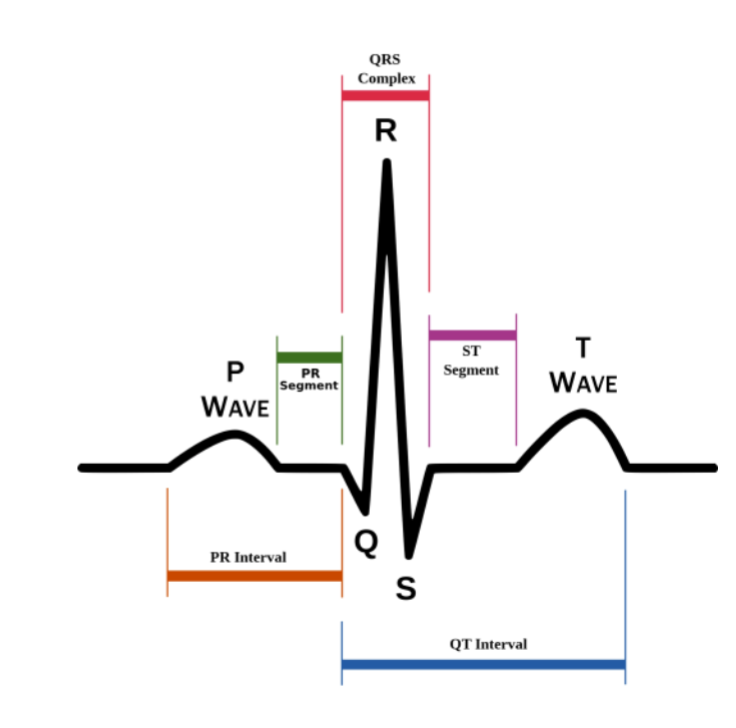
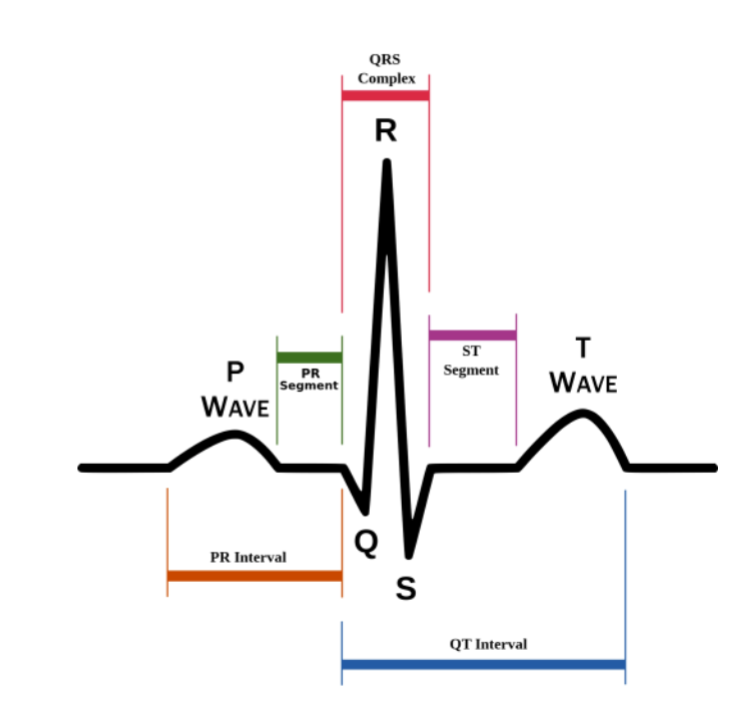
the recoding of all the electrical activity of the heart, which is picked up by electrodes that are placed on the surface of the body, giving information about the heal of the heart


isometrical line
The straight line where no electrical activity is talking place

P wave
an upward deflection from the baseline and represents the depolarization of the atria


QRS complex
Represents ventricular depolarization as significant electrical activity
Repolarization of the atria occurs here but is masked by these complex bc its not that big


T wave
the repolarization of the ventricles


PR segment
AV nodal delay, the phase that allows the atria to fully contract their blood into the ventricles


ST segment
the plateau, where the L-type Ca2+ channel opens


QT interval
The beginning of the QRS complex to the end of the T- wave
represents when the ventricles are contracting and are in systole to the end of ventricular repolarization

R-R interval
The time between each heartbeat, the HR distance will be short; the slow HR distance will be long

where is atria depolarization ?
in the QRS complex, but it’s not really seen bc its not a significant electrical event

Explain this EKG
A node fires, creating the P wave and spreading to both atria
AP hits the AV node (isoelectric line before the QRS complex; brief delay ) during the PR segment; no electrical activity, but the atria are contracting
QRS complex: the AP moves down the AV node to the bundle, then to the Purkinje fibers
Atria are also repolarizing
Ventricles complete their depolarization but are still contracting
T-waves: ventricles are starting to repolarize
T wave ends, and repolarization is complete, and we return to the isoelectric line (ventricular diastole)
Giving the SA node time to send an AP


partial AV block
AP to the ventricle doesn’t get conducted properly; a block at the AV node causes some of the AP to not be conducted and spread properly into the ventricle
Atria depolarize but something goes missing


complete AV block
The AV node doesn’t conduct APs at all, so the AV bundle ( aka the bundle of His) has to take over and cause the ventricle to contract
HR becomes very slow and is very dangerous bc it might not be able to pump enough blood around the body


Normal HR is ..
70 BPM


arrhythmia
an aabnomal HR


bradycardia
slow rhythm ( 60 BPM )
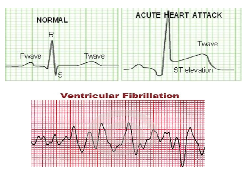

Tachycardia
fast rhythm ( 100 BPM )


Asystole
very dangerous, means the ventricles are not going into systole and are not contracting, causing a quick death
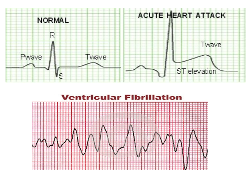

When cardiac cells are injured, they release …
creatin kinases & troponin into the blood, which can be checked in the blood to confirm a heart attack

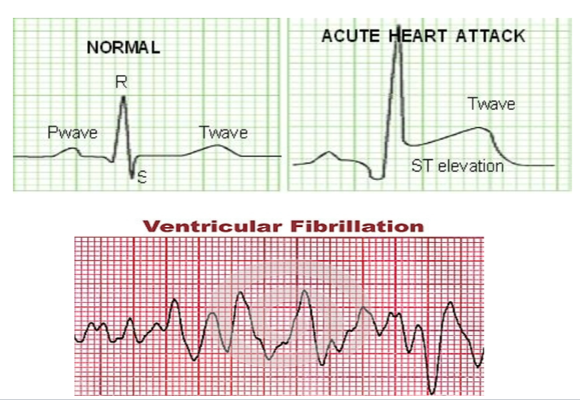
fibrillation
an uncontrolled and irregular depolarization of the heart muscles

atria fibrillation
will cause multiple fibrillation of the P waves, but the atria will still be able to pump blood into the ventral bc its downstream and doesn’t require a lot of force, and the ventricles are working properly


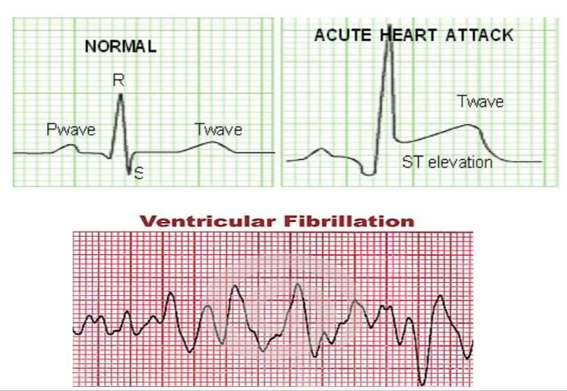
ventricle fibrillation
Depolarization of the ventricle becomes very erratic and can cause death

cardiac cycle
The events of a single heartbeat ( 0.8)
ventricular relaxation allowing the ventricle to receive blood
ventricular contraction allowing the ventricle to eject its blood
When atrial pressure is high, then the pressure in the ventricles
the AV valve opens
At the end of ventricular filling, the ventricular pressure is high, then the arterial pressure leading to
the semilunar valves opening

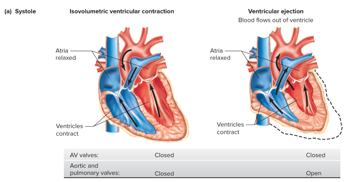
isovolumetric phase
the time they are just starting to contract, the tension in the muscle is starting to develop, but as soon as the pressure goes slightly above the atrial pressure, the AV valves will shut, but the pressure within the ventricle is not enough to open the semilunar valves
All the valves are closed at this point, and no blood is leaving or coming in

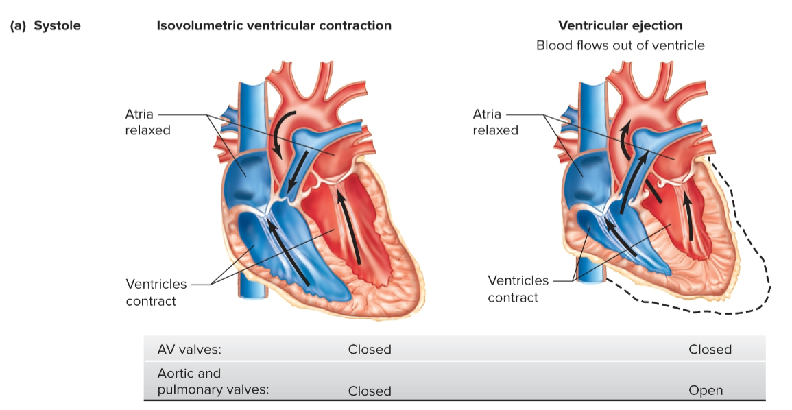
Ventricular ejection
When the pressure of the ventricle is higher than the atria, keeping the AV valves closed and higher than the atria, opening the semilunar valves

isovolumetric relaxation
As soon as the ventricle starts relaxing, in early diastole the pressure in the ventricle starts to decrease, and the semilunar valves are going to shut; AV valves still shut bc the ventricular pressure is still higher than the atrial pressure
All valves are closed


Ventricular fillingves are closed
as pressure continues to decrease, long phase of the cardiac cycle
In early filling, the ventricles are in diastole and are filling with blood, and at the same time blood is returning into the atria. Blood fills bc BP in the atria is less than BP in the veins ( inferior vena cava); the valves are opening due to the beginning part of ventricular filling, but the semilunar vale

80% of the blood moving through the atria passively flows down into the open AV valves into the ventricles; the last 20% of blood from the atria will...
move actively because that when the atria squeeze and release that last 20% of blood within them, ensuring the ventricles are completely full

pressure in the atria via pressure in the ventricle during a cardiac cycle
As soon as the atria go into diastole, the pressure on them decreases (to its lowest ), causing the ventricle to enter systole and increase its pressure, leading to a contraction opening the semilunar valves, allowing blood to be ejected into the arteries
As blood leaves the ventricle, BP drops; most of the blood gets ejected. When the ventricular pressure drops below atrial pressure again the AV valves open again


ventricle fluctuates between 110 compared to arterial pressure, which doesn’t fluctuate that much because
they dont need to generate that much force as most of the blood in them passively moves into the ventricles
